Signals of MLCK and ROCK Pathways Triggered via Lymphotoxin β Receptor are Involved in Stress Fiber Change of Fibroblastic Reticular Cells
Dae Sik Kim1 and Jong-Hwan Lee1,2,3*
1Department of Biotechnology and Bioengineering, College of Engineering, Dong-eui University, Busan 47340, Korea
2Biomedical Engineering and Biotechnology Major, Division of Applied Bioengineering, College of Engineering, Dong-eui University, Busan 47340, Korea
3Department of Smart-Biohealth, College of Engineering, Dong-eui University, Busan 47340, Korea Received October 26, 2018 /Revised December 8, 2018 /Accepted December 10, 2018
Lymphotoxin β receptor (LTβR), a member of the tumor necrosis factor receptor family, plays an im- portant role in lymphoid tissue’s architecture and organogenesis. In contrast, MLCK and ROCK play critical roles in the regulation of stress fiber (SF) formation in cells. To determine whether LTβR stim- ulation in fibroblastic reticular cells (FRCs) is involved in these signaling pathways, myosin light chain kinase inhibitor-7 (ML-7) was used to inhibit them. ML7-treated FRCs completely blocked SFs and showed retraction and shrinkage processes comparable to those observed in agonistic anti-LTβR anti- body-treated cells. The inhibition of ROCK activity with Y27632-induced changes in actin cytoskeleton organization and cell morphology in FRCs. Actin bundles rearranged into SFs, and phospho-myosin light chain (p-MLC) co-localized in FRCs. We checked the level of Rho-guanosine diphosphate (Rho- GDP)/guanosine triphosphate (GTP) exchange activity using FRC lysate. When LTβR was stimulated with agonistic anti-LTβR antibodies, Rho-GDP/GTP exchange activity was markedly reduced. Regarding LTβR signaling with a focus on MLCK inhibition, we showed that the phosphorylation of MLCs was reduced by LTβR stimulation in FRCs. Cytoskeleton components, such as tubulin, b-actin, and phos- pho-ezrin proteins acting as membrane-cytoskeleton linkers, were produced in de-phosphorylation, and they reduced expression in agonistic anti-LTβR antibody-treated FRCs. Collectively, the results suggested that MLCK and ROCK were simultaneously responsible for SF regulation triggered by LTβ R signaling in FRCs.
Key words : FRC (Fibroblastic reticular cell), LTβR (Lymphotoxin β receptor), MLCK, ROCK, SF (stress fiber)
*Corresponding author
Tel : +82-51-890-2280, Fax : +82-505-182-6897 E-mail : [email protected]
This is an Open-Access article distributed under the terms of the Creative Commons Attribution Non-Commercial License (http://creativecommons.org/licenses/by-nc/3.0) which permits unrestricted non-commercial use, distribution, and reproduction in any medium, provided the original work is properly cited.
Journal of Life Science 2019 Vol. 29. No. 2. 256~264 DOI : https://doi.org/10.5352/JLS.2019.29.2.256
Introduction
The primary role of the lymph node (LN) is to coordinate immune reactions to antigens trafficking from peripheral microenvironments. LNs are specialized to trap anti- gen-bearing dendritic cells and to facilitate the initiation of adaptive immune response. During immune responses, LN carries out the rapid and extensive expansion [1]. LN is com- posed of aggregations of lymphocytes in a framework of non-leukocyte stromal cells, which provide both the basic structural organization of the tissue and survival signals to help sustain the life of the lymphocytes [17]. LN stromal
cells are being recognized as key organizers of the LN expansion. The stromal cells of LN subsets assemble the ar- chitecture and network necessary to guide cellular traffick- ing and organization, facilitate antigen presentation to circu- lating naïve T and B cells and thereby promote immune sur- veillance against infection [14]. Fibroblastic reticular cells (FRC) are LN-resident stromal cells that secrete and remodel extracellular matrix to construct a complicated reticular mesh that filter draining lymph [21]. In addition, FRCs are responsible for the production and presentation of chemo- kines such as CCL19/CCL21 to coordinate lymphocyte traf- ficking into and throughout the LN [8] and presentation of pro-survival cytokines such as interleukin (IL)-7 and IL-15 to T cells [5, 12] and B-cell activating factor of the tumor necrosis factor (TNF) family (BAFF) to B cells [9].
Mammalian cells can generate traction forces against solid extra cellular supports by assembling contractile stress fibers (SFs), which are bundles of filamentous actin (F-actin), ac- tin-binding proteins, and nonmuscle myosin II (NMMII)
[10]. These traction forces are crucial to a variety of funda- mental cellular properties and behaviors, including motility, mechanosensing, shape stability, polarity, and fate determi- nation [10]. The assembly and mechanics of actomyosin SFs depend on myosin regulatory light chain (RLC) phosphor- ylation, which is driven by myosin light chain kinase (MLCK) and Rho-associated kinase (ROCK) [15]. MLCK is encoded by one gene and exists in two forms: long MLCK (~211 kDa) and short MLCK (~150 kDa) which lacks the N-terminal extension thought to be associated with actin [2].
Both MLCK forms directly phosphorylate RLC. On the other hand, ROCK promotes RLC phosphorylation either by direct phosphorylation of RLC or by phosphorylating and in- activating RLC phosphatase [15]. Precisely how these kin- ases differentially contribute to RLC phosphorylation re- mains unclear, with the few studies focused on this question producing differing results depending on the cellular system and method of perturbation.
Lymphotoxins (LTs) are cytokines related to TNFα, and function in organizing and maintaining lymphoid organs [11]. There are two LT subunits, soluble α and mem- brane-bound β, primarily found as a soluble homotrimer of α (LTα3) that binds TNF receptors, or a membrane-bound heterotrimer (LTα1β2) that interacts with the LT β receptor (LTβR) [13]. LTα1β2 is expressed on activated T, B and natu- ral killer cells [6, 13] and interacts with LTβR on dendritic cell (DC), monocyte lineage cells and stromal cells including FRC [11]. Thus, LTβR plays an important role in the remod- eling and maintenance of function of LNs [4, 5]. Treg LTαβ rapidly modulates cytoskeletal and membrane structure of lymphatic endothelial cells dependent on LTβR [3]. However, there are no reports which one is important factors involved on cytoskeletal alteration linked to LTβR in FRC. So far, many immunologic parameters have investigated in im- mune interaction, but little attention has focused on the SF alteration aspects of FRC triggered via LTβR. To understand these tasks, FRC was stimulated via LTβR with agonistic anti- LTβR antibody [16, 20, 24]. Here, in order to investigate SF regulation when FRC was treated with agonistic anti-LTβ R antibody, the cytoskeleton alteration, with particular em- phasis on SF, was studied in FRCs. And, our results point to a model in which MLCK and ROCK regulate SF elastic properties through phosphorylation of RLC, offering con- nections between kinase activity, RLC phosphorylation, and SF elasticity in FRC.
Materials and Methods
Cell culture
FRC was established as described previously [16]. FRC was maintained in 10% fetal calf serum–dulbeco's modified eagle's medium (DMEM) supplemented with streptomycin and penicillin.
Reagent and antibodies
The following antibodies or fluorescent probes were used in this study: Inhibitor Y27632 (Sigma-Aldrich, St. Louis, MO, US), rhodamine phalloidin (Cytoskeleton, Denver, CO, US), SuperSignal West Pico Chemiluminescent Substrate for detecting signal in Western blot (Thermo Scientific, Wal- tham, MA, US), pMLC (phospho Thr18/Ser19) primary anti- body (Cell Signaling, Danvers, MA, US), anti-myosin light chains (anti-MLCs) antibody (Cell Signaling), anti-ezrin (Upstate Biotechnology, Lake Placid, NY, US), anti–p-ERM (Cell Signaling), anti-rabbit and anti-goat peroxidase-con- jugated secondary antibody for Western blot (Cell Signal- ing). RhoA activation assay kit was from Cytoskeleton, Inc.
Polyclonal goat antibody against LTβR extracellular domain and normal IgG were purchased from R&D Systems.
Immunofluorescence microscopy
FRCs were treated with agonistic anti-LTβR antibody (100 ng/ml), as described above, were gently rinsed, fixed in 4%
formaldehyde in PBS for 5 min, washed with PBS, and stained with rhodamine-labeled phalloidin (5 μg/ml) for 45 min. For ROCK and MLCK inhibition, cells were pretreated with 5 mM Y-27632 (Calbiochem, San Diego, CA, US) or 10 mM ML-7 (Sigma), respectively, for 1 h before imaging. For immunofluorescence staining, cells were fixed with 3% PFA in PBS and permeabilized with 0.2% Triton X-100/PBS or 0.5% saponin/PBS. To detect the intracellular localization of p-ezrin we performed a TCA fixation method, which in- activates phosphatases and maintains the level of p-ERM proteins during sample processing [19], with a slight mod- ification. In brief, cells were fixed in suspension with 10%
TCA solution for 15 min at room temperature, and were rinsed three times with PBS. After being blocked with 5%
BSA-PBS, cells were stained with antibodies for 30~60 min.
The nucleus was visualized by staining with 0.1 μg/ml DAPI. Stained cells were mounted with Permafluor aqueous mounting medium (Immunotech, Monrovia, CA, US) and examined with a Zeiss photomicroscope equipped for fluo- rescence microscopy. Digital images were processed by
Rhodamine phalloidin DAPI MERGE
Fig. 1. FRC preserves cortical contractile ring and central stress fibers. Representative images of rhodamine-phalloidin staining for F-actin (red), and DAPI staining (blue) of FRC. Actin cytoskeletons in FRC are indicated with white arrows and head-arrow. Bar is 10 μm.
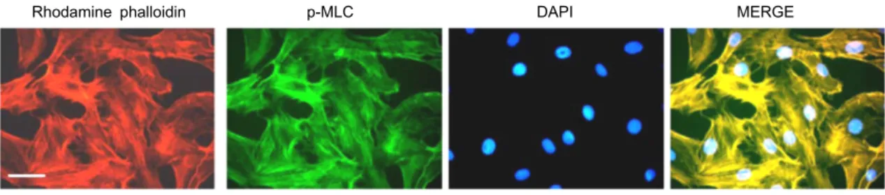
Rhodamine phalloidin p-MLC DAPI MERGE
Fig. 2. pMLC and actin co-localized in normal FRC. FRCs were treated with phalloidin and anti-pMLC antibody for 1 hr and examined for changes in F-actin distribution (phalloidin staining) and p-MLC. Bar is 10 μm.
Adobe Photoshop software.
Rho A pull-down assay
More than 3×10⁷ cells were lysed with a 2 ml RIPA buffer.
The amount of Rho-GTP in the reaction solution measured by a pull-down method based on the specific binding to Rhotekin-RBD followed by Western blotting using an an- ti-Rho antibody.
Immunoblot
FRCs were pretreated with agonistic anti-LTβR antibody (100 ng/ml) and were lysed in a 5× sodium dodecyl sulfate (SDS) sample buffer. After the samples were boiled, equal amounts of total lysates were separated by SDS-PAGE and transferred onto polyvinylidene difluoride membranes. The membranes were soaked in a blocking solution (5% skim milk and 0.2% Tween 20-PBS) for 1 hr, and then incubated with primary antibodies for 1 hr. After being washed with Tween 20-PBS, membranes were incubated with appropriate horse radish peroxidase (HRP)-conjugated secondary anti- bodies for 1 hr. Specific bands were visualized by an en- hanced chemiluminescence (ECL) method (ECL+; Amersham Biosciences, Piscataway, NJ, US).
Results
FRC preserves cortical contractile rings and central stress fibers
The ability of a cell to distribute contractile stresses across the extracellular matrix in a spatially heterogeneous fashion underlies many cellular behaviors, including motility and tissue assembly. It is possible to use the mushroom-derived fluorescinated toxins, phalloidin, to label F-actin of the SF, as is described in this article. The SFs containing F-actin can be broadly separated into two morphological shapes: thick and dense stress fibers, which are located in the peripheral portion of the cell (‘cortical actin’), and SFs, which are lo-
cated in the central portion of the cell (‘central SF’). To con- firm this subcellular compartmentalization of SF, we cul- tured FRCs on collagen-coated glass, and used immuno- fluorescence to examine the spatial distribution of SFs.
Phalloidin staining revealed that FRC contained a cortical contractile ring (Fig. 1, arrow) and central stress fibers (Fig.
1, head-arrow).
Co-localization of actin filaments and pMLC in SF structure of FRC
In cultured mammalian cells, SF structures are composed of antiparallel arrays of F-actin stabilized by actin-binding proteins and interleaved with nonmuscle myosin II (NMMII), contribute to cytoskeletal prestress by anchoring into cell-ECM adhesions and permitting the cell to generate trac- tion against the ECM [10]. To confirm the localization of pMLC and actin filaments in FRC, we used immuno-staining with anti-pMLC antibody. Fig. 2 illustrated the SF distribu- tion in a control monolayer as viewed by fluorescence microscopy. SFs formed thick cables and connected to the nuclear periphery with the processes of these flat FRCs (Fig.
2). Moreover, pMLC and actin filaments were co-localized into SF in normal FRC (Fig. 2).
Rhodamine DAPI MERGE phalloidin
Noraml IgG (36 hr)
Agonictic anti LTβR antibody (25 μg/ml)
Agonictic anti LTβR antibody (25 μg/ml)
Fig. 3. Agonistic anti-LTβR antibody disrupted the formation of SF in FRC. FRCs on chamber slides were treated with agonistic anti-LTβR antibody (25, 50 mg/ml) for 24 hr and examined for changes in F-actin distribution (phal- loidin staining). Bar is 10 μm.
Rhodamine phalloidin DAPI MERGE
ML7 (0 hr)
ML7 (12 hr)
ML7 (24 hr)
ML7 (36 hr)
Fig. 4. LTβR controls FRC spreading, elongation, and actomyo- sin contractility. FRC were treated with 5 μM ML7 in- cubated for indicated times. Actin SF completely abro- gated in ML7 treated FRC. Bar is 10 μm.
Stress fiber alteration in FRC via LTbR stimulation with agonistic anti-LTbR antibody
The delicate microstructure of the LN, which is largely supported by a reticular network of FRC and extracellular matrix, is essential for immune response. FRCs in the LN ensheath the thin strands of ECM, while they are also con- tinually in contact with immune cells [16]. Thus, FRCs are able to induce T cell chemotaxis and adhesion to the FRC surface [16]. The characteristic network made by FRCs ap- pears to be optimal for making spaces inside the LN for mo- tile T cells to move through. The membrane heterotrimeric form of LTa1b2 is expressed in T cells and dendritic cells, which is an important for lymphoid tissue architecture and organogenesis [18]. LTa1b2 activates a signaling pathway via LTbR [18]. It is known that several monoclonal antibodies raised against the extracellular part of LTbR mimic receptor engagement and are able to induce downstream signals [16].
We used an agonistic anti-LTβR antibody which is a com- mercially available polyclonal antibody against the extracel- lular part of LTβR to trigger signal cascades. Treatment with agonistic anti-LTβR antibody led to a significant decrease in the quantity of actin polymer compared with that in the control group and had a dose-independent effect on actin distribution and morphology (Fig. 3). In addition, when ago- nistic anti-LTβR antibody was treated to the flat FRC mono- layer, we could not detect the appearance of SFs, and FRC showed a striking morphological change with a circular arc and shrinked shape of their processes and empty space was made among FRCs, whereas no effect was induced by the
addition of a control antibody (Fig. 3). Those findings sug- gest that signaling via LTβR activation plays a critical role in the inhibition of actin cytoskeleton formation and mor- phological change in FRC.
Effects of a MLCK inhibitor, ML7, on SF disruption in FRCs.
There are two kinases playing a critical role on SF for- mation in mammalian cells; myosin light-chain kinse (MLCK) and Rho GTPase kinase (ROCK). To evaluate the involvement of the MLCK pathway on SF change and mor- phology alteration in FRC, we treated ML7, a specific in- hibitor of MLCK, in FRCs. After stimulation, cells were fixed and stained with rhodamine-conjugated phalloidin to visual- ize F-actin. The SF formation found in the control FRCs. In contrast, cellular alterations were readily apparent after 12 hr, 24 hr and 36 hr of treatment with ML7. ML7-treated FRCs were elongated and extended membrane protrusions by time dependent pattern. Each protrusion extending from the cell body of ML7-treated FRCs was markedly longer than those of wild-type FRCs. With retraction and shrink of proc- esses similar to those observed in agonistic anti-LTβR anti- body-treated FRCs, both of the central and peripheral SFs in ML7-treated FRCs were blocked completely and were not seen (Fig. 4). This result suggests that LTβR signal pathway is linked to MLCK inhibition for SF disruption in FRC.
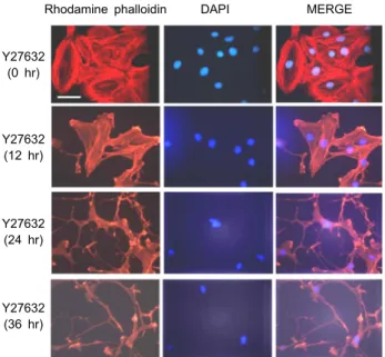
Rhodamine phalloidin DAPI MERGE
Y27632 (0 hr)
Y27632 (12 hr)
Y27632 (24 hr)
Y27632 (36 hr)
Fig. 5. Roles of ROCK on SF alignment in FRCs. Representative images of FRCs adhered on slide chamber subjected for 12 h, 24 hr and 36 hr after treatment with 10 μM Y27632.
Bar is 10 μm.
A B C
Fig. 6. Effect of LTβR signal on MLC phosphorylation and RhoA activation. (A) ML7, MLCK inhibitor, decreased MLC phosphor- ylation (p-MLC) over the control, which was abolished by 100 ng/ml agonistic anti-LTβR antibody (C). (B) LTβR stimulation induces a significant decrease in active RhoA and p-MYPT compared with control. MLC, total RhoA and MYPT were used as a loading control.
Effects of a ROCK inhibitor, Y27632, on SF dis- ruption in FRCs
Next, in order to evaluate the involvement of the ROCK pathway on SF change and morphology alteration in FRC, we treated Y27632, a specific inhibitor of ROCK, in FRCs.
After stimulation, cells were fixed and stained with rhod- amine-conjugated phalloidin to visualize F-actin. Y27632- treated FRCs were elongated and extended membrane pro- trusions by time dependent pattern. Each protrusion extend- ing from the cell body of Y27632-treated FRCs was markedly longer than those of wild-type FRCs (Fig. 5). However, Y27632 treated FRC membrane was in shreds, whereas ago- nistic anti-LTbR antibody and ML-7 treated FRC membrane
were soft and smooth. ROCK promotes actin–myosin-medi- ated contractile force generation by phosphorylating a varie- ty of downstream target proteins. This result suggests that LTβR signal pathway is linked to ROCK inhibition for SF disruption in FRC.
Rho GTPase activity after agonistic anti-LTβR antibody We observed that ML7 treatment of FRC led to complete attenuation of SFs. MLC is phosphorylated at Thr18 and Ser19 by MLCK. To further elucidate the precise mechanisms underlying ML7 function, MLC phosphorylation were exam- ined using immunoblot with p-MLC (Thr18/Ser19) antibody detects endogenous levels of myosin light chain only when dually phosphorylated at Thr18 and Ser19. Immunoblot of the ML-7 treated-cell lysate with anti-pMLC antibodies showed that the phosphorylation of MLC was completely abolished as compared with that in the control group (Fig.
6A). Rho A is activated by inflammatory factors, and an ac- tive signal is delivered to the effector ROCK, which phos- phorylates downstream targets including myosin phospha- tase target subunit 1 (MYPT1), which leads to actin-myosin crosslinking, microfilament sliding and contraction of the cells, resulting in SF alteration and morphological change of the several cells [7, 23]. To explore the expression of RhoA and p-MYPT1 in FRCs induced by agonistic anti-LTβR anti- body, we examined the activation of the small GTPase RhoA.
Agonistic anti-LTβR antibody caused about a half-fold de- crease in GTP-bound RhoA, however, RhoA-GTP levels re- mained not completely diminished from agonistic anti-LTβR antibody treated FRC (Fig. 6B). Next, the expression of p-MYPT1 in the cells stimulated by agonistic anti-LTβR anti- body was decreased obviously compared with normal cells.
To further explore the mechanisms underlying anti-LTβR
Fig. 7. Protein level of p-ezrin, f-actin, and tubulin as cytoskele- tal markers were significantly reduced in agonistic an- ti-LTβR antibody treated-FRCs. FRC was incubated with anti-LTβR antibody for 24 hr. After incubation, cell was lyzed with RIPA buffer and protein concentration of FRC lysate was measured by BCA method. FRCs were treated with mouse anti-cytoskeletal marker antibody followed by anti-LTβR antibody for 24 hr. The expression degree of p-ezrin, β-actin, a-tubulin and GAPDH was detected by Western blot. GAPDH was used as a loading control.
antibody stimulation, MLC phosphorylation were examined using immunoblot with p-MLC (Thr18/Ser19) antibody.
Immunoblot of the anti-LTβR antibody treated-cell lysate with anti-pMLC antibodies showed that the phosphorylation of MLC was completely abolished as compared with that in the control group (Fig. 6C). Our results indicate that LTβR stimulation simultaneously activates MLCK and RhoA-ROCK signal pathway.
LTbR signal interacts with cytoskeletal proteins In addition to diminished SF of FRC by fluorescence data when LTβR was stimulated, immunoblot data indicated LTβ R signal associated with cytoskeletal structures and proteins.
To determine which components of the cytoskeleton interact with LTβR signal, change of several cytoskeletal markers was examined by immunoblot. P-ezrin as a cross-linker be- tween membrane or transmembrane proteins and cytoskele- ton, actin for actin-based filaments or tubulin for micro- tubules were investigated for cytoskeletal factors. In ago- nistic anti-LTβR antibody treated-FRC, p-ezrin, β-actin, and tubulin level were significantly reduced compared to normal IgG treated-FRCs (Fig. 7). These data suggested that LTβR stimulation altered the components of the cytoskeleton.
Discussion
The effective adaptive immune response ensures an en-
counter between antigen-specific T cells or B cells and anti- gen presenting innate immune cells that infiltrate the drain- ing LN. LNs are encountering places for T lymphocytes and antigen presenting DCs. LNs are maintained by a compli- cated three dimensional framework of stromal cells and col- lagen rich RFs. These stromal cells provide the scaffold with- in which the immune cells of the LN can localize and dy- namically interact. These stromal connections contain the FRCs which produce the FRC conduit network ensheathed with the RFs and ECM components [25], and form a con- tinuous network among cells in the T cell zones of LN. T cell - DC contact is supported by FRCs that produces and ensheathes extracellular matrix components. Additionally, FRC networks provide physical routes for leucocyte migra- tion. Interaction with FRCs promotes chemokinesis in DCs facilitating their migration within LNs [18]. SFs are com- posed of F-actin, cross-linking proteins such as α-actinin, ez- rin and in some cases, the force-generating motor protein nonmuscle myosin II (NMMII). Myofibroblast SFs are me- chanically associated to the ECM through their large focal adhesions and the contractile forces generated through this linking lead to the arrangement of collagen fiber, one of fi- brous components of the ECM, in the wound area [26].
Moreover, the fibroblasts in the connective tissues are em- bedded in the ECM [22]. FRC ensheaths the reticular fiber, fibrous components of the ECM, to make the stromal retic- ulum at T zone in LN [16]. Thus, FRC stress fiber may link to stromal reticulum remodeling in LN. In this report, we demonstrated that LTβR was involved in the ROCK and MLCK pathway in FRC. LTα1β2 of T cell are important for lymphoid tissue architecture and organogenesis [16]. Thus, we propose a model that LTβR signaling function as docking sites for several downstream directional signaling molecules.
In the present study, Figure 1 illustrates the SF distribution in a control monolayer as viewed by fluorescence micro- scopy. LTβR activation by agonistic anti-LTβR antibody in- duced the abrogation of stress fiber connecting the processes of the FRC. Extreme changes of FRC morphology in prepara- tions treated with agonistic anti-LTβR antibody were ob- served (Fig. 3). Those changes ranged from partial retraction and shrink of cell processes to a dendritic configuration. This great plasticity of FRCs is thought to be taken advantage of in the supply of the proper character upon sensing im- mune responses. Various reports indicate that RhoA-ROCK pathway signals the reorganization of the cytoskeleton, which induces the formation of stress fibers. As shown in
Figure 6B, when FRC was stimulated with agonistic anti-LTβ R antibody, the level of RhoA-GDP/GTP exchange activity markedly decreased. ROCK, an important effector of RhoA, contributes to the inhibition of myosin phosphatase and is involved in the assembly of stress fiber. The inhibition of myosin phosphatase appears to account for an increase in MLC phosphorylation and induce stress fiber. Phosphorylated MLC was decreased in the presence of agonistic anti-LTβR antibody. These results revealed that stress fiber formation through LTβR signaling was involved in RhoA-ROCK and MLCK mediated cytoskeletal remodeling in FRC. MLC is phosphorylated at multiple sites. Among them, T18 and S19 are the phosphorylation sites associated with an increase in myosin ATPase activity, the formation of actin filaments such as SFs. MLCK is the kinase that was identified to phos- phorylate T18 and S19. MLCK phosphorylates MLC with preference for S19 over T18; therefore, the phosphorylation of S19 and T18 takes place in a sequential manner. Thus, our results indicate that LTβR signal linked to closer MLCK pathway than ROCK signal pathway for SF alteration in FRC. However, whether MLCK and ROCK play any differ- ential roles in SF regulation remains to be investigated in detail. The cytoskeleton is a three-dimensional grid structure composed of a variety of structural and contractile proteins in a particular pattern. The cytoskeleton exists in three main forms including microfilaments, microtubules, and inter- mediate filaments. RhoA is one of Rho GTPases that has been well studied and regarded as a main regulator of the function of cytoskeletal architecture. Ezrin proteins have been suggested to participate in Rho activation. F-actin, regulated by RhoA, has been shown to participate in LTβR signaling. Ezrin belongs to the family of closely related pro- teins, ezrin, radixin and moesin (ERM), which tether the ac- tin cytoskeleton to the plasma membrane. We therefore ex- amined phosphorylation of ezrin proteins, along with b-actin and a/b tubulin, which are components of cytoskeletal pro- teins, in mammalian cells. In contrast to agonistic anti-LTβR antibody untreated cells, LTβR activated cells displayed low levels of p-ezrin, b-actin and a/b tubulin proteins. Our stud- ies have revealed that the LTbt signaling pathway in FRC is involved in cytoskeleton regulation including SF change in FRC. In summary, our studies showed that the ligation of LTβR on FRCs is linked to marked alterations in stress fiber through RhoA/ROCK and MLCK pathway, appear to have morphological change, and it makes the supporting en- vironment that functions as an active component of the im-
mune system.
Acknowledgment
This research was supported by the Basic Science Research Program through the National Research Foundation of Korea (NRF) funded by the Ministry of Education, Science and Technology (Grant #: 2017R1D1A1B03028537).
References
1. Acton, S. E., Farrugia, A. J., Astarita, J. L., Mourão-Sá, D., Jenkins, R. P., Nye, E., Hooper, S., van Blijswijk, J., Rogers, N. C., Snelgrove, K. J., Rosewell, I., Moita, L. F., Stamp, G., Turley, S. J., Sahai, E. and Reis e Sousa, C. 2014. Dendritic cells control fibroblastic reticular network tension and lymph node expansion. Nature 514, 498-502.
2. Blue, E. K., Goeckeler, Z. M., Jin, Y., Hou, L., Dixon, S. A., Herring, B. P., Wysolmerski, R. B. and Gallagher, P. J. 2002.
220- and 130-kDa MLCKs have distinct tissue distributions and intracellular localization patterns. Am. J. Physiol. Cell Physiol. 282, C451-460.
3. Brinkman, C. C., Iwami, D., Hritzo, M. K., Xiong, Y., Ahmad, S., Simon, T., Hippen, K. L., Blazar, B. R. and Bromberg, J. S. 2016. Treg engage lymphotoxin beta receptor for affer- ent lymphatic transendothelial migration. Nat. Commun. 7, 12021. doi: 10.1038/ncomms12021
4. Browning, J. L., Allaire, N., Ngam-Ek, A., Notidis, E., Hunt, J., Perrin, S. and Fava, R. A. 2005. Lymphotoxin-β receptor signaling is required for the homeostatic control of HEV dif- ferentiation and function. Immunity 23,539-550.
5. Chai, Q., Onder, L., Scandella, E., Gil-Cruz, C., Perez-Shi- bayama, C., Cupovic, J., Danuser, R., Sparwasser, T., Luther, S. A., Thiel, V., Rülicke, T., Stein, J. V., Hehlgans, T. and Ludewig, B. 2013. Maturation of lymph node fibroblastic reticular cells from myofibroblastic precursors is critical for antiviral immunity. Immunity 38, 1013-1024.
6. Chiang, E. Y., Kolumam, G. A., Yu, X., Francesco, M., Ivelja, S., Peng, I., Gribling, P., Shu, J., Lee, W. P., Refino, C. J., Balazs, M., Paler-Martinez, A., Nguyen, A., Young, J., Barck, K. H., Carano, R. A., Ferrando, R., Diehl, L., Chatterjea, D.
and Grogan, J. L. 2009. Targeted depletion of lymphotox- in-alpha-expressing TH1 and TH17 cells inhibits auto- immune disease. Nat. Med. 15, 766-773.
7. Chirino, Y. I., García-Cuellar, C. M., García-García, C., Soto- Reyes, E., Osornio-Vargas, Á. R., Herrera, L. A., López- Saavedra, A., Miranda, J., Quintana-Belmares, R., Pérez, I.
R. and Sánchez-Pérez, Y. 2017. Airborne particulate matter in vitro exposure induces cytoskeleton remodeling through activation of the ROCK-MYPT1-MLC pathway in A549 epi- thelial lung cells. Toxicol. Lett. 272, 29-37.
8. Denton, A. E., Roberts, E. W., Linterman, M. A. and Fearon, D. T. 2014. Fibroblastic reticular cells of the lymph node are required for retention of resting but not activated CD8+
T cells. Proc. Natl. Acad. Sci. USA. 111, 12139-12144.
9. Dubey, L. K., Karempudi, P., Luther, S. A., Ludewig, B. and Harris, N. L. 2017. Interactions between fibroblastic reticular cells and B cells promote mesenteric lymph node lym- phangiogenesis. Nat. Commun. 8, 367. doi: 10.1038/s41467- 017-00504-9.
10. Elson, E. L. and Genin, G. M. 2013. The role of mechanics in actin stress fiber kinetics. Exp. Cell Res. 319, 2490-2500.
11. Fritz, J. H. and Gommerman, J. L. 2010. Cytokine/stromal cell networks and lymphoid tissue environments. J. Interfer- on Cytokine Res. 31, 277-289 .
12. Gil-Cruz, C., Perez-Shibayama, C., Onder, L., Chai, Q., Cup- ovic, J., Cheng, H. W., Novkovic, M., Lang, P. A., Geuking, M. B., McCoy, K. D., Abe, S., Cui, G., Ikuta, K., Scandella, E. and Ludewig, B. 2016. Fibroblastic reticular cells regulate intestinal inflammation via IL-15-mediated control of group 1 ILCs. Nat. Immunol. 17, 1388-1396.
13. Gommerman, J. L. and Browning, J. L. 2003. Lymphotoxin/
light, lymphoid microenvironments and autoimmune dis- ease. Nat. Rev. Immunol. 3, 642-655.
14. Girard, J. P., Moussion, C. and Förster, R. 2012. HEVs, lym- phatics and homeostatic immune cell trafficking in lymph nodes. Nat. Rev. Immunol. 12, 762-773.
15. Kassianidou, E., Hughes, J. H. and Kumar, S. 2017. Activa- tion of ROCK and MLCK tunes regional stress fiber for- mation and mechanics via preferential myosin light chain phosphorylation. Mol. Biol. Cell 28, 3832-3843.
16. Katakai, T., Hara, T., Sugai, M., Gonda, H. and Shimizu, A. 2004. Lymph node fibroblastic reticular cells construct the stromal reticulum via contact with lymphocytes. J. Exp.
Med. 200, 783-795.
17. Kedl, R. M. and Tamburini, B. A. 2015. Antigen archiving by lymph node stroma: A novel function for the lymphatic endothelium. Eur. J. Immunol. 45, 2721-2729.
18. Kumar, V., Dasoveanu, D. C., Chyou, S., Tzeng, T. C., Rozo, C., Liang, Y., Stohl, W., Fu, Y. X., Ruddle, N. H. and Lu, T. T. 2015. A dendritic-cell-stromal axis maintains immune
responses in lymph nodes. Immunity 42, 719-730.
19. Lee, J. H., Katakai, T., Hara, T., Gonda, H., Sugai, M. and Shimizu, A. 2004. Roles of p-ERM and Rho-ROCK signaling in lymphocyte polarity and uropod formation. J. Cell Biol.
167, 327-337.
20. Lee, J. S., Kim, Y. H. and Lee, J. H. 2013. Involvement of RhoA/ROCK signaling for alteration of stress fiber via lym- photoxin β receptor stimulation in fibroblastic reticular cell isolated from lymph node. Anim. Cells Syst. 17, 421-428.
21. Malhotra, D., Fletcher, A. L. and Turley, S. J. 2013. Stromal and hematopoietic cells in secondary lymphoid organs:
partners in immunity. Immunol. Rev. 251, 160-176.
22. Qin, Z., Fisher, G. J., Voorhees, J. J. and Quan, T. 2108. Actin cytoskeleton assembly regulates collagen production via TGF-β type II receptor in human skin fibroblasts. J. Cell Mol.
Med. 22, 4085-4096.
23. Ramachandran, C., Patil, R. V., Combrink, K., Sharif, N. A.
and Srinivas, S. P. 2011. Rho-Rho kinase pathway in the actomyosin contraction and cell-matrix adhesion in im- mortalized human trabecular meshwork cells. Mol. Vis. 17, 1877-1890.
24. Scarzello, A. J., Jiang, Q., Back, T., Dang, H., Hodge, D., Hanson, C., Subleski, J., Weiss, J. M., Stauffer, J. K., Chai- saingmongkol, J., Rabibhadana, S., Ruchirawat, M., Ortaldo, J., Wang, X. W., Norris, P. S., Ware, C. F. and Wiltrout, R.
H. 2016. LTβR signalling preferentially accelerates onco- genic AKT-initiated liver tumours. Gut 65, 1765-1775.
25. Sobocinski, G. P., Toy, K., Bobrowski, W. F., Shaw, S., Anderson, A. O. and Kaldjian, E. P. 2010. Ultrastructural localization of extracellular matrix proteins of the lymph node cortex: evidence supporting the reticular network as a pathway for lymphocyte migration. BMC Immunol. 11, 42.
26. Yamaguchi, H. and Sakai, R. 2015. Direct interaction be- tween carcinoma cells and cancer associated fibroblasts for the regulation of cancer invasion. Cancers (Basel) 7, 2054-2062.
초록:FRC에서 Lymphotoxin β receptor의 자극은 MLCK와 ROCK의 이중 신호전달 경로를 통해 stress fiber 변화에 관여
김대식1․이종환1,2,3*
(1동의대학교 생명공학과, 2동의대학교 바이오응용공학부 의생명공학 전공, 3동의대학교 바이오헬스학과)
Lymphotoxin β receptor (LTβR)는 TNF 계열로 림프조직의 미세구조와 기관형성에 중요한 역할을 한다.
MLCK와 ROCK는 세포의 stress fiber 형성조절에 관여하는 주요 신호전달자이다. Fibroblastic reticular cell (FRC)에서 LTβR 자극을 통한 이런 신호전달자들의 관련성을 알아보기 위해 ML-7 (MLCK 저해제)이 사용되었다.
ML7 처리된 FRC에서 SF가 완전히 파괴되었고 anti-LTβR antibody 처리 세포와 유사하게 ML7 처리 FRC에서 응축된 세포형태를 관찰 할 수 있었다. Y27632로 ROCK를 저해 했을 때 FRC의 액틴 세포골격과 세포형태 변화가 유도 되었다. FRC에서 p-MLC가 액틴과 함께 SF 구성성분을 이루었다. FRC세포 추출물로 Rho-guanosine di- phosphate (GDP)/guanosine triphosphate (GTP) 교환활성을 확인했다. Agonistic anti-LTβR antibody로 LTβR을 자극 했을 때 Rho-GDP/GTP 교환활성이 크게 감소했다. MLCK 저해처럼 LTβR 자극은 MLC의 인산화를 감소 시켰다. Agonistic anti-LTβR antibody-treated FRC에서 세포골격 구성요소인 세포막과 세포골격 링커 역할을 하 는 p-ezrin의 인산화는 감소 되었고 b- actin, 그리고 tubulin 발현도 줄었다. 이런 결과는 FRC의 LTβR 신호전달을 통한 SF 조절에는 MLCK와 ROCK가 관여하고 있다는 것을 알 수 있었다.