Effects of an Aqueous Extract of Asparagus cochinchinensis on the Regulation of Nerve Growth Factor in Neuronal Cells
Hyun Ah Lee
1, Ji Eun Kim
1, Sung Hwa Song
1, Ji Eun Sung
1,Min Gi Jung
1, Dong Seob Kim
1, Hong Joo Son
1, Chung Yeoul Lee
2, Hee Seob Lee
3and Dae Youn Hwang
1*
1College of Natural Resources and Life Science/Life and Industry Convergence Research Institute, Pusan National University, Miryang 627-706, Korea
2Gangrim Organics, Miryang 627-706, Korea
3College of Human Ecology, Pusan National University, Busan 609-735, Korea Received January 7, 2016 /Revised March 15, 2016 /Accepted March 15, 2016
Asparagus cochinchinensis is a medical plant that has long been used to treat fever, cough, kidney dis- ease, breast cancer, inflammatory disease and brain disease in northeast Asian countries. Although several studies have been conducted on the anti-neuroinflammatory effects of A. cochinchinensis, the correlation between these effects and nerve growth factor (NGF) has not yet been examined. In this study, we investigated the effects of an aqueous extract of A. cochinchinensis (AEAC) on the secretion and action mechanism of NGF in neuronal cells. The concentration of the NGF protein in the super- natant collected from cultured cells increased significantly in B35 cells treated with AEAC in com- parison with the vehicle-treated group without any specific cytotoxicity. Furthermore, the mRNA ex- pression of NGF showed a very similar pattern to its protein concentration. To examine the bio- activity of NGF secreted from B35 cells, undifferentiated PC12 cells were cultured in an AEAC-con- ditioned medium and neuritic outgrowth was observed. The dendrite length of PC12 cells in the AEAC-treated group was significantly higher than that in the vehicle-treated group. Moreover, the level of the downstream effectors p-TrkA and p-ERK of the high-affinity NGF receptor was sig- nificantly higher in the AEAC-treated group, while the expression of the downstream effectors of the low-affinity NGF receptor was significantly lower in the same group. These results suggest that AEAC may contribute to the regulation of NGF expression and secretion in neuronal cells; it is therefore an excellent candidate for further investigation as a therapeutic drug for neurodegenerative diseases.
Key words :
Asparagus cochinchinensis, B35 cells, NGF, NGF receptor, PC12 cells
*Corresponding author
*Tel : +82-55-350-5388, Fax : +82-55-350-5389
*E-mail : [email protected]
This is an Open-Access article distributed under the terms of the Creative Commons Attribution Non-Commercial License (http://creativecommons.org/licenses/by-nc/3.0) which permits unrestricted non-commercial use, distribution, and reproduction in any medium, provided the original work is properly cited.
Journal of Life Science 2016 Vol. 26. No. 5. 509~518 DOI : http://dx.doi.org/10.5352/JLS.2016.26.5.509
Introduction
Asparagus cochinchinensis is a perennial herb belonging to the Liliaceae family that is widely distributed in China, Japan and Korea [38]. The root of A. cochinchinensis has been used as a traditional medicine in those countries for thou- sands of years [37, 38], and it is considered a therapeutic drug owing to its anti-inflammatory, diuretic, antiseptic, an- titussive, antibacterial, nervine, sialogoue, antipyretic, and stomachic effects, although there is a lack of scientific evi- dence of these effects. Moreover, it is commonly used in
combination with other herbs to treat the lungs, spleen, im- mune system and aging [37, 38].
Various substances including 19 amino acids, polysac- charides, and more than 20 multi-functional compounds have been identified in the root of A. cochinchinensis. These include β-sitosterol [19], daucosterol [25], n-ethatriaconta- noic acid [31], palmitic acid [14], 9-heptacosylene [41], smila- genin [3], diosgenin [6], sarsasapogenin-3-O-β-D-glucoside feeding grapes imidacloprid [39], 5-methoxy methyl furfural, yame sapogenin, diosgenin-3-O-β-D imidacloprid feeding glucose glycosides [29, 39], aspacochioside D [30], iso-aga- tharesinoside [18] and seven steroidal saponins [15]. Among these, some polysaccharides exhibited therapeutic effects against several diseases such as aging [15, 27, 43], inflam- matory diseases [15], tumor [11, 20, 36], diabetes [42] and cough [20, 22].
Although several extracts of A. cochinchinesis have been
applied as traditional medicines to treat various diseases, sci-
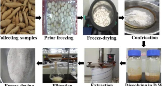
Fig. 1. Preparation scheme of AEAC. AEAC was obtained from the roots of A. cochinchinensis using aqueous extraction under the conditions described in the materials and methods.
entific evidence is only available for their anti-inflammatory activities. Specifically, these effects and related mechanisms were observed in several models, including Lipopolysac- charide (LPS)/substance P-stimulated mouse astrocytes [10], LPS-stimulated BV-2 microglial cells [12] and cockroach allergen-induced mice [9]. However, the underlying mecha- nism by which A. cochinchinensis influences NGF secretion ability has not yet been clearly identified, even though the effects of Liriope platyphylla, another plant in the Liliaceae family, have been thoroughly investigated in neuronal cells and animal models [2, 3, 24].
In this study, we investigated the effects of AEAC on the NGF secretion ability and NGF receptor signaling pathway in B35 neuroblastoma and PC12 cells to provide scientific evidence of the therapeutic effects of A. cochinchinensis on neurodegenerative disorders.
Materials and Methods
Preparation of AEAC
The roots of A. cochinchinensis used in this study were collected from plantations in the Go-Chang area of Korea and dried in a drying machine (Ilshinbiobase Co., Dongducheon, Korea) at 60°C. Voucher specimens of A. co- chinchinensis (WPC-14-003) were deposited in the functional materials bank of the PNU-Wellbeing RIS Center at Pusan National University. Dry roots of A. cochinchinensis were re- duced to powder using a pulverizer (Hanil Electric Co., Seoul, Korea), after which AEAC was purified from 75 g of A. cochinchinensis roots for 45 min at 121°C using circulat- ing extraction equipment (IKA Labortechnik, Staufen, Germany) after adding 500 ml of distilled water (Fig. 1A).
In addition, a solution of the extracts was concentrated and
subsequently passed through a 0.4 μm filter, after which the pellets were dried in a rotary evaporator (EYELA, Tokyo, Japan) and stored at -80°C until needed.
Analysis of total flavonoids and phenolics
Total phenolics in AEAC were measured by the Folin- Ciocalteu method, with slight modification [32]. Briefly, 1 ml of AEAC solution was mixed with 5 ml of Folin- Ciocalteu reagent (Sigma-Aldrich Co., St. Louis, MO, USA), then incubated at room temperature for 5 min. This mixture was subsequently added to 15 ml of 20% Na
2CO
3and vor- texed for 30 sec, after which the absorbance was repeatedly measured at 765 nm using a Versa-max plate reader (Molecular Devices, Sunnyvale, CA, USA). A standard cali- bration curve was made using different concentrations of gallic acid (Sigma-Aldrich Co., St. Louis, MO, USA), after which the concentration of total phenolic contents in AEAC was presented as mg gallic acid equivalent of extract.
The flavonoid contents in AEAC were also measured as previously described [44]. Briefly, 200 μl of several different concentrations of AEAC were mixed with 60 μl of 5% NaNO
2(Sigma-Aldrich Co.) and 60 μl of 10% AlCl
3(Sigma- Aldrich Co.). Following incubation at 25°C for 5 min, the mixture was added to 400 μl of 1 M NaOH and the absorbance was repeatedly measured at 510 nm using a Versa-max plate reader (Molecular Devices). A standard calibration curve was then made using different concentrations of catechin (Sigma-Aldrich Co.). The flavonoid contents of the AEAC were presented as mg catechin equivalent of extract.
Cell culture
The B35 cell line used in this study is a neuroblastoma
that originated from the central nervous system of rats
(Rattus norvegicus). This cell line was kindly provided by the
Korean Cell Line Bank (Seoul, Korea). They were grown
with Dulbecco Modified Eagle's Medium (DMEM, Welgene,
Gyeongsan-si, Korea) containing 10% fetal bovine serum
(FBS, Welgene, Gyeongsan-si, Korea), L-glutamine, pen-
icillin, and streptomycin (Thermo Scientific, Waltham, MA,
USA) in a humidified incubator at 37°C under 5% CO
2and
95% air. The PC12 cell line, which is a pheochromocytoma
that originated from the adrenal medulla of rats, was also
used in this study and provided by the Korean Cell Line
Bank (Seoul, Korea). PC12 cells were grown in Roswell Park
Memorial Institute medium (RPMI, Welgene, Gyeongsan-si,
Korea) containing 10% FBS (Welgene), L-glutamine, pen-
icillin, and streptomycin (Thermo Scientific, Waltham, MA, USA) in a humidified incubator at 37°C under 5% CO
2and 95% air.
Cell viability assay
The viability of B35 cells was determined using the tetra- zolium compound 3-[4,5-dimethylthiazol-2-yl]-2,5-diphenyl- tetrazolium bromide (MTT) (Sigma-Aldrich Co.). To de- termine the cell viability, B35 cells were seeded at a density of 5×10
4cells/0.2 ml and grown for 24 hr in a 37°C incubator. When the cells attained 70-80% confluence, they were either treated with vehicle (1x PBS) or pretreated with 100 μg/ml of AEAC dissolved in 1× PBS for 24 hr. After discarding the supernatants, 0.2 ml of fresh MEM media and 50 μl of MTT solution (2 mg/ml in PBS) were added to each well. The cells were then incubated at 37°C for 4 hr.
Formazan precipitate was dissolved in DMSO, after which the absorbance at 570 nm was read directly in wells using a Molecular Devices VERSA max plate reader (Sunnyvale, CA, USA). The morphological features of B35 cells in each treated group were also observed using a microscope at 200×
magnification (Leica Microsystems, Switzerland).
Western blot
Total protein extracted from PC12 cells using Pro-Prep Protein Extraction Solution (iNtRON Biotechnology, Seong- nam, Korea) was collected by the centrifugation at 15,000 rpm for 15 minutes, then quantified using a SMARTTM BCA Protein Assay Kit (Thermo Scientific, Waltham, MA, USA) for Western blot analysis. Briefly, these proteins were sepa- rated by 4-20% sodium dodecyl sulfate-polyacrylamide gel electrophoresis (SDS-PAGE) for 2 hr, after which resolved proteins were transferred to nitrocellulose membranes for 2 hr at 40 V. Each membrane was then incubated separately at 4°C with the following primary antibodies overnight: an- ti-TrkA antibody (Cell Signaling Technology, Beverley, MA, USA), anti-p-TrkA antibody (Cell Signaling Technology), an- ti-Akt antibody (Cell Signaling Technology), anti-p-Akt anti- body (Cell Signaling Technology), anti-ERK antibody (Santa Cruz Biotechnology, Santa Cruz, CA, USA), anti-p-ERK anti- body (Santa Cruz Biotechnology), anti-p75
NTRantibody (Cell Signaling Technology), anti-JNK antibody (Cell Signaling Technology), anti-p-JNK antibody (Cell Signaling Technol- ogy) and anti-actin antibody (Sigma-Aldrich Co.). Next, the membranes were washed with washing buffer (137 mM NaCl, 2.7 mM KCl, 10 mM Na
2HPO
4, and 0.05% Tween 20),
then incubated with 1:1,000 diluted horseradish peroxidase (HRP)-conjugated goat anti-rabbit IgG (Invitrogen, Carlsbad, CA, USA) at room temperature for 1 hr. Finally, membrane blots were developed using Amersham ECL Select Western Blotting detection reagent (GE Healthcare, Little Chalfont, UK). The chemiluminescence signals that originated from specific bands were detected using FluorChemi
®FC2 (Alpha Innotech Co., San Leandro, CA, USA).
Enzyme-linked immunosorbent assay (ELISA) of NGF
The levels of NGF in culture supernatant from B35 cells treated with AEAC were measured using a NGF ELISA kit (Chemicon International Inc., Temecula, CA, USA). Briefly, the samples and standards were incubated overnight on an- tibody-coated plates in a plate shaker at 100-150 rpm and 2-8°C. The wells were then washed four times with washing buffer, after which 100 μl of anti-mouse NGF monoclonal antibody was added to each well. Plates were subsequently incubated in a shaker for 2 hr at room temperature, after which 100 μl of peroxidase conjugated donkey anti-mouse IgG polyclonal antibody was added to each well and sam- ples were incubated at room temperature for an additional 2 hr. After washing, 100 μl of TMB substrate was added to each well and the plate was incubated at room temperature for 15 min. The reaction was then quenched by the addition of 100 μl of stop solution, after which the plate was analyzed using a SoftMax Pro5 spectrophotometer (Molecular Devices, Sunnyvale, CA, USA).
Dot blot analysis
Total protein prepared from B35 cells treated with AEAC was transferred to a nitrocellulose membrane using a SlotBlot kit (Pharmacia Biotech, CA). The membrane was in- cubated separately with primary rabbit polyclonal anti-NGF antibody (Cell Signaling Technology, Beverley, MA, USA) at 2 μg in blocking buffer at room temperature for 3 hr, then washed in washing buffer and incubated with secondary an- tibody, horseradish peroxidase-conjugated goat anti-rabbit IgG (GenTest, MA), at 1:1,000 for 1 hr at room temperature.
NGF proteins were detected using Amersham ECL Select
Western Blotting detection reagent (GE Healthcare, Little
Chalfont, UK). The chemiluminescence signals that origi-
nated from specific slot were detected using FluorChemi
®FC2 (Alpha Innotech Co).
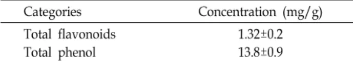
Table 1. The concentration of total flavonoids and phenol was assayed in a mixture containing different concen- trations of AEAC
Categories Concentration (mg/g)
Total flavonoids Total phenol
1.32±0.2 13.8±0.9
RT-PCR analysis of NGF transcripts
The relative quantities of NGF transcripts were measured by RT-PCR as previously described [28]. First, total RNA molecules were purified by removing media from each cul- tured sample and then lysing B35 cells in RNAzol (Tel-Test Inc., Friendswood, USA). The isolated RNA was sub- sequently measured by UV spectroscopy, after which the ex- pression of NGF genes was assessed using RT-PCR with 5 μg of total RNA from cells of each group. Next, 500 ng of oligo-dT primer (Invitrogen, Carlsbad, CA, USA) were an- nealed at 70°C for 10 min. The complementary DNA (cDNA), which was used as the template for further amplifi- cation, was synthesized by the addition of deoxyadenosine triphosphate (dATP), deoxycytidine triphosphate (dCTP), deoxyguanosine triphosphate (dGTP), and deoxythymidine triphosphate (dTTP) with 200 units of reverse transcriptase (Superscript II, Invitrogen, 200 U/μl). Next, 10 pmol of the sense and antisense primers were added, and the reaction mixture was subjected to 28-32 cycles of amplification.
Amplification was conducted in a Perkin- Elmer Thermal Cycler using the following cycles: 30 sec at 94°C, 30 sec at 62°C, and 45 sec at 72°C. The primer sequences for target gene expression identification were as follows: NGF, sense primer: 5'-CAT GTT GTT CTA CAC TCT GAT CAC-3', an- ti-sense primer: 5'-CTC CTT GCC CTT GAT GTC TGT GG-3’; β-actin, sense primer: 5'-GTG GGG CGC CCC AGG CAC CAG GGC-3', anti-sense primer: 5'-CTC CTT AAT GCT ACG CAC GAT TTC-3'. The experiment was repeated three times, and all samples were analyzed in triplicate. The final PCR products were separated on 1% agarose gel and then visualized by ethidium bromide staining.
Analysis of neuritic outgrowth
The neuritic outgrowth of PC12 cells was measured as previously described [3, 24]. After incubation of PC12 cells treated with AEAC-conditioned medium for 24 hr, the mor- phology of PC12 cells was observed under a microscope at 200× magnification (Leica Microsystems, Switzerland). The length of dendrite of PC12 cells was analyzed using the Leica Application Suite (Leica Microsystems, Switzerland).
Cell cycle assay
The cell cycle was measured using a Muse
™Cell Cycle Kit (Millipore Co., Billerica, MA, USA) according to the manu- facturer’s instructions. Briefly, PC12 cells were divided into 100 mm
2dishes (2.5×10
6cells/dish), then treated with
AEAC-conditioned medium for 24 hr. Total cells from each group were harvested by centrifugation at 3,000 ×g for 5 minutes, then fixed with 70% EtOH at -20°C for 3 hr. The fixed cells were subsequently washed with 1× PBS, then added to 200 μl of Cell Cycle Reagent. Following incubation at 37°C in a CO
2incubator for 30 min, cell cycles were ana- lyzed using FACS (Millipore Co., Billerica, MA, USA).
Statistical analysis
One-way ANOVA was used to identify significant differ- ences between vehicle and AEAC treated groups (SPSS for Windows, Release 10.10, Standard Version, Chicago, IL, USA). All values are reported as the means ± standard devi- ation (SD) and a p value of <0.05 was considered significant.
Results
Bioactive components of AEAC
To quantify the bioactive components in AEAC, total phe- nolic contents and total flavonoids were analyzed using the method suggested in previous studies. As shown in Table 1, AEAC contained high concentrations of two important an- tioxidants, flavonoids (1.32±0.2 mg/g) and phenolics (13.8±0.9 mg/g). These findings suggest that AEAC can have high antioxidative activity.
Toxicity of AEAC
To determine the toxicity of AEAC, cell viability was measured in B35 cells treated with 100 μg/ml using an MTT assay. No significant alterations in cell viability or morpho- logical features were observed in B35 cells treated with AEAC alone for 24 hr (Fig. 2). These findings indicate that AEAC showed no toxicity at less than 100 μg/ml.
Effects of AEAC on NGF secretion and biosynthesis
To investigate the effects of AEAC on the synthesis and
secretion of NGF in B35 cells, the levels of NGF protein and
mRNA were measured in the culture supernatant and cells
after AEAC treatment for 24 hr. The concentration of NGF
protein was significantly higher in the supernatant of B35
Fig. 2. Toxicity of AEAC. After incubation of B35 cells with 100 μg/ml of AEAC for 24 hr, their morphologies were observed under a microscope at 200× magnification. The viability of B35 cells treated with AEAC was determined by MTT assay in triplicate. The data shown represent the means ± SD of three replicates.
A
B
C
Fig. 3. Secretion and synthesis of NGF. The protein concen- tration of NGF was detected in the supernatant and cell homogenate of B35 cells treated with AEAC for 24 hr using ELISA (A) and dot blot (B). The level of NGF transcripts was measured in B35 cells using RT-PCR (C).
The β-actin signal was used as the endogenous control, and the transcript (540 bp) indicates the RNA loading.
The density of the transcript was quantified using a Kodak Electrophoresis Documentation and Analysis System 120. The values of data shown are the means±
SD of three experiments. *, p<0.05 compared to the ve- hicle treated group.
cells treated with AEAC compared with the vehicle treated group (Fig. 3A, Fig. 3B). A similar increase was detected in the mRNA levels of the same group (Fig. 3C). These findings suggest that AEAC treatment can increase NGF secretion and synthesis in B35 neuroblastoma cells.
To verify the activity of secreted NGF, AEAC-conditioned media were inoculated with undifferentiated PC12 cells and their dendritic outgrowth was measured based on micro- scopic analysis. Dendrites were longer in the AEAC-con- ditioned medium treated goups than the vehicle-conditioned medium treated group (Fig. 4). These results show that NGF secreted from B35 cells after AEAC treatment can success- fully induced the differentiation of PC12 cells.
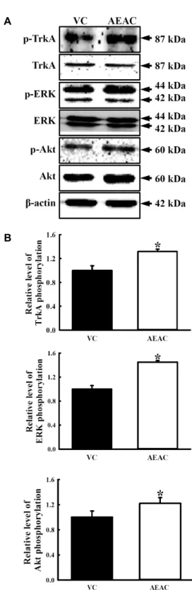
Effects of AEAC on NGF receptor signaling pathway Secreted NGF can transduce signals related to survival, proliferation and death into the cytosol through binding of two different types of NGF receptors located on the cell membrane [1]. Therefore, we investigated whether the in- crease in NGF concentration induced by AEAC treatment could affect the two NGF receptor signaling pathways in PC12 cells. Analysis of the high affinity receptor revealed that the level of p-TrkA was slightly higher in the AEAC- conditioned medium treated group than the vehicle-con- ditioned medium treated group. A similar enhancement was
observed in the level of p-ERK and p-Akt among down-
stream members of the high affinity receptor. Following
treatment with the AEAC-conditioned medium, their level
rapidly increased to 22% and 44%(Fig. 5).
A
B
Fig. 4. Dendritic outgrowth of PC12 treated with AEAC-con- ditioned medium. After treatment of B35 cells with AEAC for 24 hr, the AEAC-conditioned media were transferred to undifferentiated PC12 cells. (A) The cel- lular morphology of PC12 was viewed at 200× magnifi- cation. (B) The dendritic outgrowth of PC12 cells was measured in specific area (43.9 μm2). The values shown are the means±SD of three experiments. *, p<0.05 com- pared to the vehicle treated group.
Conversely, in the case of low affinity receptor, the level of p75
NTRand p-JNK expression was lower in the AEAC-con- ditioned medium treated group than the vehicle-conditioned medium treated group (Fig. 6). Taken together, these results showed that NGF secreted by AEAC treatment may stim- ulate proliferation and inhibit apoptosis of PC12 cells through regulation of the NGF receptor TrkA and p75
NTRsignaling pathways.
Effects of AEAC on cell cycle regulation
Finally, to examine the effects of AEAC-conditioned me- dium on the cell cycle of PC12 cells, the number of cells in each stage of the cell cycle was counted in subset groups.
After treatment with the AEAC-conditioned medium, the number of cells in the G0/G1 stage was decreased, while those in the S and G2/M stage increased (Fig. 7). These re- sults suggest that AEAC treatment can stimulate progression from the G0/G1 stage to the S and G2/M stage.
A
B
Fig. 5. Effects of AEAC treatment on the high affinity NGF re- ceptor signaling pathway of PC12 cells. Total tissue ly- sates were prepared from PC12 cells treated with vehicle or AEAC conditioned medium as described in the Materials and Methods section. Fifty micrograms of pro- tein per sample were immunoblotted with antibodies for each protein. Three samples were assayed in triplicate by Western blotting. Data shown are the means ± SD.
*, p<0.05 relative to the vehicle treated group.
A
B
Fig. 6. Effects of AEAC treatment on the low affinity NGF re- ceptor signaling pathway of PC12 cells. Total tissue ly- sates were prepared from PC12 cells treated with vehicle or AEAC conditioned medium as described in the Materials and Methods section. Fifty micrograms of pro- tein per sample were immunoblotted with antibodies for each protein. Three samples were assayed in triplicate by Western blotting. Data shown are the means ± SD.
*, p<0.0 relative to vehicle treated group.
A
B
Fig. 7. Cell cycle analysis. The cell cycle distribution was de- termined by flow cytometric analysis of the DNA con- tent of nuclei of PC12 cells following staining with pro- pidium iodide. After treatment with AEAC-conditioned medium, the number of cells in the G0/G1, S and G2/M stage was determined. Data shown are the means ± SD.
*, p<0.05 relative to the vehicle treated group.
Discussion
Nerve growth factor has received recent consideration as a potential treatment regulator for human central and pe- ripheral nervous system related disorders because it has been reported to be an important molecule that regulates neuronal survival and differentiation [1, 34, 35]. Many stud- ies have focused on the identification and development of a novel NGF stimulator to provide therapeutic drugs for the treatment of neurodegenerative disorders. In an effort to identify candidate natural products for the treatment of such diseases and verify their mechanism of action, we inves- tigated the NGF stimulatory effects of AEAC purified from the roots of A. cochinchinensis in B35 cells and PC12 cells.
Our results showed that AEAC could significantly stimulate
the secretion ability and synthesis of NGF in B35 cells, and that secreted NGF induced the differentiation of PC12 cells through regulation of the high affinity and low affinity re- ceptor signaling pathway.
Many natural products have long been considered a
source of leads for the development of therapeutic drugs
available for the treatment of human diseases because they
provide some active principles that can be used as the back-
bone for synthesis of new drugs [6, 25]. However, a large
number of candidates have been discarded because they
could not overcome several essential problems. Among
these, toxicity has long been considered one of the most im-
portant problems during preclinical investigations of natural
products [5]. In this study, we investigated the toxicity of
AEAC in B35 cells to determine the optimal concentration
of AEAC. As shown in Fig. 1, no significant toxicity was
detected in B35 cells treated with 100 μg/ml of AEAC. These
results were similar to those of previous studies. Three dif-
ferent concentrations (1, 10 and 100 μg/ml) of aqueous ex-
tract of A. cochinchinensis did not induce significant toxicity
in the human hepatoma cell line HepG2 [13]. Furthermore,
the viability of A549 cells was maintained at a constant level
in 100, 300 and 500 μg/ml of 70% ethanol extract of A. cochin-
chinensis [15].
Nerve growth factor was secreted from various cell types including neurons, inflammatory cells (lymphocytes or mast cells), and structural cells (fibroblast, epithelial cells and smooth muscle cells) after stimulation with cytokines and treatment with natural products [7]. Korea white ginseng ex- tract and ginsenoside Rb2 stimulated an increase of NGF mRNA and protein in a steroid-induced polycystic ovary murine model and SD rats [27, 29]. Moreover, NGF-medi- ated neuritic outgrowth was significantly upregulated by treatment with Picrohizae Rhizoma methanol extracts [18].
Furthermore, some extracts (butanol extract, spicatoside A, LP-E, LP-M, LP-M50 and LP2E17PJ) from L. platyphylla in- duced the secretion and synthesis of NGF in B35 cells [9, 10, 24]. In this study, we investigated the effects of A. cochin- chinensis on NGF secretion and synthesis ability of neuro- blastoma cells. The results presented herein provide addi- tional evidence that AEAC purified from A. cochinchinensis can be considered a novel NGF stimulator.
Nerve growth factor can transfer the signal for the surviv- al, proliferation and apoptosis of target cells through the binding of high affinity and low affinity NGF receptors [35].
These signal pathways are regulated by various natural products. Trk phosphorylation of the high affinity receptor was rapidly enhanced by spicatoside A and LP-M from L.
platyphylla in neuronal cells and brain hippocampus cells [10, 24], while the expression of p75
NTRand RhoA in low affinity receptor was lower in LP-M treated animal cells [24]. A sim- ilar result was observed in AEAC-conditioned medium treated PC12 cells as shown Fig. 5 and 6.
Finally, the results of the present study suggest that AEAC may stimulate NGF synthesis and secretion from neu- roblastoma cells without any significant toxicity. Moreover, secreted NGF can successfully induce survival, proliferation and differentiation of undifferentiated PC12 cells through regulation of the NGF receptor signaling pathway.
Acknowledgements
This study was supported by grants to Dr. Dae Youn Hwang from the Korea Institute of Planning Evaluation for Technology of Food, Agriculture, Forestry and Fisheries (114034-03-1-HD030).
References
1. Barde, Y. A. 1989. Trophic factors and neuronal survival.
Neuron 2, 1525-1534.
2. Choi, S. I., Goo, J. S., Kim, J. E., Hwang, I. S., Lee, H. R., Lee, Y. J., Son, H. J., Lee, J. S. and Hwang, D. Y. 2012. Effects of Red Liriope platyphylla on NGF seretion ability, NGF re- ceptor signaling pathway and γ-secretase components in NSE/hAPPsw transgenic mice expressing Alzheimer’s disease. Lab. Anim. Res. 28, 155-163.
3. Choi, S. I., Park, J. H., Her, Y. K., Lee, Y. K., Kim, J. E., Nam, S. H., Goo, J. S., Jang, M. J., Lee, H. S., Son, H. J., Lee, C. Y. and Hwang, D. Y. 2010. Effects of water extract of Liriope platyphylla on the mRNA expression and protein secretion of nerve growth factors. Kor. J. Med. Crop Sci. 18, 291-297.
4. Cong, P. Z. and Keman, S. 2000. Handbook of analytical Chemistry—Mass Volume, pp. 296-298, 2nd ed., Chemical Industry Publishing House: Beijing, China.
5. Cragg, G. M. and Newman, D. J. 2005. Drug discovery and development from natural products: the way forward. The 11th NAPRECA Symposium Book of Proceedings. August 9-12.
Antananarivo, Madagascar.
6. Efange, S. M. N. 2002. Natural products: a continuing source of inspiration for the medical chemist, pp. 61-69. In: Iwu, M. M., Wootton, J. C. (eds), Ethnomedicine and drug discov- ery: Advances in Phytomedicine. Elsevier Science: Amster- dam, The Netherlands.
7. Freund-Michel, V. and Frossard, N. 2008. The nerve growth factor and its receptors in airway inflammatory diseases.
Pharmacol. Ther. 117, 52-76.
8. Gong, Y. H. 1986. 13C NMR chemical shifts of natural or- ganic compounds, pp. 252, 2nd ed., Yunnan Science and Technology Publishing House: Kunming, China
9. Hur, J. Y., Lee, P. J., Kim, J. M., Kim, A. J., Kim, H. C. and Kim, S. Y. 2004. Induction of nerve growth factor by butanol fraction of Liriope platyphylla in C6 and primary astrocyte cells. Biol. Pharm. Bull. 27, 1257-1260.
10. Hur, J. Y., Lee, P. J., Moon, E. J., Kang, I. S., Kim, S. H., Oh, M. S. and Kim, S. Y. 2009. Neurite outgrowth induced by spicatoside A, a steroidal saponin, via the tyrosine kinase A receptor pathway. Eur. J. Pharmacol. 620, 9-15.
11. Jung, K. H., Choi, H. L., Park, S. J., Lee, G. H., Kim, M.
R., Min, J. K., Min, B. I. and Bae, H. S. 2014. The effects of the standardized herbal formula PM014 on pulmonary inflammation and airway responsiveness in a murine model of cockroach allergen-induced asthma. J. Ethnopharmacol.
155, 113-122.
12. Kim, H. M., Lee, E. H., Lim, T. K., Jung, J. A. and Lyu, Y. S. 1998. Inhibitory effect of Asparagus cochinchinensis on tumor necrosis factor-alpha secretion from astrocytes. Int.
J. Immunopharmacol. 20, 153-162.
13. Koo, H. N., Jeong, H. J. and Choi, J. Y. 2000. Inhibition of tumor necrosis factor-a-induced apoptosis by Asparagus cochlnchinensis in HepG2 cells. J. Ethnopharmacol. 73, 137- 143.
14. Lee, D. Y., Choo, B. K., Yoon, T. S., Cheon, M. S., Lee, H.
W., Lee Y. A. and Kim, H. K. 2009. Anti-inflammatory ef- fects of Asparagus cochinchinensis extract in acute and chronic
cutaneous inflammation. J. Ethnopharmacol. 121, 28-34.
15. Lee, J. H., Lim, H. J., Lee, C. W., Son, K. H., Son, J. K., Lee, S. K. and Kim, H. P. 2015. Methyl protodioscin from the roots of Asparagus cochinchinensis attenuates airway in- flammation by inhibiting cytokine production. Evid. Based Complement Alternat. Med. 2015, 640846.
16. Liang, Z. Z., Aquino, R., De Simone, F., Dini, A., Schettino, O. and Pizza, C. 1988. Oligo furo stanosides from Asparagus cochinchinensis. Planta Med. 54, 344–346.
17. Li, M., Fei, Y. and Wang, J. K. 2005. Studies on pharmaco- logic effects of Radix asparagi. LiShiZhen Med. Mater Med.
Res. 16, 580-582.
18. Li, P., Matsunaga, K. and Ohizumi, Y. 2000. Nerve growth factor-potentiating compounds from Picrorhizae rhizoma.
Biol. Pharm. Bull. 23, 890-892.
19. Li, X. N., Chu, C., Cheng, D. P., Tong, S. Q. and Yan, J.
Z. 2012. Norlignans from Asparagus cochinchinensis. Na Prod.
Commun. 7, 1357-1358.
20. Liu, Y. Z., Qu, F. Y. and Zhang, P. X. 2001. Effect of chloro- form extract of Tiandong on the brain antioxidation of D-galatose-induced senile mice. Heilongjiang Med. Pharm. 24, 7-8.
21. Luo, J., Long, Q. D., Li, C. X., Li, L. and Huang, N. H. 2000.
Inhibitory effects of ALWB and ACM on mice bearing tumor. J. GuiYang Med. Coll. 25, 15-16.
22. Luo, J., Long, Q. D., Li, C. X., Li, L., Huang, N. H., Nie, M. and Tang, P. X. 1998. Comparison of antitussive, ex- pectorant and anti-asthmatic effect between ALWB and ACM. J. GuiYang Med. Coll. 23, 132-134.
23. Lv, B. and Liu, W. Z. 2004. Aspartate treatment of hemodial- ysis patients with hypertension in 22 cases. J. Tradit Chin.
Med. 19, 43-44.
24. Nam, S. H., Choi, S. I., Goo, J. S., Kim, J. E., Lee, Y. K., Hwang, I. S., Lee, H. R., Lee, Y. J., Lee, H. G., Choi, Y. W.
and Hwang, D. Y. 2011. LP-M, a novel butanol-extracts iso- lated from Liriope platyphylla, could induce the neuronal cell survival and neuritic outgrowth in hippocampus of mice through Akt/ERK activation on NGF signal pathway. J. Life Sci. 21. 1234-1243.
25. Newman, D. J. 2008. Natural products as leads to potential drugs: an old process or the new hope for drug discovery?
J. Med. Chem. 51, 2589-2599.
26. Ni, J. M., Zhao, R. and Wang, R. 1992. Comparison on amino acid content in prepared and unprepared Asparagus cochin- chinensis. Chin. Tradit. Herb Drugs 23, 182-183.
27. Park, S. C., Kim, S. E., Oh, D. M., Shim, K. M., Jeong, M.
J., Lim, S. C., Nah, S. Y., Park, S. H., Kang, S. S., Moon, C. J., Kim, J. C., Kim, S. H. and Bae, C. S. 2009. Effect of Korean red ginseng extract in a steroid-induced polycystic ovary murine model. Arch. Pharm. Res. 32, 347-352.
28. Qu, F. Y., Wei, X. D., Li, S. L., Wang, Y. M. and Bai, S.
G. 1999. Experimental study of Asparagus cochinchinensis de- lay aging. Acta Chin. Med. Pharm. 2, 68-70.
29. Salim, K. N., McEwen, B. S. and Chao, H. M. 1997.
Ginsenoside Rb1 regulates ChAT, NGF and trkA mRNA ex- pression in the rat brain. Brain Res. Mol. Brain Res. 47, 177-
182.
30. Shen, Y., Chen, H. S. and Wang, Q. 2007. Studies on chem- ical constituents of Asparagus cochinchinensis(II). J. Second Med. Univ. 28, 1241-1244.
31. Shen, Y., Xu, C. l., Xuan, W. D., Li, H. L., Liu, R. H., Xu, X. K. and Chen, H. S. 2011. A new furostanol saponin from Asparagus cochinchinensis. Arch. Pharm. Res. 34. 1587-1591.
32. Singleton, V. L. and Rossi, J. A. 1965. Colorimetry of total phenolics with phosphomolybdic-phosphotungstic acid reagents. Am. J. Enol. Vitic. 16, 144-158.
33. Tenji, K. and Junzo, S. 1979. Studies on the constituents of Asparagi Radix. I. On the structures of furostanol oligosides of Asparagus cochinchinensis (Lour.) Merr. Chem. Pharm. Bull.
27, 3086-3094.
34. Theonen, H., Bandtlow, C. and Heuman, R. 1987. The phys- iological function of nerve growth factor in the central nerv- ous system: comparison with the periphery. Rev. Physiol.
Biochem. Pharmacol. 109, 146-178.
35. Tsui-Pierchala, B. A. and Ginty, D. D. 1999. Characterization of an NGF-P-TrkA retrograde-signaling complex and age-dependent regulation of TrkA phosphorylation in sym- pathetic neurons. J. Neurosci. 19, 8207-8218.
36. Tuszynski, M. H., Gabriel, K., Gage, F. H., Shur, S., Meyer, S. and Rosetti, A. 1996. Nerve growth factor delivery by gene transfer induces differential outgrowth of sensory, mo- tor and noradrenergic neurites after adult spinal cord injury.
Exp. Neurol. 137, 157-173.
37. Wen, J. Y., Li, Y., Ding, S. S. and Li, Q. H. 1993. Nine Pharmacological screening of medicinal plants of China Liliaceae asparagus. J. Acta Acad Med. Shanghai 20, 107-111.
38. Xiao, P.G. 2002. Modern chinese material medica, pp. 150, 2nd ed., Chemical Industry Press: Beijing, China.
39. Xiong, D. S., Yu, L. X., Yan, X., Guo, C. and Xiong, Y. 2011.
Effects of root and stem extracts of Asparagus cochinchinensis on biochemical indicators related to aging in the brain and liver of mice. Am. J. Chin. Med. 39, 719-726.
40. Xu, C. L., Chen, H. S. and Tan, X. Q. 2005. Studies on the active constituents of Asparagi radix. Nat. Prod. Res. Dev. 17, 128-130.
41. Yang, M. H. 1981. Steroidal sapogenins of dioscorea. Chin Tradit Herb Drugs. 12, 43-44.
42. Yang, Y. C., Huang, S. Y. and Shi, J. G. 2002. Two new furostanol glycosides from Asparagus cochinchinensis. Chin.
Chem. Lett. 13, 11850-11880.
43. Yu, F. R., Lian, X. Z. and Guo, H. Y. 2006. Effect of lucid asparagus extract on the regulation of blood sugar. Chin.
J. Clin. Rehabil. 10, 57-59.
43. Zhao, Y. J., Meng, X. L., Li, X. L. and Qu, F. Y. 2005.
Influence of Radix asparagi nano-pharmaceutics on NOS, NO, LPF of aging mice. Chin. Wild. Plant Resour. 24, 49-51.
44. Zhishen, J., Mengcheng, T. and Jianming, W. 1999. The de- termination of flavonoid contents in mulberry and their scavenging effects on superoxide radicals. Food Chem. 64, 555-559.
45. Zhu, G. L., Hao, Q., Li, R. T. and Li, H. Z. 2014. Steroidal saponins from the roots of Asparagus cochinchinensis. Chin.
초록:신경세포에서 신경성장인자(nerve growth factor)의 조절에 미치는 천문동( Asparagus co- chinchinensis ) 열수추출물의 영향
이현아
1․김지은
1․송성화
1․성지은
1․정민기
1․김동섭
1․손홍주
1․이충열
2․이희섭
3․황대연
1*
(1부산대학교 생명자원과학대학, 2㈜강림오가닉, 3부산대학교 생활환경대학)
천문동(Asparagus cochinchinensis)은 북아시아 지역에서 열병, 감기, 신장질환, 유방암, 염증질환, 뇌질환 등의 치 료에 오랫동안 사용되어온 약용식물(medicinal plant)이다. 비록, 천문동의 항염증(ani-inflammatory) 효능에 대한 일부 연구들이 수행되었지만, 신경세포에서 항염증작용과 신경성장인자(nerve growth factor, NGF)의 연관성에 대한 연구는 수행된바 없다. 따라서, 본 연구에서는 신경세포에서 신경성장인자의 분비와 작용기전에 대한 천문동 열수추출물(aqueous extract from A. cochinchinensis, AEAC)의 영향을 연구하였다. AEAC로 처리된 B35세포의 배 양액에 NGF단백질의 농도는 대조물질(vehicle) 처리군에 비하여 유의적으로 증가하였으며, 특별한 독성은 관찰되 지 않았다. 또한, NGF mRNA의 발현도 단백질의 농도변화와 유사한 양상을 나타내었다. 더불어, B35세포로부터 분비된 NGF의 생리활성을 확인하기 위해, AEAC-조정배지(conditioned medium)를 미분화된 PC12세포에 처리 한 후 이들 세포의 신경염성 성장(neuritic outgrowth)을 관찰하였다. PC12세포의 수상돌기 길이(dendritic length) 는 vehicle처리군에 비하여 AEAC-조정배지처리군에서 유의적으로 증가하였다. 또한, High affinity NGF 수용체 의 하위신호전달에 포함된 p-TrkA와 p-ERK의 발현은 AEAC-조정배지처리군에서 높았지만, low affinity NGF 수 용체의 하위신호전달에서는 낮은 수준으로 관찰되었다. 이러한 결과는 AEAC가 신경세포에서 NGF발현과 분비 의 조절에 기여하기 때문에 신경퇴행성질환(neurodegenerative disease) 치료제로서 우수한 후보물질임을 제시하 고 있다.
J. Nat. Med. 12, 213-217.
46. Freund-Michel, V. and Frossard, N. 2008. The nerve growth
factor and its receptors in airway inflammatory diseases.
Pharmacol. Ther. 117, 52-76.