• 검색 결과가 없습니다.

Inhibition of osteoclast formation by putative human cementoblasts

N/A
N/A
Protected

Academic year: 2021

Share "Inhibition of osteoclast formation by putative human cementoblasts"

Copied!
4
0
0

로드 중.... (전체 텍스트 보기)

전체 글

(1)

International Journal of Oral Biology, Vol. 33, No. 3, September 30 2008, p. 113~116 Copyright ⓒ 2008, The Korean Academy of Oral Biology

113

International Journal of Oral Biology

Inhibition of osteoclast formation by putative human cementoblasts

Mi Ri Kim

1

, Won Kyung Yang

1

, Wojciech Grzesik

2

, and Hyun Jung Ko*

1

1

Department of Dentistry, Asan Medical Center, University of Ulsan.

2

Department of Anatomy and Cell Biology, School of Dental Medicine, University of Pennsylvania (Received September 18, 2008 ; Revised September 23, 2008 ; Accepted September 26, 2008)

Cementum is the mineralized tissue of the tooth. It is similar to bone in several aspects but it differs from bone.

Human bone marrow stromal cells (BMSC) and human cementum derived cells (HCDC) (10,000 cells/cm

2

) were plated in 6 well plates as feeder cells. The next day, mouse bone marrow cells (1.5 million cells/cm

2

) were added. One group of these plates were incubated in serum-free conditioned medium (SFCM) generated from BMSC or HCDC supple- mented with 2% FBS, parathyroid hormone (PTH), 1, 25 dihydroxyvitamin D

3

(Vit. D

3

) and dexamethasone, or plain medium with the same supplements. Another group of plates were cocultured with BMSC or HCDC in plain medium supplemented with 2% FBS, PTH, Vit. D

3

and dexamethasone. Plates grown without SFCM or coculture were used as controls. After 10 days, the cells were stained for tartrate-resistant acid phosphatase (TRAP). BMSC were found to support osteoclast formation under normal conditions. This was inhibited however by both SFCM generated from HCDC and also by coculture with HCDC.

In addition, HCDC themselves did not support osteoclast formation under any conditions. Our results thus indicate that HCDC do not support osteoclast formation in vitro and that soluble factor (s) from HCDC may inhibit this process.

In addition, we show that this inhibition also involves an active mechanism that is independent of osteoprotegerin, a feature that may distinguish cementoblasts from other cells present in periodontium.

Key words : BMSC (bone marrow stromal cells), HCDC (human cementum derived cells), osteoclast, TRAP

(Tartrate resistant acid phosphatase) assay

Introduction

Cementum is a mineralized tissue of the tooth anchoring the periodontal ligament to the root surface. The principal cells are cementoblasts lining the surface of the tooth root, and cementocytes, which are embedded in the mineralized matrix in a manner similar to osteocytes (Freeman, 1994;

Bosshardt and Schroeder, 1996). Although the structure and matrix composition of cementum resembles bone, there are several morphological and functional differences between these tissues. Cementum lacks innervation, is avascular and does not exhibit clear lamellar organization as mature human bones do. In contrast to bone, mineralized tissues of teeth-cementum, dentin and enamel do not undergo remodeling as a matter of course.

The resorption of mineralized matrix (both physiological and pathological) is carried out by specialized multi- nucleated cells termed osteoclasts (in bone), odontoclasts (dentin) and cementoclast (cementum). At the cellular and molecular level all these three cell types are considered to be similar, if not identical, and the nomenclature depicts only the specific location where the cell is found.

Osteoclastogenesis is a reasonably well understood, multi- step and finely tuned process of differentiation of bone- marrow derived precursor cells of macrophage-monocyte lineage. Numerous factors have been identified that play role in this process. Of these, three have been shown to be of highest significance: macrophage colony stimulating fractor-1 (CSF-1); osteoprotegerin ligand (OPGL, also termed receptor activator of nuclear factor kappa B (NF- κB) ligand-RANKL) and osteoprotegerin (OPG). CSF-1 and

*Corresponding author: Hyun Jung Ko, Dept. of Dentistry, Asan Medical Center, 388-1 Pungnap-Dong, Songpa-Gu, Seoul, Korea.

Tel.: 02-3010-3850; Fax.: 02-3010-6967; E-mail: hyunjko@gmail.com

(2)

114 Mi Ri Kim, Won Kyung Yang, Wojciech Grzesik, and Hyun Jung Ko RANKL have been shown to be indispensable for osteo-

clastogenesis, while osteoprotegerin (OPG) is a potent inhibitor of osteoclast formation. OPG is soluble decoy receptor that binds and neutralizes of OPGL (RANKL) thereby preventing its binding to RANK (Suda et al., 1999;

Bell, 2003; Cha et al., 2007). Most of the other factors that can influence osteoclast formation [such as transforming growth factor- β (TGF-β) interleukins, 1,25 dihydroxy- vitamin D

3

, etc.] are thought to play modulatory roles, mostly (but not exclusively) by regulating the expression/

secretion of CSF-1, RANKL and/or OPG (Roux and Orcel, 2000; Bell, 2003).

As mentioned above, in normal conditions (with the exception of the physiological root resorption of deciduous teeth), tooth tissues do not contain specialized cells capable of resorbing mineralized matrix. Tooth root resorption is always a pathological event, an effect of injury/chemical insult, or a side effect (at times progressing to serious complication) of dental procedure. Despite the clinical relevance, little information is available about the mecha- nisms controlling/regulating the resorption process in dental tissues, especially in humans. Although still a not completely resolved issue, it is generally thought that mineralized matrices of dental tissues (dentin and cementum) do not exhibit significantly higher resistance to resorption than bone matrix (Jones et al., 1995). Taken together, available evidence suggests that periodontal cells (cementoblasts and/or PDL cells) in close proximity to the root surface may play a more important role in protecting root from resorption than the matrix.

In our previous studies, we have developed a method to culture cells from human cementum (human cementum- derived cells, HCDC) (Grzesik et al., 1998; 2000). These cells can be easily expanded in culture using standard media and conditions. HCDC, when attached to synthetic hydroxy- apatite/tricalcium phosphate ceramic and transplanted into immunodeficient mice, form histologically proven cementum- like tissue.

Consistent with the expected cementoblastic phenotype, the mineralized tissue formed by transplanted HCDC was devoid of osteoclasts (Grzesik et al., 2000). In the same transplantation system, osteoblastic human bone marrow stromal cells (BMSC) formed lamellar bone, with osteoclasts and bone marow (both of mouse origin).

The results from the in vivo studies indicated that cementoblast do not provide a proper environment for osteoclastogenesis. In this study, as a first step toward investigating the possible role played by cementoblasts in the protection of tooth root from osteoclastic resorption, we have focused on establishing whether cementoblasts are passively (i.e., they do not provide proper environment) or actively (i.e., inhibit) involved in this process. We have established that cementoblastic cells, as a feeder, do not support osteoclast formation in vitro and, in addition, soluble factors from cementoblastic cells actively inhibit

osteoclastogenesis. Interestingly, this effect was not solely dependent on osteoprotegerin.

Materials and Methods

Cell Culture

The cultures of HCDC were established from cementum explants as described previously (Grzesik et al., 1998, 2000). Since primary cultures of HCDC form colonies that are separated by a large distance, cells from individual colonies were isolated and subcultured (single colony derived strains SCDS). Cells that remained on the original plate were allowed to grow until semiconfluent and then passaged, providing multicolony-derived strains (MCDS).

BMSC cultures were established from bone marrow content of normal spine bone fragments as previously described (Krebsbach et al., 1997, Kuznetsov et al., 1997, Bianco et al., 1998). “Human subjects” protocol approved by the Committee on Investigations Involving Human Subjects, School of Dental Medicine, University of Pennsylvania (Philadelphia, PA, U.S.A).

All cellular strains used in these studies have been tested for cementum (or bone) formation in an in vivo assay in the course of our previous studies (Grzesik et al. 1998, 2000).

Mouse bone marrow cells (MBMC) were obtained from bone marrow flushed from mouse long bones with alpha- MEM (Invitrogen, Life Technologies, Carlsbad, CA).

MBMC were washed 4-5 times and re-suspended in growth medium at the density of 2 × 10

6

cells/ml. The animal experi- mental protocol was approved by the Animal Research Committee of Asan Medical Center (Seoul, Korea).

Assay of Osteoclast Formation HCDC as a feeder

In vivo bone forming BMSC or cementum forming HCDC were plated in 24-well plates at a density of 2 × 10

4

cells/

cm

2

, and incubated in a humidified atmosphere of 95% air and 5% CO

2

at 37

o

C. After 24 hour incubation, freshly prepared MBMC in suspension were added to the wells at a density of 2 × 10

6

cells/cm

2

. The cultures were maintained in the growth medium (supplemented with 10

−8

M dexamethasone, 10

−7

M PTH and 10

−8

M Vit. D

3

) for 10 days.

After 10 days, cultures were stained for tartrate-resistant acid phosphatase (TRAP) using a commercial kit (Sigma Diagnostics, St.Louis, MO), according to manufacturer’s instructions.

The influence of soluble factors of HCDC on osteoclast formation

The cell culture inserts seeded with HCDC or BMSC were prepared (6 × 10

4

cells/insert), incubated for 24 hours and placed in the wells that BMSC and MBMC were cultured.

Meanwhile, serum free conditioned medium (SFCM)

from HCDC or BMSC added to the wells that BMSC and

(3)

Inhibition of osteoclast formation by putative human cementoblasts 115 MBMC were cultured. SFCM was generated by incubating

relevant confluent cultures for 24 hrs in the plain alpha- MEM supplemented with antibiotics only.

After 10 days, TRAP staining was performed.

Immunoblotting

Western blot analysis was used to assess the levels of OPG protein in SFCM of BMSC and HCDC. OPG was detected by anti-OPG antibody from Santa Cruz Biotechnology (Santa Cruz, CA). Blots were developed by AP-conjugated secondary antibody and visualized by Immun-Star Chemi- luminescent Protein Detection Systems (Bio-Rad, Hercules, CA).

Results

When HCDC or BMCS were cultured with MBMC in the presence of dexamethasone, PTH and Vit. D

3

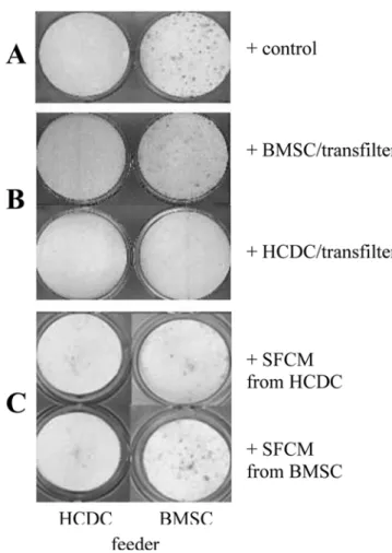
, cementum- forming HCDC did not support osteoclast formation in the in vitro assay, whereas bone-forming osteoblastic BMSC did (Fig. 1A). Overall, HCDC obtained from 3 different patients were employed, yielding comparable results. In total 3 MCDS and 4 SCDS of HCDC were tested.

Using the co-culture experiments, we have established that osteoclast formation capacity of BMSC was abolished when HCDC were present in cell culture inserts (Fig. 1B), while BMSC in the inserts supported osteoclast formation.

Employing SFCM from HCDC instead of cells in the transfilter also had a moderate inhibitory effect, although it did not reach the same level as the HCDC in the inserts (Fig.

1C). Thus, concentration, stability of the molecule(s) and/or continuous supply of the secreted HCDC products can be important for maintenance of this inhibitory effect.

To test if OPG was the major factor responsible for this inhibitory activity of HCDC on osteoclast formation, we investigated the levels of OPG protein in the SFCM from BMSC or HCDC by immunoblotting and established that the levels of OPG protein secreted into the medium by HCDC and BMSC were comparable (Fig. 2).

Discussion

In this study, we have established that putative human cementoblasts (cementum-forming HCDC) do not support osteoclast formation in vitro. This corroborated the earlier evidence obtained from the in vivo transplantation where cementum-like tissue formed by transplanted cells was devoid of TRAP-positive osteoclasts. Furthermore, using the co-culture system, we have also established that HCDC secrete a potent inhibitor of osteoclastogenesis. Our initial experiments aiming at identifying the molecular mechanisms of this inhibition suggested that the prime candidate for such an activity, OPG may not be the solely responsible factor.

The available data on the dental phenotype of OPG- deficient mice (Oshiro et al., 2002) are also consistent with the hypothesis that OPG may not be the sole factor actively Fig. 1. The influence of HCDCs on osteoclast formation. Repre- sentative wells stained with TRAP. A. Mouse bone marrow cells(2 × 10

6

cells/cm

2

) were cultured with HCDC(2 × 10

4

cells/

cm

2

)(Left) or BMSC(2 × 10

4

cells/cm

2

)(Right). B. The mouse bone marrow cells were cultured with HCDCs(Left) or BMSCs(Right) in the 24 well plates and then the cell culture inserts seeded by HCDCs(6 × 10

4

cells/insert) (Lower) or BMSCs(6 × 10

4

cells/

insert)(Upper) were placed on the wells. C. Feeder cells (BMSCs or HCDCs) were co-cultured with mouse bone marrow cells in the presence of SFCM generated from BMSCs (Lower) or HCDCs (Upper).

Fig. 2. Western blot for OPG. Western blot analysis was used to

assess the levels of OPG protein in serum-free conditioned medium

(SFCM) of BMSC and HCDC.

(4)

116 Mi Ri Kim, Won Kyung Yang, Wojciech Grzesik, and Hyun Jung Ko suppressing osteoclast formation in the dental tissues. In

OPG deficient mice no gross abnormalities within cementum (or tooth root) were evident, while alveolar bone was negatively affected; these differences became especially pronounced following a challenge with an orthodontic force.

Periodontal ligament (PDL), apart from its mechanical function, also serves as an important physical and functional barrier separating the non-remodeling mineralized matrices of tooth from that of alveolar bone. Thus, previous studies were focused on the cells residing in PDL proper (or cells obtained for dental follicle) in the context of regulating the formation of resorptive cells. Several reports have shown that cells isolated from PDL are both capable of supporting as well as inhibiting osteoclast formation, somewhat depending on the conditions employed (Wise et al., 2002; Hatakeyama et al., 2003). The common theme emerging from these studies was the clear involvement of the OPG/RANKL/

RANK axis. It is difficult to directly compare the results of these studies to ours, since different animal models, techniques for isolation and expansion of cells as well as modifications of the in vitro osteoclast formation assay were employed. To our knowledge, however, ours is the first study showing direct experimental evidence that cemen- toblasts are actively inhibiting the formation of cells capable of resorbing mineralized matrix. In addition, we show that this inhibition also involves an active mechanism that is independent of osteoprotegerin, a feature that may distinguish cementoblasts from other cells present in periodontium (Hasegawa et al., 2002; Oshiro et al., 2002).

Acknowledgement

This study was supported by a grant (2007-426) from the Asan Institute for Life Sciences, Seoul, Korea.

References

Bell N. RANK ligand and modulation of skeletal remodeling.

J Clin Invest. 2003;111:1120-1122.

Bianco P, Kuznetsov SA, Riminucci M, Fisher LW, Spiegel AM, Robey P. A miniature of human fibrous dysplasia of bone reproduced in immunodeficient mice by trans- plantation of mosaics of normal and Gs alpha-mutated marrow stromal fibroblasts. J Clin Invest. 1998;101:1737- 1744.

Bosshardt DD, Schroeder HE. Cementogenesis reviewed : A comparison between human premolars and rodent molars.

Anat Rec. 1996;245:267-292.

Cha Ji-Hun, Ryoo HM, Woo KM, Kim GS, Baek JH. Dlx3

Plays a Role as a Positive Regulator of Osteoclast Differen- tiation. Int J Oral Biol. 2007;32(3):85-91

Freeman E. Periodontium. In:Ten Cate AR(ed.) Oral Histology, 4th Ed., pp.276-312, Mosby-Year Book, Inc., St. Louis, MO, U.S.A., 1994.

Grzesik WJ, Kuznetsov SA, Uzawa K, Mankani M, Gehron Robey P, Yamauchi M. Normal human cementum-derived cells: isolation, clonal expansion and in vitro and in vivo characterization. J Bone Miner Res. 1998;13:1547-1554.

Grzesik WJ, Cheng H, Oh JS, Kuznetsov SA, Mankani MH, Uzawa K, Robey PG, Yamauchi M. Cementun-forming cells are phenotypically distinct from bone-forming cells. J Bone Miner Res. 2000;15:52-59.

Hasegawa T, Yoshimura Y, Kikuiri T, Yawaka Y, Takeyama S, Matsumoto A, Oguchi H, Shirakawa T. Expression of receptor activator of NF-kappa B ligand and osteoprotegerin in culture of human periodontal ligament cells. J Periodontal Res. 2002;37(6):405-11.

Hatakeyama J, Sreenath T, Hatakeyama Y, Thyagarajan T, Shum L, Gibson CW. The receptor activator of nuclear factor-kappa B ligand-mediated osteoclastogenic pathway is elevated in amelogenin-null mice. J Biol Chem. 2003;12;

278(37): 35743-8.

Jones SJ, Arora M, Boyde A. The rate of osteoclastic destruction of calcified tissues is inversely proportional to mineral density. Calcif Tissue Int. 1995;56(6):554-8.

Katz RW, Hollinger JO, Reddi AH. The functional equivalence of demineralized bone and tooth matrices in ectopic bone induction. J Biomed Mater Res. 1993;27(2):239-45, Krebsbach PH, Kuznetsov SA, Satomura K, Emmons RVB,

Rowe DW, Robey PG. Bone formation in vivo comparison of osteogenesis by transplanted mouse and human marrow stromal fibroblasts. Transplantation. 1997;63:1059-1069.

Kuznetsov SA, Krebsbach PH, Satomura K, Kerr J, Rimmi- nucci M, Benayahu D, Robey PG. Single colony derived strains of human marrow fibroblasts form bone after transplantation in vitro. J Bone Miner Res. 1997;12:1335- 1347.

Oshiro T, Shiotani A, Shibasaki Y, Sasaki T. Osteoclast induction in periodontal tissue during experimental movement of incisors in osteoprotegerin-deficient mice. Anat Rec.

2002;1;266(4):218-25.

Roux S and Orcel P. Bone loss factors that regulate osteoclast differentiation. : an update. Arthritis Res. 2000;2;451-456.

Suda T, Takahashi N, Udagawa N, Jimi E, Gillespie MT, Martin TJ. Modulation of osteoclast differentiation and function by the new members of the tumor necrosis factor receptor and ligand families. Endocr Rev. 1999;20(3): 345-57.

Wise GE, Yao S, Zhang Q, Ren Y. Inhibition of osteo-

clastogenesis by the secretion of osteoprotegerin in vitro by

rat dental follicle cells and its implications for tooth

eruption. Arch Oral Biol. 2002;47(3):247-54.

수치

Fig. 2. Western blot for OPG. Western blot analysis was used to assess the levels of OPG protein in serum-free conditioned medium (SFCM) of BMSC and HCDC.

참조

관련 문서

It also suggest that cell proliferation inhibits by a novel signal transduction for adenosine effect in human FaDu hypopharynx squamous cell carcinoma cells....

Frequency of conidial germination (A) and appressorium formation (B) of Colletotrichum orbiculare and callose formation of plant cells (C) at the

Effects of pulse frequency of low-level laser thrapy (LLLT)on bone nodule formation in rat calvarial cells.. Low-level laser therapy stimulats

Determining optimal surface roughness of TiO₂blasted titanium implant material for attachment, proliferation and differentiation of cells derived from

Histologic evaluation of early human bone response to different implant surfaces2. Histologic evaluation of human bone integration on machined and

Results: The human breast cancer cell subline MCF-7/MX5 cells selected in the presence of 5 µg/ml mitoxantrone (MX) were more resistant to MX (15.7... Western blot and

Approved clinical use of bone marrow stem cells for myocardial infarction treatment... Cardiac

4 > Effects of Taro on COX-2 expression and iNOS expression(hot water) in human thyroid cancer cells. The cells were pretreated for 48hours with either