http://dx.doi.org/10.11620/IJOB.2017.42.4.149 pISSN 1226-7155, eISSN 2287-6618
Cyclooxygenase-2 (COX-2)-mediated prostaglandin E
2(PGE
2) plays a key role in development and progression of inflammatory responses and Porphyromonas gingivalis is a common endodontic pathogen. In this study, we investigated induction of COX-2 and PGE
2by P. gingivalis in human dental pulp cells (HDPCs). P. gingivalis increased expression of COX-2, but not that of COX-1. Increased levels of PGE
2were released from P. gingivalis-infected HDPCs and this PGE
2increase was blocked by celecoxib, a selective COX-2 inhibitor. P. gingivalis activated all three types of mitogen-activated protein kinases (MAPKs). P.
gingivalis-induced activation of nuclear factor-κB (NF-κB)
was demonstrated by the results of phosphorylation of NF-κ B p65 and degradation of inhibitor of κB-α (IκB-α).
Pharmacological inhibition of each of the three types of MAPKs and NF-κB substantially attenuated P. gingivalis- induced PGE
2production. These results suggest that P.
gingivalis should promote endodontic inflammation by
stimulating dental pulp cells to produce PGE
2.
Key words: Human dental pulp cells, prostaglandin E
2, cyclooxygenase-2, Porphyromonas gingivalis
Introduction
Endodontic infections refer to those that occur within the tooth pulp, root canal system or at the root apex. Normally the pulp and root canal system are sterile. However, bacteria may enter through cracks around restorations, areas of exposed dentin and possibly microfracture, or through trauma to the tooth. Endodontic infections have a polymicrobial nature, with obligate anaerobic bacteria dominating the microbiota in primary infections. The most prevalent cultivable bacteria from root canals are Fusobacterium nucleatum, Porphyromonas gingivalis, Pseudoramibacter alactolyticus, Parvimonas micra, Streptococcus mitis, S. intermedius, and other streptococci [1-4].
It is noteworthy that the well-known periodontal pathogen, P.
gingivalis, is one of the commonest bacteria detected in endodontic infections and it can participate in the pathogenesis of apical periodontitis [5].
The main cellular components of the pulp are peripherally located odontoblasts and stromal fibroblasts. There are also undifferentiated mesenchymal and immune cells. Cells in human dental pulp that express Toll-like receptors contribute to trigger immune responses to bacteria [6,7]. Increased expression of pro-inflammatory mediators are found in inflamed pulp, including cytokines, chemokines, adhesion molecules, and
Induction of Prostaglandin E
2by Porphyromonas gingivalis in Human Dental Pulp Cells
So-Hee Kim
1, Yun-Woong Paek
2and In-Chol Kang
1,*1
Department of Oral Microbiology, School of Dentistry, Chonnam National University, Gwangju, 61186, Republic of Korea
2
Department of Physical Therapy, Gwangju Health University, Gwangju, 62287, Republic of Korea (received November 20, 2017; revised December 12, 2017; accepted December 13, 2017)
*Correspondence to: In-Chol Kang, Department of Oral Microbiology, School of Dentistry, Chonnam National University, Gwangju, 61186, Republic of Korea
Tel: +82-62-530-4851, E-mail: [email protected] ORCID : 0000-0002-5993-8728
This is an Open-Access article distributed under the terms of the Creative Commons Attribution Non-Commercial License (http://creati- vecommons.org/licenses/by-nc/3.0) which permits unrestricted non- commercial use, distribution, and reproduction in any medium, pro- vided the original work is properly cited.
149
eicosanoids. COX-1 is constitutively expressed in most cells and plays a role in basal physiological functions in several cells and tissues. COX-2, on the other hand, is usually expressed at low or undetectable levels in most tissues and cells, but is significantly induced by inflammatory stimuli [10].
In spite of the importance of P. gingivalis and PGE
2in endodontic pathogenesis, there have been no reports that determined PGE
2production by P. gingivlais in pulp cells.
Therefore, the aim of this study was to investigate the production of PGE
2by P. gingivalis and the involved mechanisms in human dental pulp cells (HDPCs).
Materials and Methods
Reagents
PD98059, SB203580, SP600125, GF109203X, and U73122 were purchased from Calbiochem (San Diego, CA, USA).
Wortmannin, genistein, and SC-514 were purchased from Sigma (St. Louis, MO,USA). Antibodies to phospho-extracellular signal-regulated kinase (ERK), phospho-p38, phospho-c-Jun N-terminal kinase (JNK) were from Cell Signaling Technology (Beverly, MA, USA). Antibodies to phospho-NF-κB p65 and inhibitor of κB-α (IκB-α) were also from Cell Signaling Technology. Anti-glyceraldehyde-3-phosphate dehydrogenase (GAPDH) was from Sigma.
Bacterial culture
P. gingivalis 381 was grown in Trypticase soy broth supplemented with yeast extract (1 mg/ml), hemin (5 µg/ml), and menadione (1 µg/ml). The bacteria were incubated anaerobically (85% N
2, 10% H
2, and 5% CO
2) at 37°C.
HDPCs culture
HDPCs were kindly provided by professor Ji-Yeon Jung (Department of Oral Physiology, Chonnam National University Dental School). HDPCs were grown in minimum essential medium α (Life Technologies, Grand Island, NY, USA)
(Invitrogen, Carlsbad, CA, USA) as specified by the manufacturer and was quantified spectctrophotometrically. First-strand cDNA was synthesized from 1 µg of RNA using random primers (Promega, Madison, WI, USA) and Molony murine leukemia virus reverse transcriptase (Promega). 2 μl of cDNA products were amplified in 25 μl volumes under a layer of mineral oil using a GeneAmp 2700 thermal cycler (Applied Biosystems, Foster City, CA, USA). Each PCR reaction mixture contained 50 mM KCl, 10 mM Tris-HCl, 1.5 mM MgCl
2, 0.2 µM each dNTP, 1 U Taq DNA polymerase, and 0.5 μM of each primer.
Each cycle consisted of denaturation at 94℃ (30 s), annealing at 57℃ (30 s), and extension at 72℃ (60 s). The sequences of primers were 5′-TTCAAATGAGATTGTGGGAAAATTGCT- 3′, 5′-AGTTCATCTCTGCCTGAGTATCTT-3′ for COX-2 (305 bp); 5′-GAGTCTTTCTCCAACGTGAGC-3′, 5′-ACCGGTAC TTGAGTTTCCCA-3′ for COX-1 (350 bp); and 5′-AGCGGGAAA TCGTGCGTG-3′, 5′-CAGGGTACATGGTGGTGCC-3′ for β -actin (300 bp). The PCR products of 10 µl were fractionated on 1.2% (w/v) agarose gels containing RedSafe (Intron Biotechnology, Korea), visualized by UV transillumination, and photographed.
ELISA
The HDPCs were seeded in 12-well plates (3×10
5cells/well).
The next day, the cells were stimulated with P. gingivalis for various times. Cell culture supernatants were sampled and centrifuged at 100 ×g for 10 min for clarification of debris.
The levels of PGE
2was quantified using commercial ELISA kits (R&D Systems, Minneapolis, MN, USA) according to the manufacturer’s directions.
Western blot
HDPC cells were plated onto 10-cm dishes (2×10
6cells/dish).
The next day, the cells were stimulated with P. gingivalis for
various times. The cells were harvested and lysed with 300
µl of Cell Lysis Buffer (Cell Signaling Technology). 30-50 μg
of each boiled sample was resolved by SDS-PAGE (10%) and
transferred to a polyvinylidene difluoride membrane (Bio-Rad,
Hercules, CA, USA). The membrane was probed with rabbit polyclonal antibodies against phospho-ERK, phosphor-p38, phosphor-JNK, phosphor-p65, or IκBα (1:1000, Cell Signaling Technology) and a 1:1500 dilution or horseradish peroxidase- conjugated goat anti-rabbit IgG secondary antibody (Cell Signaling Technology). Immunoreactive proteins were detected by enhanced chemiluminescence (LumiGLO, Cell Signaling Technology). The same membrane was stripped and reprobed with anti-GAPDH (1:2000)
Statistical analysis
Our experiments were conducted in three independent experiments to confirm the reproducibility of the results. The data are presented as means with standard deviations (SD).
Statistical analysis of one-way analysis of variance (ANOVA) with Tukey-Kramer multiple comparisons test was performed using GraphPad InStat (GraphPad Software, La Jolla, CA, USA). A p-value < 0.05 was considered statistically significant.
Results
Induction of COX-2 mRNA expression
First, we determined whether P. gingivalis could induce COX-2 and COX-1 mRNAs in HDPCs. HDPCs were infected with increasing MOIs of P. gingivalis for 3 h. Total RNA was isolated and levels of COX-1 and COX-2 mRNA were determined by RT-PCR. Expression of COX-2 mRNA was induced by P. gingivalis in a multiplicity of infection (MOI)-dependent manner. A relatively low MOI of 1:10 could induce COX-2 mRNA. In contrast, COX-1 mRNA was constitutively expressed and not altered by P. gingivalis infection (Fig. 1 A). In order to determine the time course of COX-2 mRNA expression, HDPCs were infected with P.
gingivalis (MOI=1:100) for various time periods from 2 h to
16 h. Strong COX-2 expression was demonstrated at 2-4 h postinfection but the COX-2 message did not appear at 8-h and 16-h time points (Fig. 1 B).
Production of PGE
2To determine whether the induction of COX-2 mRNA leads to increased production of PGE
2, PGE
2concentrations of the culture supernatants were measured by ELISA. P. gingivalis stimulated HDPCs to produce PGE
2. Three to four-fold increase of PGE
2production was demonstrated in P. gingivalis-infected cells at both time points of 12 h and 24 h. Moreover, this PGE
2increase was completely blocked by celecoxib, a selective COX-2 inhibitor (Fig.2).
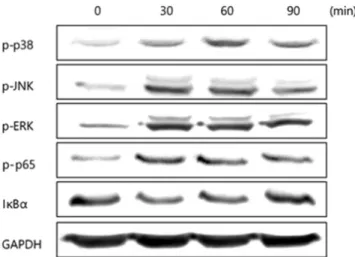
Activation of MAPKs and NF-κB pathways
As activation of mitogen-activated kinases (MAPKs)and NF- κB plays important roles in the induction of COX-2, we determined whether MAPKs and NF-κB pathways are activated by P. gingivalis in HDPCs. Western blot analysis demonstrated that P. gingivalis induced phosphorylation of all three types
Fig. 2. PGE2 production by HDPCs in response to P.
gingivalis. HDPCs were pretreated with celecoxib (10 μM) for 1 h and then infected with P. gingivalis (1:100) for 12 or 24 h.
PGE2 concentrations of the culture supernatants were measured by ELISA. Data are the means±S.D. of a representative experiment performed in triplicate. The asterisks indicate significant differences .
Fig. 1. COX-2 mRNA expression by HDPCs in response to P. gingivalis. (A) HDPCs were infected with increasing MOIs of P.
gingivalis for 3 h. (B) HDPCs were infected with P. gingivalis (1:100) for various times. Total RNA was isolated and levels of COX-1 and COX-2 mRNA were determined by RT-PCR.
Fig. 3. Activation of MAPKs and NF-κB in P. gingivalis- infected HDPCs. HDPCs were infected with P. gingivalis (1:100) for the indicated time periods. Cell lysates were prepared and Western blot analysis was performed for phospho-p38, phospho-ERK, phospho-JNK, phospho-p65, or IκB-α.
of MAPKs (ERK, p38, and JNK) through the time course of 30-90 min. Phosphorylation of p65 NF-κB and degradation of IκB-α were also demonstrated (Fig. 3).
Effects of various signaling inhibitors
In order to evaluate the relative importance of various signaling pathways in P. gingivalis-induced PGE
2production in HDPCs, specific pharmacological inhibitors were used.
HDPCs were pretreated with GF109203X (proteinkinase C, 1 µM), wortmannin (phosphatidylinositol 3-kinase, 100 nM),
Fig. 4. Effect of various signaling inhibitors on P. gingivalis- stimulated PGE2 in HDPCs. HDPCs were pretreated for 1 h with GF109203X (1 µM), wortmannin (100 nM), U73122 (10 µM), genistein (50 µM), PD98059 (50 µM), SB203580 (10 µM), SP600125 (10 µM), or SC-514 (30 µM) and then infected with P.
gingivalis (1:100) for 12 h. PGE2 concentrations of the culture supernatants were measured by ELISA. Data are the means±S.D.
of a representative experiment performed in triplicate. The asterisks indicate significant differences compared to P. gingivalis stimulation without inhibitors.
blocked PGE
2production stimulated by P. gingivalis. In contrast, PGE
2production was significantly elevated in the presence of U73122 and wortmannin.
Discussion
The present study demonstrated that P. gingivalis stimulates HDPCs to express COX-2 and to produce PGE
2without affecting COX-1 expression. The induction of PGE
2production was COX-2-mediated, as it was completely blocked by celecoxib, a selective COX-2 inhibitor. The use of selective COX-2 inhibitors has resulted in pain relief after endodontic treatment [11].
This study demonstrated the importance of MAPKs and NF-κ B in P. gingivalis-stimulated PGE
2production by HDPCs.
Increased phosphorylation of ERK, p38, and JNK was observed in P. gingivalis-infected HDPCs and pharmacological inhibition of MAPKs blocked the PGE
2production. COX-2 is the primary COX controlling PGE
2synthesis in response to inflammatory stimuli. Transcription of COX-2 gene requires binding of transcription factors, including NF-κB, C/EBP, and CREB, to the promoter region of COX-2 gene [12]. As MAPKs regulate these transcription factors, activated MAPKs may positively regulate the activity of the transcription factors in P.
gingivalis-stimulated HDPCs [13]. The present study showed involvement of NF-κB activation in P. gingivalis-induced PGE
2production. The NF-κB activation was demonstrated by the results of p65 phosphorylation and IκB-α degradation [14].
Activation of NF-κB mainly occurs via phosphorylation and subsequent degradation of IκB-α. Optimal induction of NF-κB target genes also requires phosphorylation of NF-κB proteins, such as p65 [15].
The present study showed that genistein strongly inhibited
the PGE
2induction, implying involvement of protein tyrosine
kinases. A previous report showed that genistein treatment
exerted a significant inhibitory effect on NF-κB activation,
leading to downregulation of COX-2 in gastric cancer cells [16].
Therefore, the inhibitory action of genistein in our study may also be mediated by inhibition of NF-κB activation. Interestingly, PGE
2production by P. gingivalis was significantly increased by inhibition of phospholipase C or phosphatidylinositol 3-kinase. Further studies are needed to define the role of these signaling pathways in PGE
2production by HDPCs.
The present study showed for the first time that P.
gingivalis stimulated HDPCs to express COX-2, leading to induced production of PGE
2. These results suggest that P.
gingivalis should promote endodontic inflammation, at least in part, by stimulating dental pulp cells to produce PGE
2.
Acknowledgments
This study was supported by a grant (CRI15001-1) from Chonnam National University Hospital Biomedical Research Institute.
Conflict of interest
The authors declare no conflict of interest.
References
1. Fouad AF. Endodontic Microbiology and Pathobiology:
Current State of Knowledge. Dent Clin North Am. 2017;
61:1-15. doi: 10.1016/j.cden.2016.08.001.
2. Narayanan LL and Vaishnavi C. Endodontic microbiology. J Conserv Dent. 2010;13:233-239. doi: 10.4103/0972-0707.
73386.
3. Peciuliene V, Maneliene R, Balcikonyte E, Drukteinis S and Rutkunas V. Microorganisms in root canal infections: a review. Stomatologija. 2008;10:4-9.
4. Siqueira JF, Jr., Rocas IN, Ricucci D and Hulsmann M.
Causes and management of post-treatment apical periodontitis. Br Dent J. 2014;216:305-312. doi: 10.1038/
sj.bdj.2014.200.
5. Siqueira JF, Jr., Rocas IN and Silva MG. Prevalence and clonal analysis of Porphyromonas gingivalis in primary endodontic infections. J Endod. 2008;34:1332-1336. doi:
10.1016/j.joen.2008.08.021.
6. Hirao K, Yumoto H, Takahashi K, Mukai K, Nakanishi T and Matsuo T. Roles of TLR2, TLR4, NOD2, and NOD1 in pulp fibroblasts. J Dent Res. 2009;88:762-767. doi: 10.1177/
0022034509341779.
7. Maltos KL, Menezes GB, Caliari MV, Rocha OA, Santos JM, Alves DL, Duarte ID and Francischi JN. Vascular and cellular responses to pro-inflammatory stimuli in rat dental pulp.
Arch Oral Biol. 2004;49:443-450. doi: 10.1016/j.archoralbio.
2004.01.004.
8. Martinho FC, Chiesa WM, Leite FR, Cirelli JA and Gomes BP. Antigenicity of primary endodontic infection against macrophages by the levels of PGE2production.JEndod.
2011;37:602-607.doi:10.1016/j.joen.2010.12.005.
9. Coon D, Gulati A, Cowan C and He J. The role of cyclooxygenase-2 (COX-2) in inflammatory bone resorption.
J Endod. 2007;33:432-436. doi: 10.1016/j.joen.2006.12.001.
10. Caughey GE, Cleland LG, Penglis PS, Gamble JR and James MJ. Roles of cyclooxygenase (COX)-1 and COX-2 in prostanoid production by human endothelial cells: selective up-regulation of prostacyclin synthesis by COX-2. J Immunol. 2001;167:2831-2838.
11. Barden J, Edwards JE, McQuay HJ and Moore RA. Single dose oral celecoxib for postoperative pain. Cochrane Database Syst Rev. 2003;(2):CD004233. doi: 10.1002/
14651858.cd004233.
12. Chun KS and Surh YJ. Signal transduction pathways regulating cyclooxygenase-2 expression: potential molecular targets for chemoprevention. Biochem Pharmacol. 2004;68:
1089-1100. doi: 10.1016/j.bcp.2004.05.031.
13. Zhang YL and Dong C. MAP kinases in immune responses.
Cell Mol Immunol. 2005;2:20-27.
14. Viatour P, Merville MP, Bours V and Chariot A.
Phosphorylation of NF-κB and IκB proteins: implications in cancer and inflammation. Trends Biochem Sci. 2005;30:
43-52. doi: 10.1016/j.tibs.2004.11.009.
15. Lawrence T. The nuclear factor NF-κB pathway in inflammation. Cold Spring Harb Perspect Biol. 2009;1:
a001651. doi: 10.1101/cshperspect.a001651.
16. Li YS, Wu LP, Li KH, Liu YP, Xiang R, Zhang SB, Zhu LY and Zhang LY. Involvement of nuclear factor κB (NF-κB) in the downregulation of cyclooxygenase-2 (COX-2) by genistein in gastric cancer cells. J Int Med Res. 2011;
39:2141-2150. doi: 10.1177/147323001103900610.