ABSTRACT
Background: Cervical cancer is the fourth common cancer in women worldwide. The Papanicolau test is the primary screening procedure to detect abnormal cervical cells.
Colposcopy is the main procedure for discriminating high-grade cervical lesions. The study aimed at clarifying the discrepancy between cervical cytology and colposcopic biopsy histology as well as confounding factors.
Methods: Eligible patients visited thirteen tertiary hospitals for colposcopic biopsy following cervical cytology and human papillomavirus (HPV) genotypes between January and
December 2018. Baseline characteristics including age, body mass index (BMI), and parity were collected.
Results: In our study, 3,798 eligible patients were included. Mean age of patients was 42.7 (19–88) years and mean BMI was 22.5 (16.9–34.1) kg/m
2. The referred cervical cytologic findings consisted of 495 normal, 1,390 atypical squamous cells of undetermined significance, 380 atypical squamous cells cannot exclude high-grade squamous intraepithelial lesion, 792 low-grade squamous intraepithelial lesion, 593 high-grade
squamous intraepithelial lesion, 79 atypical glandular cells, 46 squamous cell carcinoma, and 23 adenocarcinoma. HPV-positive findings were found in 3,008 (79.2%) patients and were not detected in 914 (24.1%) cases. The risk of unexpected low-grade lesions from histology
Original Article
Received: Feb 17, 2021 Accepted: May 24, 2021 Address for Correspondence:
Kyung-Jin Min, MD
Department of Obstetrics and Gynecology, Korea University Ansan Hospital, 123 Jeokgeum-ro, Danwon-gu, Ansan 15355, Republic of Korea.
E-mail: [email protected]
© 2021 The Korean Academy of Medical Sciences.
This is an Open Access article distributed under the terms of the Creative Commons Attribution Non-Commercial License (https://
creativecommons.org/licenses/by-nc/4.0/) which permits unrestricted non-commercial use, distribution, and reproduction in any medium, provided the original work is properly cited.
ORCID iDs Yung-Taek Ouh
https://orcid.org/0000-0001-5887-4497 Ji Jeong Park
https://orcid.org/0000-0003-1905-5821 Minjoo Kang
https://orcid.org/0000-0003-1658-3389 Miseon Kim
https://orcid.org/0000-0002-5118-9275 Jae Yun Song
https://orcid.org/0000-0002-2752-5638 So Jin Shin
https://orcid.org/0000-0002-7432-6025 Seung-Hyuk Shim
https://orcid.org/0000-0001-8043-2257 Heon Jong Yoo
https://orcid.org/0000-0003-4808-3450 Maria Lee
https://orcid.org/0000-0002-8017-3176
Yung-Taek Ouh ,
1,2Ji Jeong Park ,
3Minjoo Kang ,
3Miseon Kim ,
4Jae Yun Song ,
5So Jin Shin ,
6Seung-Hyuk Shim ,
7Heon Jong Yoo ,
8Maria Lee ,
9Sung-Jong Lee ,
10Whan Shin ,
11,12Gun Oh Chong ,
13Min Chul Choi ,
14Chel Hun Choi ,
15and Kyung-Jin Min
161
Department of Obstetrics and Gynecology, Korea University Guro Hospital, Seoul, Korea
2
Department of Obstetrics and Gynecology, Graduate School of Medicine, Kangwon National University, Chuncheon, Korea
3
National Evidence-based Healthcare Collaborating Agency (NECA), Seoul, Korea
4
Department of Obstetrics and Gynecology, CHA Gangnam Medical Center, Seoul, Korea
5
Department of Obstetrics and Gynecology, Korea University Anam Hospital, Seoul, Korea
6
Department of Obstetrics and Gynecology, Keimyung University Dongsan Medical Center, Daegu, Korea
7
Department of Obstetrics and Gynecology, Konkuk University Hospital, Seoul, Korea
8
Department of Obstetrics and Gynecology, Chungnam National University Hospital, Daejeon, Korea
9
Department of Obstetrics and Gynecology, Seoul National University Hospital, Seoul, Korea
10
Department of Obstetrics and Gynecology, Seoul St. Mary's Hospital, College of Medicine, The Catholic University of Korea, Seoul, Korea
11
Department of Obstetrics and Gynecology, Yonsei University Severance Hospital, Seoul, Korea
12
Department of Obstetrics and Gynecology, Dankook University Hospital, Dankook University, Cheonan, Korea
13
Department of Obstetrics and Gynecology, Kyungpook National University Chilgok Hospital, Daegu, Korea
14
Comprehensive Gynecologic Cancer Center, CHA Bundang Medical Center, Seongnam, Korea
15
Department of Obstetrics and Gynecology, Samsung Medical Center, Seoul, Korea
16
Department of Obstetrics and Gynecology, Korea University Ansan Hospital, Ansan, Korea
Discrepancy between Cytology and
Histology in Cervical Cancer Screening:
a Multicenter Retrospective Study (KGOG 1040)
Obstetrics & Gynecology
Sung-Jong Lee
https://orcid.org/0000-0002-6077-2649 Whan Shin
https://orcid.org/0000-0003-1050-2648 Gun Oh Chong
https://orcid.org/0000-0003-4887-4017 Min Chul Choi
https://orcid.org/0000-0003-4509-6731 Chel Hun Choi
https://orcid.org/0000-0002-0199-6669 Kyung-Jin Min
https://orcid.org/0000-0002-5783-4968 Funding
This study was part of the health technology assessment project (No. NECA-A-19-007) funded by the National Evidence-based Healthcare Collaborating Agency (NECA) in Korea.
Disclosure
The authors have no potential conflicts of interest to disclose.
Data Availability Statement
All data relevant to the study are included in the article or uploaded as supplementary information. The data that support the findings of this study are available from the corresponding author upon reasonable request.
Author Contributions
Conceptualization: Ouh YT, Park JJ, Kang M, Min KJ. Data curation: Ouh YT, Song JY, Shin SJ, Shim SH, Yoo HJ, Lee M, Lee SJ, Shin W, Chong GO, Choi MC, Choi CH, Min KJ. Formal analysis: Park JJ, Kang M, Min KJ.
Investigation: Ouh YT, Song JY, Shin SJ, Shim SH, Yoo HJ, Lee M, Lee SJ, Shin W, Chong GO, Choi MC, Choi CH, Min KJ. Methodology: Park JJ, Kang M, Min KJ. Validation: Ouh YT, Park JJ, Kang M, Min KJ. Visualization: Park JJ, Kang M, Min KJ. Writing - original draft: Ouh YT, Min KJ. Writing - review & editing: Ouh YT, Park JJ, Kang M, Kim M, Song JY, Shin SJ, Shim SH, Yoo HJ, Lee M, Lee SJ, Shin W, Chong GO, Choi MC, Choi CH, Min KJ.
was higher in patients > 45 years (odds ratio [OR], 2.137; 95% confidence intervals [CIs], 1.475–3.096). In contrast, the risk of unexpected high-grade lesions from colposcopic biopsy was lower in patients ≥ 45 years (OR, 0.530; 95% CI, 0.367–0.747) and HPV 16/18 infection was higher than other HPV (OR, 1.848; 95% CI, 1.385–2.469).
Conclusion: Age and HPV genotypes were responsible for the discrepancies between
cytology and histology. Precautions should be taken for women over the age of 45 in triage for colposcopy in order to avoid unnecessary testing.
Keywords: HPV; Cervical Cytology; Colposcopy; Discrepancy; Histology
INTRODUCTION
Cervical intraepithelial neoplasia (CIN) is known to progress to invasive cervical cancer in some cases.
1Patients with more progressive disease have the higher risk for malignant transformation. The overall prevalence of high-risk human papillomavirus (hrHPV) was calculated at 12% in normal cytology while hrHPV was positive in 89% of cervical cancer cases.
2In Korea, human papillomavirus (HPV) testing is currently recommended to triage women with abnormal cytology and the combined test of cervical cytology with HPV genotyping has reduced the incidence rate of cervical cancer.
3Colposcopy is the most common procedure performed in patients referred for cervical cytologic abnormalities. It facilitates detailed localization of the suspected cervical lesion, determines its severity, and eases biopsy.
4Colposcopic biopsy is primarily used to discriminate high-grade from low-grade lesions, in order to limit unnecessary surgical excision of the cervix.
5In addition, it differentiates CIN lesions from invasive cervical cancer and enables ablation treatment.
6A number of patients undergoing colposcopic biopsy after cervical cancer screening were diagnosed as low-grade intraepithelial lesions.
7Low-grade lesions (CIN1) were unlikely to progress to invasive carcinoma (CIN3).
8In fact, the risk of progression to a high-grade lesion was not significantly different in no CIN as well as CIN1 patients.
Inconsistencies between cervical cytology and colposcopic biopsy histology can lead to confusion in diagnosis and clinical management.
9Investigating this discrepancy and factors influencing it, are needed to avoid excessive unnecessary testing and underdiagnoses of high- grade cervical lesions. Therefore, we aimed at investigating the discrepancy between cytology and histology in cervical cancer screening.
METHODS
Study population
A multicenter retrospective analysis was performed in this study. Patients with both
cervical cytology and HPV test followed by colposcopic biopsy of cervix between January
and December 2018 were identified at thirteen certified tertiary hospitals (Supplementary
Data 1). The inclusion criteria was for patients who underwent HPV test of HPV genotypes
including HPV 16/18, so patients who did not have data for HPV genotypes such as Hybrid
Capture II were excluded.
Data collection
Baseline characteristics including age, height, weight, and parity were obtained from patients. Cytological, histologic, and HPV-genotype results were recorded in database.
Patients without data on cytology or HPV DNA tests were excluded from the analyses.
All patients with abnormal cytology and/or positive for HPV were referred participating institutions.
To analyze the risk of discordance of cytology and histology, body mass index (BMI) over 25 is defined as overweight. In addition, the risk was analyzed before and after 45 years old, the age of generally entering perimenopause.
10The patients' group as HPV 16/18 was representative to not only positive for HPV 16 and/or 18, but also to co-infected with other hrHPV groups. HPV others included patients who were positive for hrHPV genotypes except for HPV 16 and 18.
Definition of correlation
Overcall was defined as the patients whose cervical smear result of atypical squamous cells cannot exclude high-grade squamous intraepithelial lesion (ASC-H)/high-grade squamous intraepithelial lesion (HSIL) or higher grade lesion followed by cervical biopsy showing negative or CIN1.
11Undercall was defined as the patients whose cervical smear result of negative for
intraepithelial lesion or malignancy, atypical squamous cells of undetermined significance (ASCUS) followed by cervical biopsy showing CIN2 or higher lesion.
Statistical analysis
Baseline data are presented as mean ± standard deviation for continuous variables or frequency (%) for categorical variables. A logistic regression model was used to evaluate the odds ratio (OR) and 95% confidence intervals (CIs) to predict the results associated with epidemiological characteristics. All P values less than 0.05 were considered statistically significant. Data were analyzed using SPSS software (version 22.0; SPSS Inc., Chicago, IL, USA).
Ethics statement
The Institutional Review Board of the National Evidence-based Healthcare Collaborating Agency (NECA) in Korea approved the study (IRB No. NECAIRB19-015-2). The IRB of each institute approved the collection of data within a database of each institution. Information obtained from patients was coded for analysis, so the requirement for informed consent or parental permission was waived.
RESULTS
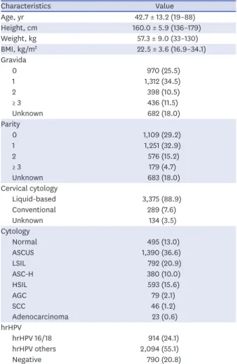
A total of 3,798 patients tested for hrHPV and cervical cytology, followed by colposcopic biopsy met the inclusion criteria. The baseline characteristics of the patients are displayed in Table 1. The mean age was 42.7 (19–88) years and mean BMI was 22.5 (16.9–34.1) kg/m
2. Nine-hundred seventy (25.5%) patients were nulligravida and 1,109 (29.2%) were nulliparity.
The most prevalent cytological abnormalities referred for colposcopic biopsy was ASCUS
(1,390 patients, 36.6%). In addition, 495 patients (13.0%) were normal, 380 (10.0%) were
ASC-H, 792 (20.9%) were low-grade squamous intraepithelial lesion (LSIL), and 593 (15.6%)
were HSIL. HPV was positive for 3,008 (79.2%) and HPV 16/18 was found in 914 (24.1%) patients. Various types of HPV tests were used (Supplementary Table 1), of which Anyplex II HPV 28 (Seegene), the most common, was performed in 1,358 (35.8%) of all patients.
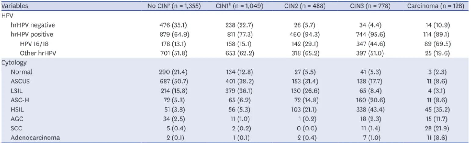
Distribution of hrHPV positivity and cytologic findings in relation to histologic results were shown in Table 2. In patients with CIN, the positive rate of hrHPV was 64.9%, and HPV 16/18 was positive in 13.1%. On the other hand, in patients with CIN3, the positive rate of hrHPV was 95.6%, and the 69.5% of patients were positive for HPV 16/18. In particular of 128 patients with carcinoma on histologic finding, 10.9% of patients were negative for hrHPV.
Among the patients with no CIN, ASCUS in the cytology result were most common (50.7%), followed by normal cytology (21.4%). The preceding cytology findings of patients with CIN1 were most common in the order of ASCUS (38.2%) and LSIL (36.1%), and in patients with CIN2, ASCUS (31.4%) and LSIL (26.6%) were the most common. In CIN3, HSIL was the most common with 43.4%, followed by ASC-H with 20.6%.
Table 1. Overview of patients' baseline characteristics (n = 3,798)
Characteristics Value
Age, yr 42.7 ± 13.2 (19–88)
Height, cm 160.0 ± 5.9 (136–179)
Weight, kg 57.3 ± 9.0 (33–130)
BMI, kg/m
222.5 ± 3.6 (16.9–34.1)
Gravida
0 970 (25.5)
1 1,312 (34.5)
2 398 (10.5)
≥ 3 436 (11.5)
Unknown 682 (18.0)
Parity
0 1,109 (29.2)
1 1,251 (32.9)
2 576 (15.2)
≥ 3 179 (4.7)
Unknown 683 (18.0)
Cervical cytology
Liquid-based 3,375 (88.9)
Conventional 289 (7.6)
Unknown 134 (3.5)
Cytology
Normal 495 (13.0)
ASCUS 1,390 (36.6)
LSIL 792 (20.9)
ASC-H 380 (10.0)
HSIL 593 (15.6)
AGC 79 (2.1)
SCC 46 (1.2)
Adenocarcinoma 23 (0.6)
hrHPV
hrHPV 16/18 914 (24.1)
hrHPV others 2,094 (55.1)
Negative 790 (20.8)
Values are presented as mean ± standard deviation (range) or number (%).
BMI = body mass index, ASCUS = atypical squamous cells of undetermined significance, LSIL = low-grade
squamous intraepithelial lesion, ASC-H = atypical squamous cells cannot exclude high-grade squamous
intraepithelial lesion, HSIL = high-grade squamous intraepithelial lesion, AGC = atypical glandular cells, SCC =
squamous cell carcinoma, hrHPV = high-risk human papillomavirus.
We investigated the overcall defined as unexpected low-grade results as ≤ CIN1 from colposcopic biopsy from patients referred for abnormal cytology (≥ ASC-H). This amounted to 299 patients in the study. To investigate the associated factors, we performed a logistic regression analysis of variables. The OR of overcall was higher in age ≥ 45 (OR, 2.137; 95%
CI, 1.475–3.096) as shown in Table 3. Multiparous women were a higher risk group at overcall compared to nulliparous women, but insignificant in multivariate analysis (OR, 1.090; 95%
CI, 0.775–1.535). Overweight (BMI ≥ 25) infection was not associated with overcall while HPV 16/18 were at lower risk of overcall compared to other hrHPV or HPV negative (P < 0.05).
Conversely, the findings of undercall defined as ≥ CIN2 following referral was ≤ ASCUS in 373 patients (Table 4). Patients aged over 45 years (OR, 0.530; 95% CI, 0.376–0.747) were at lower risk while HPV 16/18 positive cases were at higher risk for undercall compared to other hrHPV or HPV negative (P < 0.01). On the other hand, BMI or parity did not affect the results of colposcopy.
Table 2. Distribution of hrHPV types and cytologic findings in relation to histology lesions (n = 3,798)
Variables No CIN
a(n = 1,355) CIN1
b(n = 1,049) CIN2 (n = 488) CIN3 (n = 778) Carcinoma (n = 128)
HPV
hrHPV negative 476 (35.1) 238 (22.7) 28 (5.7) 34 (4.4) 14 (10.9)
hrHPV positive 879 (64.9) 811 (77.3) 460 (94.3) 744 (95.6) 114 (89.1)
HPV 16/18 178 (13.1) 158 (15.1) 142 (29.1) 347 (44.6) 89 (69.5)
Other hrHPV 701 (51.8) 653 (62.2) 318 (65.2) 397 (51.0) 25 (19.6)
Cytology
Normal 290 (21.4) 134 (12.8) 27 (5.5) 41 (5.3) 3 (2.3)
ASCUS 687 (50.7) 401 (38.2) 153 (31.4) 138 (17.7) 11 (8.6)
LSIL 214 (15.8) 379 (36.1) 130 (26.6) 65 (8.4) 4 (3.1)
ASC-H 72 (5.3) 65 (6.2) 72 (14.8) 160 (20.6) 11 (8.6)
HSIL 51 (3.8) 56 (5.3) 103 (21.1) 338 (43.4) 45 (35.2)
AGC 34 (2.5) 11 (1.0) 1 (0.2) 18 (2.3) 15 (11.7)
SCC 5 (0.4) 2 (0.2) 0 (0.0) 11 (1.4) 28 (21.9)
Adenocarcinoma 2 (0.1) 1 (0.1) 2 (0.4) 7 (1.0) 11 (8.6)
Values are presented as number (%).
hrHPV = high-risk human papillomavirus, CIN = cervical intraepithelial neoplasia, HPV = human papillomavirus, ASCUS = atypical squamous cells of undetermined significance, LSIL = low-grade squamous intraepithelial lesion, ASC-H = atypical squamous cells cannot exclude high-grade squamous intraepithelial lesion, HSIL = high-grade squamous intraepithelial lesion, AGC = atypical glandular cells, SCC = squamous cell carcinoma.
a
No CIN included inflammatory changes as well as normal;
bCIN1 included koilocytic changes.
Table 3. Logistic regression results for predicting the ≤ CIN1 following ≥ ASC-H (overcall) (n = 299)
Variables At risk cases Univariate Multivariate
OR (95% CI) P value OR (95% CI) P value
Age, yr < 0.010 < 0.010
≥ 45 196/299 (65.6) 2.795 (2.181–3.581) 2.137 (1.475–3.096)
< 45 103/299 (34.4) 1.000 1.000
BMI, kg/m
20.065 0.620
≥ 25 52/299 (17.4) 1.366 (0.981–1.901) 1.090 (0.775–1.535)
< 25 156/299 (52.2) 1.000 1.000
Parity < 0.010 0.260
Multiparous 221/299 (73.9) 2.373 (1.749–3.219) 1.277 (0.834–1.956)
Nulliparous 55/299 (18.4) 1.000 1.000
HPV types 0.007 0.028
hrHPV 16/18 61/299 (20.4) 1.000 1.000
Other hrHPV 155/299 (51.8) 1.118 (0.822–1.519) 1.294 (0.897–1.867)
HPV negative 83/299 (27.8) 1.642 (1.162–2.319) 1.804 (1.168–2.785)
Values are presented as number (%).
CIN = cervical intraepithelial neoplasm, ASC-H = atypical squamous cells cannot exclude high-grade squamous intraepithelial lesion, OR = odds ratio, CI =
confidence interval, BMI = body mass index, HPV = human papillomavirus, hrHPV = high-risk human papillomavirus.
DISCUSSION
This study investigated the current overall status of colposcopic biopsy and analyzed the associated factors for the discrepancy between cytology and colposcopic biopsy histology in Korea. The most common cytological finding referred for colposcopy was ASCUS, followed by LSIL, HSIL, and normal cytology. Age and HPV-genotypes affected the discrepancy between cytology and colposcopic biopsy histology. The unexpected colposcopic biopsy histology in negative or CIN1 findings following cytology of ASC-H or higher grade was found in 299 patients. Patients over 45 years and hrHPV others (compared to HPV 16/18) were at higher risk for initially overestimated before cervical biopsy. In contrast, patients under 45 years were at higher risk for underestimated before cervical biopsy. As expected, HPV 16/18 were at higher risk for high-grade lesions than hrHPV others. When targeting for colposcopic biopsy following cervical cancer screening, HPV genotypes, age, and cytology should be considered.
Immature squamous metaplasia or air-drying artifacts have been known as factors influencing the discrepancy between cytology and histology.
12In addition, infectious cause such as bacteria or fungi will reduce the accuracy of cytology. Unlike our findings were patients' age was associated with the accuracy of cytology, previous studies have demonstrated the accuracy of Pap smear in diagnosing cervical intraepithelial lesions by comparing the characteristics of cervical dysplasia versus non-dysplasia.
13Age, smoking, number of abortions, age at first delivery, and number of sexual partners were not
significantly different across both groups. HPV infection is the most important factor, though the genotypes were not investigated.
In our study, histology results tended to be of lower grade compared to those of combined cytology with HPV, in patients over 45 years. First, this can be explained by the fact that the squamous-columnar junction of menopausal women was deeper in the cervical canal than that of pre-menopausal women, thus rendering colposcopic examination unsatisfactory.
14Unfortunately, the results of cervical biopsy (loop electrosurgical excisional procedure [LEEP] or conization) were not included in our data, so further analysis was limited. Second, atrophic changes in postmenopausal women (caused by estrogen deficiency) could lead to confusion in the cytological diagnosis of Pap smear because they may simulate high-grade Table 4. Logistic regression results for predicting the ≥ CIN2 following ≤ ASCUS (undercall) (n = 373)
Variables At risk cases Univariate Multivariate
OR (95% CI) P value OR (95% CI) P value
Age, yr < 0.010 < 0.010
≥ 45 100/373 (26.9) 0.465 (0.366–0.590) 0.530 (0.376–0.747)
< 45 272/373 (73.1) 1.000 1.000
BMI, kg/m
20.348 0.701
≥ 25 40/373 (10.7) 0.843 (0.590–1.205) 0.930 (0.643–1.346)
< 25 186/373 (49.9) 1.000 1.000
Parity < 0.010 0.211
Multiparous 159/373 (42.6) 0.632 (0.495–0.806) 1.234 (0.887–1.716)
Nulliparous 133/373 (35.7) 1.000 1.000
HPV types < 0.010 < 0.010
hrHPV 16/18 146/373 (39.1) 1.000 1.000
Other hrHPV 208/373 (55.8) 0.580 (0.462–0.728) 0.541 (0.405–0.722)
HPV negative 19/373 (5.1) 0.130 (0.080–0.211) 0.233 (0.133–0.408)
Values are presented as number (%).
CIN = cervical intraepithelial neoplasm, ASCUS = atypical squamous cells of undetermined significance, OR = odds ratio, CI = confidence interval, BMI = body
mass index, HPV = human papillomavirus, hrHPV: high-risk human papillomavirus.
lesions.
15The cervical mucosa develops a morphological diversity with age, while dysplasia decreases with age.
16HPV has genotype-specific risk for high-grade lesions and cancer. HPV 16, 18, and 58 are associated with significant risk of CIN3 and invasive cancer.
17HPV testing was approved as a primary screening method by Food and Drug Administration (FDA) in April 2014. We found out that the ratio of HPV 16/18 to hrHPV others was higher in ≥ CIN3 lesions compared to
≤ CIN2 lesions. Colposcopy is recommended for HPV 16/18 infected patients, regardless of cytology findings in the Australian Society for Colposcopy and Cervical Pathology.
18Furthermore, HPV 16/18 infections can be prevented by vaccination fortunately.
Various assays for detecting and genotyping HPV have been introduced in Korea. Among them, the Hybrid Capture 2 HPV test was the first assay detecting 13 hrHPV, which has been approved by the U.S. FDA. Subsequently, various assays to detect hrHPV genotypes including HPV 16 or 18 by real-time polymerase chain reaction were introduced, and the Roche Cobas HPV assay was approved by the U.S. FDA in 2011. Various HPV genotype assays which were investigated in our study are currently used in Korea, and their usefulness has been proved in several literatures.
19-24CIN1 is not considered a precursor of CIN3 or invasive carcinoma. CIN1 is not a target for screening and should not be managed but recommended for observation. However, the accuracy and reproducibility of cervical cytology are limited.
13Cytology diagnosis accuracy mainly depends on the pathologists' experience and the characteristics of patients.
25Recently, several studies show that a reduction in unnecessary colposcopy and induce objective cytology results. MicroRNAs (miRNAs) expression underlies the carcinogenesis.
26Cervical exfoliated cells can be used for miRNA detection in the diagnosis of cervical lesions.
In fact, miRNAs expression as a cervical cancer screening tool, not only induces accurate cytology, but also detects high-grade lesions that would be missed in histology.
27Killeen et al.
28evaluated whether stain for p16 and Ki-67 improve the triage of abnormal Pap smears.
Immunostaining was of value in selecting abnormal cytology and had diagnostic accuracy.
This study is the first multicenter analysis about patients who underwent colposcopic biopsy in Korea. Limitations encountered in the study include its multicenter retrospective design and the diverse indications of colposcopy across centers. In addition, final histology of cone or LEEP specimen could not be obtained. Nevertheless, the histologic results from colposcopic biopsy are critically enough to have a significant impact on the course of treatment. Although a large-scale study was conducted on patients with colposcopic biopsy, referred for abnormal cytology and/or positive hrHPV infection. A possible limitation is that our results are insufficient to investigate the sensitivity or specificity of Pap smear and/or HPV genotypes. This is due to different biopsy indications across centers and clinicians. This study includes not only cases which Pap smear and HPV test conducted at the same time, but also cases with abnormal cervical cytology followed by HPV testing. In addition, there was no central review of cervical biopsy histology specimens.
Most women underwent additional procedures such as repeated cytology or colposcopy
unnecessarily. Therefore, establishing an accurate indication of colposcopy is important for
the management of patients with abnormal cytology. Age and HPV 16/18 were responsible
for the majority of discrepancies between cytology and histology results, and should be
considered in cervical cancer screening. We recommend further research to assess efficient cervical cancer screening especially with abnormal cytology.
SUPPLEMENTARY MATERIALS
Supplementary Data 1 Thirteen institutes Click here to view
Supplementary Table 1 Proportion of HPV assay types Click here to view
REFERENCES
1. Moss E, Redman C, Arbyn M. Accuracy of colposcopy-directed punch biopsies. BJOG 2013;120(7):903.
PUBMED | CROSSREF
2. Guan P, Howell-Jones R, Li N, Bruni L, de Sanjosé S, Franceschi S, et al. Human papillomavirus types in 115,789 HPV-positive women: a meta-analysis from cervical infection to cancer. Int J Cancer 2012;131(10):2349-59.
PUBMED | CROSSREF
3. Min KJ, Lee YJ, Suh M, Yoo CW, Lim MC, Choi J, et al. The Korean guideline for cervical cancer screening.
J Gynecol Oncol 2015;26(3):232-9.
PUBMED | CROSSREF
4. Hong DG, Seong WJ, Kim SY, Lee YS, Cho YL. Prediction of high-grade squamous intraepithelial lesions using the modified Reid index. Int J Clin Oncol 2010;15(1):65-9.
PUBMED | CROSSREF
5. Moss EL, Hadden P, Douce G, Jones PW, Arbyn M, Redman CW. Is the colposcopically directed punch biopsy a reliable diagnostic test in women with minor cytological lesions? J Low Genit Tract Dis 2012;16(4):421-6.
PUBMED | CROSSREF
6. Shumsky AG, Stuart GC, Nation J. Carcinoma of the cervix following conservative management of cervical intraepithelial neoplasia. Gynecol Oncol 1994;53(1):50-4.
PUBMED | CROSSREF
7. Castle PE, Gage JC, Wheeler CM, Schiffman M. The clinical meaning of a cervical intraepithelial neoplasia grade 1 biopsy. Obstet Gynecol 2011;118(6):1222-9.
PUBMED | CROSSREF
8. Tverelv LR, Sørbye SW, Skjeldestad FE. Risk for cervical intraepithelial neoplasia grade 3 or higher in follow-up of women with a negative cervical biopsy. J Low Genit Tract Dis 2018;22(3):201-6.
PUBMED | CROSSREF
9. Joste NE, Crum CP, Cibas ES. Cytologic/histologic correlation for quality control in cervicovaginal cytology. Experience with 1,582 paired cases. Am J Clin Pathol 1995;103(1):32-4.
PUBMED | CROSSREF
10. Hong JS, Yi SW, Kang HC, Jee SH, Kang HG, Bayasgalan G, et al. Age at menopause and cause-specific mortality in South Korean women: Kangwha Cohort Study. Maturitas 2007;56(4):411-9.
PUBMED | CROSSREF
11. Gupta R, Hariprasad R, Dhanasekaran K, Sodhani P, Mehrotra R, Kumar N, et al. Reappraisal of cytology- histology correlation in cervical cytology based on the recent American Society of Cytopathology guidelines (2017) at a cancer research centre. Cytopathology 2020;31(1):53-8.
PUBMED | CROSSREF
12. Pimple SA, Amin G, Goswami S, Shastri SS. Evaluation of colposcopy vs cytology as secondary test to triage women found positive on visual inspection test. Indian J Cancer 2010;47(3):308-13.
PUBMED | CROSSREF
13. Nkwabong E, Laure Bessi Badjan I, Sando Z. Pap smear accuracy for the diagnosis of cervical precancerous lesions. Trop Doct 2019;49(1):34-9.
PUBMED | CROSSREF
14. Costa S, De Simone P, Venturoli S, Cricca M, Zerbini ML, Musiani M, et al. Factors predicting human papillomavirus clearance in cervical intraepithelial neoplasia lesions treated by conization. Gynecol Oncol 2003;90(2):358-65.
PUBMED | CROSSREF
15. Waddell CA. The influence of the cervix on smear quality. I: Atrophy. An audit of cervical smears taken post-colposcopic management of intraepithelial neoplasia. Cytopathology 1997;8(4):274-81.
PUBMED | CROSSREF
16. Halford J, Walker KA, Duhig J. A review of histological outcomes from peri-menopausal and post- menopausal women with a cytological report of possible high grade abnormality: an alternative management strategy for these women. Pathology 2010;42(1):23-7.
PUBMED | CROSSREF
17. So KA, Lee IH, Lee KH, Hong SR, Kim YJ, Seo HH, et al. Human papillomavirus genotype-specific risk in cervical carcinogenesis. J Gynecol Oncol 2019;30(4):e52.
PUBMED | CROSSREF
18. Perkins RB, Guido RS, Castle PE, Chelmow D, Einstein MH, Garcia F, et al. 2019 ASCCP risk-based management consensus guidelines for abnormal cervical cancer screening tests and cancer precursors. J Low Genit Tract Dis 2020;24(2):102-31.
PUBMED | CROSSREF
19. Lee DH, Hwang NR, Lim MC, Yoo CW, Joo J, Kim JY, et al. Comparison of the performance of Anyplex II HPV HR, the Cobas 4800 human papillomavirus test and Hybrid Capture 2. Ann Clin Biochem 2016;53(Pt 5):561-7.
PUBMED | CROSSREF
20. Choi JJ, Kim C, Park H. Peptide nucleic acid-based array for detecting and genotyping human papillomaviruses. J Clin Microbiol 2009;47(6):1785-90.
PUBMED | CROSSREF
21. Moon JH, Jeong K, Kim K, Lee C, Jin MS, Ryu HS. Comparison of clinical performance of two high- throughput liquid bead microarray assays, GeneFinder and CareGENE, for cervical screening in the general population. Arch Virol 2019;164(11):2699-706.
PUBMED | CROSSREF
22. Cai X, Guan Q, Huan Y, Liu Z, Qi J, Ge S. Development of high-throughput genotyping method of all 18 HR HPV based on the MALDI-TOF MS platform and compared with the Roche Cobas 4800 HPV assay using clinical specimens. BMC Cancer 2019;19(1):825.
PUBMED | CROSSREF
23. Park Y, Lee E, Choi J, Jeong S, Kim HS. Comparison of the Abbott RealTime High-Risk Human Papillomavirus (HPV), Roche Cobas HPV, and Hybrid Capture 2 assays to direct sequencing and genotyping of HPV DNA. J Clin Microbiol 2012;50(7):2359-65.
PUBMED | CROSSREF
24. Hesselink AT, Sahli R, Berkhof J, Snijders PJ, van der Salm ML, Agard D, et al. Clinical validation of Anyplex™ II HPV HR Detection according to the guidelines for HPV test requirements for cervical cancer screening. J Clin Virol 2016;76:36-9.
PUBMED | CROSSREF
25. Samiee Rad F, Ghaebi M, Zarabadipour S, Bajelan A, Pashazade F, Kalhor M, et al. Comparison of diagnostic methods in detection of squamous cell abnormalities in Iranian women with abnormal Pap's smear test and associated demographic and issues. Iran J Pathol 2020;15(2):106-16.
PUBMED | CROSSREF
26. Long MJ, Wu FX, Li P, Liu M, Li X, Tang H. MicroRNA-10a targets CHL1 and promotes cell growth, migration and invasion in human cervical cancer cells. Cancer Lett 2012;324(2):186-96.
PUBMED | CROSSREF
27. Ye J, Cheng XD, Cheng B, Cheng YF, Chen XJ, Lu WG. MiRNA detection in cervical exfoliated cells for missed high-grade lesions in women with LSIL/CIN1 diagnosis after colposcopy-guided biopsy. BMC Cancer 2019;19(1):112.
PUBMED | CROSSREF
28. Killeen JL, Dye T, Grace C, Hiraoka M. Improved abnormal Pap smear triage using cervical cancer biomarkers. J Low Genit Tract Dis 2014;18(1):1-7.
PUBMED | CROSSREF