Original Article
Treatment of the patients with abnormal cervical cytology:
a “see-and-treat” versus three-step strategy
HanByoul Cho, Jae-Hoon Kim
Department of Obstetrics and Gynecology, Gangnam Severance Hospital, Yonsei University College of Medicine, Seoul, Korea
Objective: To examine the correlation between cervical cytology and final histological results in patients who have undergone loop electrosurgical excision procedure (LEEP) with or without colposcopy-directed biopsy.
Methods: A retrospective review was performed of 829 patients who underwent LEEP for abnormal cervical cytology at Gangnam Severance Hospital between January 2004 and December 2008. Patients were classified to three groups according to cervical cytology and also divided into two groups based on the treatment they received: see-and-treat group and the standard three-step group. Final histological results were compared for the each study group.
Results: There were no differences in the final histological results between see-and-treat and three-step group in patients with high-grade squamous intraepithelial lesions (HSIL) cytology (N=523) (p=0.71). However, in patients with low-grade squamous intraepithelial lesions (LSIL)/atypical squamous cells of undetermined significance (ASCUS) (N=257) or normal cytology (N=49), the final histological results were significantly different between see-and-treat and three-step group (p<0.001) and the rate of overtreatment was significantly higher in the see-and-treat group (p
<0.001).
Conclusion: A see-and-treat protocol may be a viable alternative only in patients with HSIL cytology if colposcopic impression is suggestive of cervical intraepithelial neoplasia (CIN) 2 or 3 lesions.
Key Words: See-and-treat, High-grade squamous intraepithelial lesions, Loop electrosurgical excision procedure
Received June 23, 2009, Revised August 25, 2009, Accepted August 26, 2009
Correspondence to Jae-Hoon Kim
Department of Obstetrics and Gynecology, Gangnam Severance Hospital, Yonsei University College of Medicine, 146-92, Dogok- dong, Gangnam-gu, Seoul 135-720, Korea
Tel: 82-2-2019-3436, Fax: 82-2-3462-8209 E-mail: [email protected]
This work was supported in part by a grant from the National R&D Program for Cancer Control, Ministry of Health and Welfare, Republic of Korea (0620140) and a Korea Research Foundation Grant funded by the Korean Government (MOEHRD, Basic Research Promotion Fund) (KRF-2008-314-E00121).
INTRODUCTION
Cervical cancer is the second most common cancer in wom- en worldwide, with approximately 440,000 cases reported annually.1 Cervical cancer has been the most common cancer among Korean women, but a 2002 report indicated that the number of women with cervical cancer has decreased to less than 10% of the total number of cancers in women, reducing the annual rate of cervical cancer to 20 cases per 100,000 women. However, approximately 5,000 new cases still devel- op in Korea annually, and about 1,300 of these patients die of the disease.2
One problem with cervical cancer screening is the cost and time that is required for evaluation, treatment, and fol- low-up.3-7 Under the standard three-step treatment, the ap- pearance of high-grade squamous intraepithelial lesions (HSIL) on a Papanicolaou (Pap) smear usually leads to a cer- vical biopsy guided by colposcopy. If the histological result of the biopsy is cervical intraepithelial neoplasia (CIN) 2 or 3, loop electrosurgical excision procedure (LEEP) or conization is performed. It is cost-ineffective and time-consuming due to all of the visits required for diagnosis, treatment, and out- patient follow-up. Once a patient has an abnormal cervical cy- tology, she has to visit an outpatient clinic once for a colpo- scopy-directed biopsy, and once again for treatment. In addi- tion, if the colposcopy-directed biopsy cannot be performed in the appropriate area, it can affect the prognosis of the patient and make the reliability of biopsy controversial. Even further, patients may suffer from emotional anxiety while waiting for the results of the biopsy. These factors have led to new and al- ternative strategies for evaluating and treating abnormal cer- vical cytology.
When a see-and-treat is applied, LEEP can be performed im- mediately without biopsy if CIN2/3 is suspected at colpo- scopic examination. The see-and-treat provides an oppor- tunity for patients to be diagnosed and treated in a single out- patient visit. This approach should reduce overall costs, the
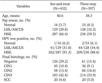
Table 1. General characteristics of study populations Variables See-and-treat
(N=432) Three-step (N=397) Age, means
Pap smear, no. (%) Normal
LSIL/ASCUS HSIL
HPV test positive, no. (%) Normal
LSIL/ASCUS HSIL
Final histology, no. (%) Negative
CIN1 CIN2 CIN3 SCC
40.6
16 (3.7) 129 (29.8) 287 (66.4)
1/16 (6.2) 41/129 (31.8) 262/287 (91.3)
126 (29.2) 45 (10.4) 58 (13.4) 183 (42.4) 20 (4.6)
38.3
33 (8.3) 128 (32.2) 236 (59.5)
2/33 (6.1) 44/128 (34.4) 209/236 (88.6)
61 (15.4) 36 (9.1) 66 (16.6) 214 (53.9) 20 (5.0) LSIL: low-grade squamous intraepithelial lesions, ASCUS: atypical squamous cells of undetermined significance, HSIL: high-grade squamous intraepithelial lesion, HPV: human papilloma virus, CIN:
cervical intraepithelial neoplasia, SCC: squamous cell carcinoma.
number of hospital visits, noncompliance, and the patient anxiety.8,9 Although advantageous in certain patient pop- ulations, this strategy may result in overtreatment because of the low specificity of Pap smears.
In this study, we evaluated the feasibility of a see-and-treat by dividing patients with abnormal cytology who have under- gone LEEP into two study groups, based on whether they were treated with or without colposcopy-directed biopsy. The final histological results of the two study groups were compared.
MATERIALS AND METHODS 1. Study subjects
We conducted a retrospective review of the medical records of 1,011 patients who underwent LEEP at the Department of Obstetrics and Gynecology at Gangnam Severance Hospital from January 2004 to December 2008. Each patient with a di- agnosis of abnormal cervical cytology initially underwent col- poscopic examination. However, colposcopic examination was also performed in the patients with normal cytology when the patient needed further evaluation due to continuous de- tection of the human papillomavirus (HPV), or the fear of cancer. “Continuous detection of the HPV” was defined as having two or more HPV DNA positive tests with more than 6 months interval between HPV tests. Study subjects were divided into two groups based on the treatment they received:
see-and-treat group (If the colposcopic impression was CIN 2/3, LEEP was immediately performed without processing a colposcopy-directed biopsy), and the standard three-step group (If there appeared a suspected lesion of CIN2/3 at colposcopic examination, colposcopy-directed biopsy was performed. All patients diagnosed with CIN 2/3 in the colposcopy-directed biopsy subsequently received LEEP procedure). We only in- cluded the cases diagnosed with CIN2/3 by colposcopy-directed biopsy, and the cases with CIN1 or negative histology by col- poscopy-directed biopsy were excluded. The patients with glandular cell abnormalities in cervical cytology and a history of cervical dysplasia/cancer were also excluded. The patients were randomly assigned to one of two treatment protocols, and the decision towards LEEP was not influenced by the ex- tent of cytologic findings.
Colposcopic examination of the cervix was performed after application of 5% acetic acid solution to the ectocervix. In or- der to have an adequate colposcopic examination, the entire transformation zone, if present, was fully visualized. The LEEP procedure was performed with an electrosurgical unit (Surgitron® FFPF EMC, Ellman International INC., Hewlett, NY, USA), using an appropriate sized wire loop (8, 15, 20, or 25 mm loop bayonet electrodes). The size of the wire loop was chosen on the basis of the colposcopic findings during the procedure. Surgical specimens were stored in a 10% formalin solution and submitted to the Department of Pathology for histopathologic evaluation. Abnormal Pap smears are re- viewed by a senior pathologist. All colposcopic examinations
and LEEP were performed at our hospital by one gynecologic oncology specialist. The classification of cervical cytology was performed according to the 2001 Bethesda system,10 and the final histological results were reviewed by two gynecologic pathologists. This study was approved by the Institutional Review Board (IRB) of Gangnam Severance Hospital.
2. Clinical data collection
For the study subjects, cervical cytology, colposcopy-directed biopsy, and final histology were recorded. To determine the human papillomavirus (HPV) infection rate in the study group, Hybrid Capture® 2 (HC2) test results were also reviewed.
3. Statistical analysis
The final histological results were classified as Negative (absence of CIN), CIN1, CIN2, CIN3, and squamous cell car- cinoma (SCC), and then analyzed using SPSS ver. 12.0 (SPSS Inc., Chicago, IL, USA). Comparisons between the groups were evaluated by chi-square (χ2) test, and p<0.05 was con- sidered to be statistically significant. In addition, the final his- tological results were divided into two groups: 1) overtreat- ment group (final histology ≤CIN1), and 2) correct treat- ment group (final histology ≥CIN2), and the rate of agree- ment between the initial and final diagnoses in the see-and- treat and three-step group was compared.
RESULTS
A total of 829 cases were finally included in this study. The clinical characteristics of the study subjects are listed in Table 1.
There were 287 HSIL, 129 low-grade squamous intraepithelial
Table 2. Histological results of LEEP specimens in patients with HSIL cytology*
Histology See-and-treat (N=287) Three-step (N=236) p-value no. (%)
Negative CIN1 CIN2 CIN3 SCC
38 (13.2) 14 (4.9) 49 (17.1) 167 (58.2) 19 (6.6)
30 (12.7) 14 (5.9) 21 (9.0) 152 (64.4)
19 (8.0)
0.71
LEEP: loop electrosurgical excision procedure, HSIL: high-grade squamous intraepithelial lesion, CIN: cervical intraepithelial neo- plasia, SCC: squamous cell carcinoma.
*All patients had HSIL on Pap smear and high-grade lesions (≥CIN2) on colposcopy-directed biopsy or suspected lesions equivalent of
≥CIN2 on colposcopy.
Table 3. Histological results of LEEP specimens in patients with LSIL/ASCUS cytology*
Histology See-and-treat (N=129) Three-step (N=128) p-value no. (%)
Negative CIN 1 CIN 2 CIN 3 SCC
81 (62.8) 25 (19.4) 7 (5.4) 15 (11.6)
1 (0.8)
28 (21.9) 19 (14.8) 31 (24.2) 49 (38.3) 1 (0.8)
<0.001
LEEP: loop electrosurgical excision procedure, LSIL: low-grade squ- amous intraepithelial lesion, ASCUS: atypical squamous cells of un- determined significance, CIN: cervical intraepithelial neoplasia, SCC:
squamous cell carcinoma.
*All patients had LSIL/ASCUS on Pap smear and high-grade lesions (≥CIN2) on colposcopy-directed biopsy or suspected lesions equi- valent of ≥CIN2 on colposcopy.
Table 4. Histological results of LEEP specimens in patients with normal cytology*
Histology See-and-treat (N=16) Three-step (N=33) p-value no. (%)
Negative CIN1 CIN2 CIN3 SCC
7 (43.7) 6 (37.5) 2 (12.6) 1 (6.2)
0 (0)
3 (9.1) 3 (9.1) 14 (42.4) 13 (39.4)
0 (0)
<0.001
LEEP: loop electrosurgical excision procedure, CIN: cervical intra- epithelial neoplasia, SCC: squamous cell carcinoma.
*All patients had normal Pap smear and high-grade lesions (≥CIN2) on colposcopy-directed biopsy or suspected lesions equivalent of
≥CIN2 on colposcopy.
Fig. 1. The rate of high-grade lesions (≥CIN2) in final histology ac- cording to treatment modalities. There were no significant differences in the rate of final diagnosis of more than CIN2 between see-and-treat and three-step group in patients with HSIL cytology (p=0.51), while there were significant differences in LSIL/ASCUS and normal cytol- ogy cases (p<0.001).
CIN: cervical intraepithelial neoplasia, SCC: squamous cell carcinoma, HSIL: high-grade squamous intraepithelial lesions, LSIL: low-grade squamous intraepithelial lesions, ASCUS: atypical squamous cells of undetermined significance.
lesions (LSIL)/atypical squamous cells of undetermined sig- nificance (ASCUS), and 16 normal cervical cytology cases in the see-and-treat group and 236 HSIL, 128 LSIL/ASCUS, and 33 normal cytology cases in the three-step group. The mean ages were 40.6±9.8 years for see-and-treat group and 38.3±
9.2 years for three-step group. There were no significant age differences between the groups (p=0.19). In the HC2 test, HPV DNA positivity was 6.1% in normal cytology, 33.1% in LSIL/ASCUS cytology, and 90.1% in HSIL cytology. There was no significant difference in HC2 positivity between the see-and-treat and three-step group (p=0.76).
The results of the final histological tests of 523 patients with HSIL cytology who were treated either by the see-and-treat or three-step protocol are presented in Table 2. There was no sig- nificant difference in final histology between the two study groups. The final histological results of 257 patients who re- ceived LEEP despite LSIL/ASCUS on cervical cytology are shown in Table 3. Patients in the three-step group were sig- nificantly more likely to have high-grade histological results
(≥CIN2) (p<0.001). In patients with normal cervical cytol- ogy, the final histological results showing high-grade lesions (≥CIN2) were also significantly more prevalent in the three- step group (p<0.001) (Table 4).
Fig. 1 demonstrates the rate of agreement between the cer- vical cytology and final histology in the study groups. In pa- tients with HSIL cytology, the final histological diagnosis was in agreement with initial cytologic diagnosis in 81.9% (235 out of 287 cases) in the see-and-treat group, and 81.4% (192 out of 236 cases) in the three-step group. The rate of agree-
ment between the two study groups was similar (p=0.51).
However, in patients with LSIL/ASCUS and normal cervical cytology, the rate of agreement was 17.8% (23 out of 129 cas- es) and 18.8% (3 out of 16 cases), respectively, in the see-and- treat group, and 63.3% (81 out of 128 cases) and 81.8% (27 out of 33 cases), respectively, in the three-step group. There were significant differences in the rate of agreement between the initial and final diagnosis (p<0.001).
DISCUSSION
See-and-treat approaches are relatively new and have been surrounded with controversy since their introduction. One of the controversies is due to the overtreatment which is partly based on the fact that colposcopy is not a perfect diagnostic test. Data from the ALTS (ASCUS/LSIL Triage Study) trial showed only 15% of women referred with ASCUS cytology had biopsy-proven CIN2/3 (85% over diagnosis). Similarly, women with LSIL cytology had a 25% rate of CIN2/3 (75%
over diagnosis).11,12
The risk of overtreatment, or unnecessary treatment, is one of the main criticisms of the see-and-treat approach. The ef- fectiveness of see-and-treat depends on colposcopic im- pression. Although most studies have found colposcopy to be reasonably accurate compared with cervical pathological diag- nosis,13 colposcopy is subject to intra- and interobserver vari- ability, and potentially leads to overtreatment. Thus, patients may be unnecessarily exposed to bleeding and infection, which are the most common complications of the LEEP procedure.
Therefore, we recommend that see-and-treat strategy is only appropriate when an experienced colposcopist can differ- entiate low-grade from high-grade lesions, and the quality of colposcopic practice should be improved by setting appro- priate standards.
Recently, strategies have limited the see-and-treat protocol to only patients with HSIL cervical cytology. To decrease the possibility of overtreatment, patients must have a high proba- bility of having CIN2/3 before undergoing the see-and-treat protocol. The use of see-and-treat strategy in a patient pop- ulation with an HSIL cervical cytology has been shown to de- crease overtreatment. Irvin and co-workers reported on a trial that included patients with a Pap smear and colposcopic diag- nosis of HSIL.14 Overtreatment was 18% if the threshold in- cluded patients with mild dysplasia and those with negative pathology results. Overtreatment decreased to 4% when the threshold was lowered to include only patients with negative pathology results.
Although there are some controversies in the see-and-treat strategy, the necessity of colposcopy-directed biopsy is also controversial. In 2006, Byrom et al.15 performed biopsies guided by colposcopy in the area that was thought to be the most appropriate in lesions of 170 patients with high-grade cytology or colposcopic findings. They simultaneously per- formed LEEP and compared their histological results.
Approximately 70% of the histological results of colpo- scopy-directed biopsies concurred with the final histological results obtained by LEEP and showed a tendency to under- evaluate the disease. Another study also indicated the dis- advantages of colposcopy-directed biopsy.16 They suggested that it did not lower the rate of false positives and therefore could not improve the accuracy of diagnosis. It also delayed the treatment, resulting in increased emotional anxiety in patients.
In the current study, we compared the final histological re- sults of see-and-treat protocol with those of the standard three-step protocol. In our data set, the rate of agreement be- tween initial and final diagnoses was not significantly different only in the HSIL cytology cases, indicating that the addition of a colposcopy-directed biopsy does not reduce the ratio of overtreatment, and has a limited predictive value for the final histological result after LEEP in patients with HSIL cytology.
In the HSIL cytology cases, 19 out of 287 patients (6.6%) in the see-and-treat group were diagnosed with invasive carcino- ma after LEEP, as shown in Table 2. All patients with invasive carcinoma were immediately treated according to National Cancer Comprehensive Network (NCCN) clinical practice guidelines the next day.
In the LSIL/ASCUS, or normal cytology cases, there were more cases of correct treatment (final histology ≥CIN2) in the three-step group (63.3% in LSIL/ASCUS cytology and 81.8% in normal cytology) than in the see-and-treat group (17.8% in LSIL/ASCUS cytology and 18.8% in normal cytol- ogy ) (p<0.001). As a result, overtreatment was much more prevalent in the see-and-treat group in patients with LSIL/
ASCUS or normal cytology. Therefore, for patients with LSIL/
ASCUS or normal cytology, colposcopy-directed biopsy be- fore LEEP can be considered effective. One literature indicates that 5% to 17% of ASCUS and 15% to 30% of LSIL found on Pap smears have associated high-grade dysplasia. In contrast, 70% to 75% of HSIL test results are associated with severe dysplasia and the reproducibility of HSIL is far greater than that of ASCUS.17 Therefore, we did not believe that it was ap- propriate to perform a LEEP without colposcopy-directed bi- opsy in patients with low-grade lesions found on cervical cytology.
The see-and-treat strategy was also associated with the low- est cost in the management of HSIL cytology result, producing a 41% cost reduction compared with conventional manage- ment of the patients with HSIL.18 In addition, the patient’s anxiety may be relieved by assurance from the physician that the lesion has been found and destroyed completely, and will be assessed histologically.9 In surveys based on confidential questionnaires, patients’ satisfaction with the see-and-treat strategy was shown to be acceptable.19,20
The current study has several potential limitations that must be considered in the interpretation. One limitation is the ret- rospective nature of this study, in which the possibility of se- lection bias increases. Another limitation is the small number
of study subjects. Despite these potential limitations, we re- port that the see-and-treat protocol may be an effective strat- egy in patients with HSIL on cervical cytology and suggestive of CIN2/3 after colposcopy. However, colposcopy-directed bi- opsy must be performed before LEEP in patients with LSIL/ASCUS or normal cytology for more accurate diagnosis and prevention of overtreatment.
REFERENCES
1. Pisani P, Bray F, Parkin DM. Estimates of the world-wide preva- lence of cancer for 25 sites in the adult population. Int J Cancer 2002; 97: 72-81.
2. Korea National Cancer Registry. Annual report of Korea na- tional cancer registry enterprise (2002.1-2002.12). Seoul: Korea Ministry of Health and Welfare; 2003.
3. Abe Y, Ito K, Okamura C, Niikura H, Terada Y, Murakami T, et al. Cervical cytologic examination during physical checkup of pregnant women: cervical cancer screening in women under the age of thirty. Tohoku J Exp Med 2004; 204: 221-8.
4. Aggarwal RK, Bacus JW. A multi-spectral approach for scene anal- ysis of cervical cytology smears. J Histochem Cytochem 1977; 25:
668-80.
5. Kuo DY, Goldberg GL. Screening of cervical cancer: where do we go from here? Cancer Invest 2003; 21: 157-61.
6. Cho H, Hong SW, Oh YJ, Kim MA, Kang ES, Lee JM, et al.
Clinical significance of osteopontin expression in cervical cancer. J Cancer Res Clin Oncol 2008; 134: 909-17.
7. Katiyar S, Thelma BK, Murthy NS, Hedau S, Jain N, Gopalkrishna V, et al. Polymorphism of the p53 codon 72 Arg/Pro and the risk of HPV type 16/18-associated cervical and oral cancer in India.
Mol Cell Biochem 2003; 252: 117-24.
8. Santos C, Galdos R, Alvarez M, Velarde C, Barriga O, Dyer R, et al.
One-session management of cervical intraepithelial neoplasia: a solution for developing countries. A prospective, randomized tri- al of LEEP versus laser excisional conization. Gynecol Oncol 1996; 61: 11-5.
9. Murdoch JB. The case for early intervention ('see and treat') in patients with dyskaryosis on routine cervical screening. Int J
STD AIDS 1995; 6: 415-7.
10. Solomon D, Davey D, Kurman R, Moriarty A, O'Connor D, Prey M, et al. The 2001 Bethesda System: terminology for re- porting results of cervical cytology. JAMA 2002; 287: 2114-9.
11. Group A-LTSA. Results of a randomized trial on the manage- ment of cytology interpretations of atypical squamous cells of undetermined significance. Am J Obstet Gynecol 2003; 188:
1383-92.
12. Solomon D, Schiffman M, Tarone R. Comparison of three man- agement strategies for patients with atypical squamous cells of undetermined significance: baseline results from a randomized trial. J Natl Cancer Inst 2001; 93: 293-9.
13. Fahey MT, Irwig L, Macaskill P. Meta-analysis of Pap test accuracy. Am J Epidemiol 1995; 141: 680-9.
14. Irvin WP Jr, Andersen WA, Taylor PT Jr, Stoler MH, Rice LW.
"See-and-treat" loop electrosurgical excision: Has the time come for a reassessment? J Reprod Med 2002; 47: 569-74.
15. Byrom J, Douce G, Jones PW, Tucker H, Millinship J, Dhar K, et al. Should punch biopsies be used when high-grade disease is suspected at initial colposcopic assessment: a prospective study. Int J Gynecol Cancer 2006; 16: 253-6.
16. Sadan O, Yarden H, Schejter E, Bilevsky E, Bachar R, Lurie S.
Treatment of high-grade squamous intraepithelial lesions: a "see and treat" versus a three-step approach. Eur J Obstet Gynecol Reprod Biol 2007; 131: 73-5.
17. Wright TC Jr, Cox JT, Massad LS, Twiggs LB, Wilkinson EJ. 2001 Consensus Guidelines for the management of women with cer- vical cytological abnormalities. JAMA 2002; 287: 2120-9.
18. Holschneider CH, Ghosh K, Montz FJ. See-and-treat in the management of high-grade squamous intraepithelial lesions of the cervix: a resource utilization analysis. Obstet Gynecol 1999;
94: 377-85.
19. Nobbenhuis MA, Walboomers JM, Helmerhorst TJ, Rozendaal L, Remmink AJ, Risse EK, et al. Relation of human papil- lomavirus status to cervical lesions and consequences for cer- vical-cancer screening: a prospective study. Lancet 1999; 354:
20-5.
20. Hallam NF, West J, Harper C, Edwards A, Hope S, Merriman H, et al. Large loop excision of the transformation zone (LLETZ) as an alternative to both local ablative and cone biopsy treat- ment: a series of 1000 patients. J Gynecol Surg 1993; 9: 77-82.