© 2012 The Korean Society of Pathologists/The Korean Society for Cytopathology pISSN 1738-1843
The significance of atypical squamous cells of undetermined significance (ASC-US) diagnosis on cervical cytologic smears and the clinical management of patients with ASC-US are un- clear. There has been a strong consensus to replace ASC-US with a new category, “atypical squamous cells (ASC)”, defined as “cytologic changes that are suggestive of squamous intraepi- thelial lesion (SIL) and are quantitatively insufficient for a de- finitive interpretation.”
1The ASC can be further subdivided into “those of undetermined significance (ASC-US)” and “those where a high-grade squamous intraepithelial lesion (HSIL) can- not be excluded (ASC-H).” The ASC-US category includes cy- tologic changes that are suggestive of SIL but lack criteria for a definitive interpretation, whereas the ASC-H one does those that are suggestive of HSIL but also lack criteria for a definitive interpretation.
As might be expected, based on the concept that the ASC- US is an equivocal category, the ASC is not an interpretation that can particularly be reproduced from either an inter- or in-
tra-observer viewpoint. This has been well verified in a number of studies. In the National Cancer Institute (NCI), atypical squamous cells of undetermined significance–low-grade squa- mous intraepithelial lesion (ASCUS-LSIL) Triage Study (ALTS), estimates of reproducibility within a well-controlled “expert”
environment show similar kappa rates (kappa value=0.46) for correlations between the observers on both cytology and histo- patholgic specimen.
2In an earlier study of Sherman et al.,
3a group of five experts showed a similar variability with a lack of the unanimity in an entire study set of putative ASC-US cases.
Variability in diagnosis agreement remains concerning the di- agnosis of ASC-US.
4In addition, treatment of patients with this diagnosis is still a major point of controversy.
5Several au- thors have addressed this issue by analyzing the cytohistological correlation with this cytological diagnosis.
6-8To date, however, information retrieval from biopsy results as well as the variabil- ity of therapeutic methods has constantly been a limiting factor.
It has been proven that a cervical biopsy is poor in predicting
Outcome of “Atypical Squamous Cells” in Cervical Cytology:
Follow-up Assessment by Loop Electrical Excision Procedure
Joon Seon Song · Ilseon Hwang
1Gyungyub Gong
Department of Pathology, Asan Medical Center, University of Ulsan College of Medicine, Seoul;
1Department of Pathology, Keimyung University Dongsan Medical Center, Keimyung University School of Medicine, Daegu, Korea
Background: We have retrospectively assessed the incidence and outcome of women diagnosed during a hospital-based cytology screening program with “atypical squamous cells (ASC)” and followed-up with loop electrical excision procedure (LEEP). Methods: We analyzed 173,947 cas- es of cervical smears’ follow-up cytology and histology findings. Previous or archival cytology with LEEP results were retrieved for 390 women with ASC of undetermined significance (ASC-US) and 112 with ASC, cannot exclude high-grade squamous intraepithelial lesion (ASC-H). Results:
On the follow-up cytology, of the 390 women initially diagnosed with ASC-US, 130 (33.3%) had no follow-up records of smears before LEEP; smears of 18 (4.6%) were negative for cytologic ab- normalities, 193 (49.5%) were ASC-US, 24 (6.2%) were ASC-H, 111 (28.5%) were low grade squamous intraepithelial lesion (SIL), and 44 (11.4%) were high grade SIL. LEEP findings in these 390 women showed that 183 (46.9%) were negative, 73 (18.7%) were graded as cervical intraep- ithelial neoplasia (CIN) 1, 25 (6.4%) as CIN 2, 102 (26.2%) as CIN 3, and 7 (1.8%) had carcinoma.
LEEP was performed in 112 women initially diagnosed with ASC-H; 36 (32.1%) were negative, 4 (3.6%) were graded as CIN 1, 7 (6.3%) as CIN 2, 60 (53.6%) as CIN 3, and 5 (4.5%) with carcino- ma. Conclusions: Patients with ASC-H smears were at increased risk of SIL or carcnoma com- pared with patients with ASC-US. Careful follow-up is required in ASC patients.
Key Words: Cervix uteri; Cytology; Loop electrical excision procedure; Atypical squamous cell Received: February 7, 2012
Revised: July 4, 2012 Accepted: July 5, 2012 Corresponding Author Gyungyub Gong, M.D.
Department of Pathology, Asan Medical Center, University of Ulsan College of Medicine, 88 Olympic-ro 43-gil, Songpa-gu, Seoul 138-736, Korea
Tel: +82-2-3010-4554 Fax: +82-2-472-7898 E-mail: [email protected]
the actual status of underlying cervical intraepithelial lesion.
Given the above background, we conducted this study by taking advantage of selected cases treated by loop electrical ex- cision procedure (LEEP) in the absence of previous biopsy to evaluate the incidence and outcome of ASC by a retrospective histopathological analysis of LEEP samples of women in a hos- pital-based cytology screening program and to correlate the histological findings with the last cytological categories pro- posed for ASC diagnosis.
MATERIALS AND METHODS
From January 2004 to December 2007, 173,947 conven- tional or liquid-based cervical smears were performed at the Obstetrics and Gynecology clinic and then sent for analysis to the cervical cytology laboratory of the Asan Medical Center.
We’ve used the liquid-based cytology (LBC) with an automated system (AutoPap, TriPath Imaging, Inc., Burlington, NC, USA) since January 2006 and a conventional cytology technique dur- ing a period ranging from January 2004 to December 2005.
These smears were immediately wet-fixed, stained using the standard Papanicolau technique (Pap), screened by cytotechni- cians and then assessed by cytopathologists. In cases of LBC with an automated screening system, the case followed the pro- tocol of AutoPap primary screening system (TriPath Imaging, Inc.).
9Atypical cases were reviewed by cytotechnologist and then referred to cytopathologist. The final cytologic abnormali- ties reported by pathologists included ASC-US, atypical glan- dular cells of undetermined significance, LSILs, HSILs and ma- lignant disease.
According to Bethesda system,
1ASC-US in conventional cy- tology are criteria as shown below:
1) Nuclei are approximately two and one half to three times the area of the nucleus of a normal intermediate squamous cell.
2) Slightly increased ratio of the nuclear to cytoplasmic area.
3) Minimal nuclear hyperchromasia and irregularity in chro- matin distribution or nuclear shape.
4) Nuclear abnormalities associated with dense orangeophilic cytoplasm.
The appearance of ASC-US is similar between the smears and liquid-based cytology. That is, cells may appear larger rather than more flat on smears.
ASC-H cells are usually sparse. Small cells have high nuclear/
cytoplasmic ratios accompanied by a crowded sheet pattern. In a LBC, ASC-H cells may appear quite small, with nuclei whose size is only two to three times greater than that of the neutro-
phil nuclei.
The cytology and histology laboratory databases of the Asan Medical Center were searched for follow-up data in patients who had been initially diagnosed with ASC-US or ASC-H. But the cytology and histology slides were not reviewed. Patients were excluded if they had been diagnosed with higher-grade cervical smears (such as LSIL and HSIL) prior to diagnosis of ASC-US or ASC-H on Pap smears, if LEEP or biopsy had been performed before diagnosis of ASC-US/ASC-H or if LEEP had not been performed.
Chi-square and Fisher’s tests were used with a 95% confi- dence interval.
RESULTS
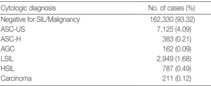
Pap smears obtained from 173,947 women were examined in our laboratory (Table 1). Of these, 7,125 (4.1%) women and 383 (0.2%) were diagnosed with ASC-US and ASC-H, respec- tively. During the same time frame (January 2004 to December 2007), the ratio of SIL to carcinoma was 17.7:1 and that of ASC to SIL was 2.01:1.
In addition, 2,810 patients underwent LEEP during the same period, 648 (23.1%) and 216 (7.7%) of whom were diag- nosed with ASC-US and ASC-H, respectively. A follow-up cy- tology with LEEP results was retrieved in 390 women with ASC-US and 112 with ASC-H.
The mean age of patients was 43.51 years (range, 22 to 73 years) in the ASC-US group and 46.94 years (range, 26 to 76 years) in the ASC-H group. Median follow-up period following the diagnosis of ASC was 24.1 months (range, 0 to 68.5 months) in the ASC-US group and 2.76 months (range, 0 to 13.1 months) in the ASC-H group. The mean length of period elapsed from ASC to LEEP was 8.07 months in the ASC-US
Table 1. Cytological diagnoses in adequate cervical smears (n= 173,947)
Cytologic diagnosis No. of cases (%)
Negative for SIL/Malignancy 162,330 (93.32)
ASC-US 7,125 (4.09)
ASC-H 383 (0.21)
AGC 162 (0.09)
LSIL 2,949 (1.68)
HSIL 787 (0.49)
Carcinoma 211 (0.12)
SIL, squamous intraepithelial lesion; ASC-US, atypical squamous cells of undetermined significance; ASC-H, atypical squamous cells, cannot ex- clude high-grade squamous intraepithelial lesion; AGC, atypical glandular cells; LSIL, low grade squamous intraepithelial lesion; HSIL, high grade squamous intraepithelial lesion.
group and 1.98 months in the ASC-H group. The mean num- ber of subsequent cervical smears was 1.69 (range, 0 to 14) in the ASC-US group and 0.3 (range, 0 to 4) in the ASC-H group.
On the second smear examination, of the 390 women who had been initially diagnosed with ASC-US, 130 (33.3%) had no follow-up records of Pap smears. In addition, the other re- maining patients had the following results: 68 (17.4%) were negative, 94 (23.8%) were again diagnosed with ASC-US, 14 (3.6%) were diagnosed with ASC-H, 64 (16.4%) were diag- nosed with LSIL, 20 (5.7%) were diagnosed with HSIL and none were diagnosed with squamous cell carcinoma. At the last cytology prior to LEEP, of the 260 patients, 18 (6.9%) were negative for epithelial abnormalities, 63 (24.2%) were diag- nosed with ASC-US, 24 (9.2%) were diagnosed with ASC-H, 111 (42.7%) were diagnosed with LSIL and 44 (16.9%) were diagnosed with HSIL, but none were diagnosed with squamous cell carcinoma.
Based on the LEEP findings, of the 390 women who had been initially diagnosed with ASC-US, 183 (46.9%) were negative, 73 (18.7%) were graded as cervical intraepithelial neoplasia (CIN) 1, 25 were graded (6.4%) as CIN 2, 102 (26.2%) were graded as CIN 3, and seven (1.8 %) had carcinoma (Table 2).
Of the 112 women who were diagnosed with ASC-H on cer- vical Pap smears, 91 (81.3%) underwent LEEP without a fol- low-up cytology. On the follow-up cytology of the 112 women who had been initially diagnosed with ASC-H, smears of four (3.6%) were negative for cytologic abnormalities, two (1.8%) were positive for ASC-US, 97 (86.6%) were positive for ASC- H, three (2.6%) were positive for LSIL, and six (5.4%) were pos-
itive for HSIL.
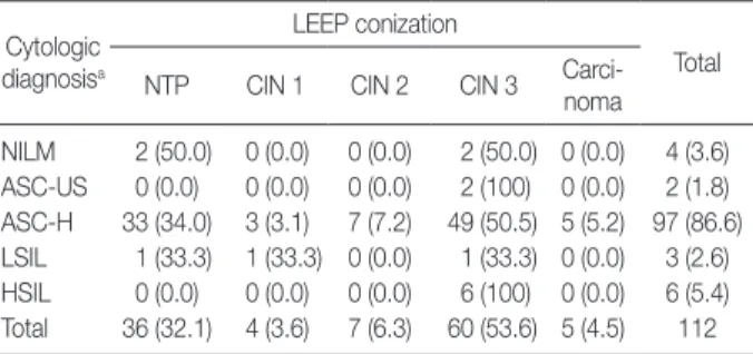
Of the 112 women who were diagnosed with ASC-H and underwent LEEP, 36 (32.1%) were negative, four (3.6%) were graded as CIN 1, seven (6.2%) were graded as CIN 2, 60 (53.6%) were graded as CIN 3, and five (4.5%) were diagnosed with carcinoma (Table 3).
DISCUSSION
Quality assurance monitoring of ASC reporting has been stressed. In screening general populations, ASC should not ex- ceed 5% of the total specimens and the ratio of ASC to SIL should not exceed 2:1 to 3:1. Furthermore, ASC-H should not account for >10% of the total ASC.
10In evaluating outcomes in women who were diagnosed with ASC-US/ASC-H on cervical smears in a single institution in Korea, these criteria were met.
Many studies have been conducted to show the rate of CIN in a follow-up interpretation of ASC.
11According to these stud- ies, there was a great variability in the rate of CIN at a follow- up and it ranged from as low as 10% to as high as 80%. Most of the studies have shown that it ranges between 30% and 60%. As might be expected, with the prepondenderance of ASC-US in the population, most cases of CIN are in the CIN 1 category, generally ranging between 60% and 95%. Based on our understanding of the biology of cervical carcinogenesis, however, more than CIN 2 is an important point for the detec- tion of the disease. Studies have shown that more than CIN 2 accounts for 0-40% of total cases of CIN detected at a follow- up. In the ALTS, the proportion of cases of more than CIN 2
Table 2. Follow-up LEEP findings for ASC-US Cytologic
diagnosisa
LEEP conization
Total NTP CIN 1 CIN 2 CIN 3 Carci-
noma
NILM 13 (72.2) 1 (5.5) 0 (0.0) 4 (22.2) 0 (0.0) 18 (4.6) ASC-US 106 (54.9) 23 (11.9) 10 (51.8) 49 (25.4) 5 (2.6) 193 (49.5) ASC-H 11 (45.8) 3 (12.5) 0 (0.0) 10 (41.7) 0 (0.0) 24 (6.2) LSIL 49 (44.1) 43 (38.7) 7 (6.3) 12 (10.8) 0 (0.0) 111 (28.5) HSIL 4 (9.1) 3 (6.8) 8 (18.2) 27 (61.4) 2 (4.5) 44 (11.3) Total 183 (46.9) 73 (18.7) 25 (6.4) 102 (26.2) 7 (1.8) 390 (100) Values are presented as number (%).
LEEP, loop electrical excision procedure; ASC-US, atypical squamous cells of undetermined significance; NTP, no tumor present which means nega- tive for cervical intraepithelial or malignancy; CIN, cervical intraepithelial neoplasia; NILM, negative for intraepithelial lesion or malignancy; ASC-H, atypical squamous cells, cannot exclude high-grade squamous intraepithe- lial lesion; LSIL, low grade squamous intraepithelial lesion; HSIL, high grade squamous intraepithelial lesion.
aThe cytologic diagnosis represents the last Pap results before LEEP con- ization.
Table 3. Follow-up LEEP findings for ASC-H Cytologic
diagnosisa
LEEP conization
Total NTP CIN 1 CIN 2 CIN 3 Carci-
noma NILM 2 (50.0) 0 (0.0) 0 (0.0) 2 (50.0) 0 (0.0) 4 (3.6) ASC-US 0 (0.0) 0 (0.0) 0 (0.0) 2 (100) 0 (0.0) 2 (1.8) ASC-H 33 (34.0) 3 (3.1) 7 (7.2) 49 (50.5) 5 (5.2) 97 (86.6) LSIL 1 (33.3) 1 (33.3) 0 (0.0) 1 (33.3) 0 (0.0) 3 (2.6) HSIL 0 (0.0) 0 (0.0) 0 (0.0) 6 (100) 0 (0.0) 6 (5.4) Total 36 (32.1) 4 (3.6) 7 (6.3) 60 (53.6) 5 (4.5) 112 Values are presented as number (%).
LEEP, loop electrical excision procedure; ASC-H, atypical squamous cells, cannot exclude high-grade squamous intraepithelial lesion; NTP, no tumor present which means negative for cervical intraepithelial or malignancy;
CIN, cervical intraepithelial neoplasia; NILM, negative for intraepithelial le- sion or malignancy; ASC-US, atypical squamous cells of undetermined significance; LSIL, low grade squamous intraepithelial lesion; HSIL, high grade squamous intraepithelial lesion.
aThe cytologic diagnosis represents the last Pap results before LEEP con- ization.
was 15.4%, which stands presently as our best benchmark.
12In our clinical series of patient, on histopatholgic examinations of the subsequent tissue specimens, there was either a low- or a high-grade dysplasia in 53.7% and 67.8% of women who had been diagnosed with ASC-US and ASC-H, respectively, on Pap smears. At a follow-up, of women who had been diagnosed with ASC-US, 11.3% had a progression to HSIL and 26.2%
did a histologically confirmed high-grade dysplasia. In addi- tion, of women who had been diagnosed with ASC-H, 5.4 % had a progression to HSIL and 53.6% did a histologically con- firmed high-grade dysplasia.
As compared with our results, previous studies have shown that 2,765 patients had an SIL rate of 28% and an HSIL rate of 11.3% at a follow-up with both cytologic and histologic exami- nations.
13According to a study that was conducted by Kim et al.
14in Korean patients, a histopathologic examination proved that 14.6% (13/89) of patients with ASC-US had an HSIL.
There are some gaps in the cytologic diagnoses between ini- tially and at a follow-up. As we have previously mentioned, the first reason is that the diagnosis of “ASC” contains a lower re- producibility and a higher variability.
2The second reason is that there is a possibility that the disease might truly progress or re- gress. In addition, the other reason might be due to sampling error.
Of note, higher SIL rates have been reported after short fol- low-up periods, which may be due to the regression of low- grade lesions.
15,16It has been shown that low-grade cervical in- traepithelial lesions are more likely to regress than to progress to HSIL or invasive carcinoma.
17Rates of regression to normal are 68% for ASC-US, 47% for LSIL, and 35% for HSIL.
18Pre- sumably, many patients with ASC-US might have SIL if biop- sies are performed several weeks after ASC-US cytology. These lesions may have regressed between the time point of the diag- nosis of ASC-US and that of a follow-up 6-12 months later.
By contrast, of women who had initially been diagnosed with ASC-H on Pap smears, 67.8% had either a low- or a high-grade dysplasia in subsequent tissue specimens. Of the patients with ASC-H, 28.6% and 53.6% had a progression to HSIL and a histologically confirmed high-grade dysplasia, respectively.
These findings are in agreement with previous reports.
18-20Duncan and Jacob
19showed that 46% of women with a smear diagnosis of ASC-H were found to have a high-grade dysplasia in tissue specimens. Consistently with previous reports,
21,22our results showed that there was a positive value of 57.7% in pre- dicting a histologically proven high-grade dysplasia in patients with ASC-H patients. This implies that ASC-H is an impor-
tant diagnosis and it warrants an immediate further evaluation by colposcopy and/or biopsy.
In the management of ASC lesions, there is a broad consensus that women should be referred for immediate colposcopic eval- uation, once they are diagnosed with ASC-H, because many of them are more likely to harbor high-grade lesions.
20,23,24It is also recommended, however, that they undergo a repeated cer- vical cytology at a certain length of intervals, receive an imme- diate colposcopy, take DNA analysis for the detection of high- risk types of human papillomavirus (HPV) or undergo a single repeat cervical cytology combined with another adjunctive method.
24In the current study, where 130 patients with ASC- US were enrolled but not followed up for cytology, we analyzed the reasons for a lack of a follow-up cytology or a punch biopsy although they directly underwent LEEP. Of them, 78 (60%) patients were referred to us for further evaluation and treat- ment. This is because they had been diagnosed with more than ASC-US at local clinic. In addition, 18 (13.8%) patients with ASC-US had a high-risk HPV and 34 (26.1%) did neither a history of ASC-US nor a high-risk HPV. In 34 patients, where a diagnosis of ASC was made, particularly including those where a diagnosis of ASC-US was initially made on the Pap cy- tology, the LEEP was not a first choice of treatment. We have therefore cautiously speculated that it is an acceptable practice in Korean patients from the viewpoints of not only the relation- ship between physicians and patients but also a fear for the ma- lignant potential. There is a great tendency that Korean pa- tients face the risk and thereby choose surgical excision for the treatment.
Recent studies have shown that the HPV typing test also had a significant effect in choosing the optimal treatment modality.
In the ALTS trial,
12HPV triage is at least as sensitive as imme- diate colposcopy in detecting CIN 3 in women with ASC-US.
In addition, a meta-analysis also showed that HPV triage is a more sensitive modality than cytology in detecting CIN 2/3.
25In the management of patients with ASC, particularly includ- ing those with ASC-US, the HPV status is a criticial clue. But one the limitations of the current study is that there were no at- tempts to identify the correlation between the HPV status and cytology of ASC. We have actually tried to identify such corre- lation, but failed to detect the statistical significance. This is because we have enrolled a smaller number of patients for the HPV test.
The other limitation is that we did not review the slides in
differentially making a pathological diagnosis of ASC from the
initial cytology or the follow-up diagnosis on the cytology or
LEEP. It was therefore impossible to rule out the discrepancy in the classification of ASC-US between the pathologists.
Then, it deserves special attention whether ASC can be elimi- nated. Of note, over the years, a number of studies have been conducted to test a hypothesis that what would happen if we eliminate the use of an atypical category and force the cytolo- gist to commit to making “normal” or “SIL” interpretations? In a study by Pitman et al.,
26100 cases of ASC-US were presented for interpretation to a group of expert cytologist. The rules of the study asked them to classify each as either negative or SIL.
Of note, there was a significant reduction in the sensitivity for SIL/HSIL with rates ranging from 100% to 39% for the former and 100% to 41% for the latter. Presumably, overall, the elimi- nation of ASC might lower the validity of the Pap test in de- tecting SIL. This is because it has been shown that the largest proportion of HSIL cases are detected initially from the ASC pool due to its high prevalence.
27Following the introduction of the Bethesda System 2001 with the elimination of the “favor reactive” sub-classification, repeated studies have been conduct- ed. The results may have an improvement in overall perfor- mance, but the current situation dictates that the equivocal cat- egory must stay for the present. In the future, as new methods of cervical screening are developed, combination of morphology and biomarkers may allow the elimination of this category.
In conclusion, there is still a controversy as to the category of ASC on the aspects of variability in the agreement between the diagnosis and treatment based on it. But we have confirmed that the positive value was relatively higher in predicting cases of more than CIN 2 at a follow-up. Additional studies are needed to better determine the actual risk of ASC in association with specific clinical parameters such as age, HPV status and viral load.
Conflicts of Interest
No potential conflict of interest relevant to this article was reported.
REFERENCES
1. Solomon D, Schiffman M, Tarone R; ALTS Study group. Compari- son of three management strategies for patients with atypical squa- mous cells of undetermined significance: baseline results from a randomized trial. J Natl Cancer Inst 2001; 93: 293-9.
2. Stoler MH, Schiffman M; Atypical Squamous Cells of Undeter- mined Significance-Low-grade Squamous Intraepithelial Lesion Triage Study (ALTS) Group. Interobserver reproducibility of cervi-
cal cytologic and histologic interpretations: realistic estimates from the ASCUS-LSIL Triage Study. JAMA 2001; 285: 1500-5.
3. Sherman ME, Schiffman MH, Lorincz AT, et al. Toward objective quality assurance in cervical cytopathology: correlation of cytopa- thologic diagnoses with detection of high-risk human papillomavi- rus types. Am J Clin Pathol 1994; 102: 182-7.
4. Barrès D, Bergeron C. Reproducibility of cytologic diagnosis: study of CRISAP Ile-de-France. Gynecol Obstet Fertil 2000; 28: 120-6.
5. Crum CR. Pathology of early cervical neoplasia: contemporary is- sue in surgical pathology. New York: Churchill-Livingstone, 1997.
6. Alanen KW, Elit LM, Molinaro PA, McLachlin CM. Assessment of cytologic follow-up as the recommended management for patients with atypical squamous cells of undetermined significance or low grade squamous intraepithelial lesions. Cancer 1998; 84: 5-10.
7. Davey DD, Nielsen ML, Naryshkin S, Robb JA, Cohen T, Kline TS.
Atypical squamous cells of undetermined significance. Current laboratory practices of participants in the College of American Pa- thologists Interlaboratory. Comparison Program in Cervicovaginal Cytology. Arch Pathol Lab Med 1996; 120: 440-4.
8. Dvorak KA, Finnemore M, Maksem JA. Histology correlation with atypical squamous cells of undetermined significance (ASCUS) and low-grade squamous intraepithelial lesion (LSIL) cytology di- agnoses: an argument to ensure ASCUS follow-up that is as aggres- sive as that for LSIL. Diagn Cytopathol 1999; 21: 292-5.
9. Wilbur DC, Norton MK. The primary screening clinical trials of the TriPath AutoPap System. Epidemiology 2002; 13 Suppl 3: S30-3.
10. Solomon D, Davey D, Kurman R, et al. The 2001 Bethesda System:
terminology for reporting results of cervical cytology. JAMA 2002;
287: 2114-9.
11. Nayar R, Tabbara SO. Atypical squamous cells: update on current concepts. Clin Lab Med 2003; 23: 605-32.
12. ASCUS-LSIL Traige Study (ALTS) Group. Results of a randomized trial on the management of cytology interpretations of atypical squamous cells of undetermined significance. Am J Obstet Gynecol 2003; 188: 1383-92.
13. DeMay RM. The art and science of cytopathology. Chicago: ASCP Press, 1996.
14. Kim HS, Kim BM, Kim YJ, Kim HS. Qualification of atypical squa- mous cells of undetermined significance: “ASCUS, R/O HSIL”. Cy- tologic features and histologic correlation. Korean J Cytopathol 2002;
13: 14-20.
15. Moscicki AB, Hills N, Shiboski S, et al. Risks for incident human papillomavirus infection and low-grade squamous intraepithelial lesion development in young females. JAMA 2001; 285: 2995-3002.
16. Nash JD, Burke TW, Hoskins WJ. Biologic course of cervical human papillomavirus infection. Obstet Gynecol 1987; 69: 160-2.
17. Holowaty P, Miller AB, Rohan T, To T. Natural history of dysplasia of the uterine cervix. J Natl Cancer Inst 1999; 91: 252-8.
18. Louro AP, Roberson J, Eltoum I, Chhieng DC. Atypical squamous cells, cannot exclude high-grade squamous intraepithelial lesion: a follow-up study of conventional and liquid-based preparations in a high-risk population. Am J Clin Pathol 2003; 120: 392-7.
19. Duncan LD, Jacob SV. Atypical squamous cells, cannot exclude a high-grade squamous intraepithelial lesion: the practice experience of a hospital-based reference laboratory with this new Bethesda system diagnostic category. Diagn Cytopathol 2005; 32: 243-6.
20. Sherman ME, Castle PE, Solomon D. Cervical cytology of atypical squamous cells-cannot exclude high-grade squamous intraepitheli- al lesion (ASC-H): characteristics and histologic outcomes. Cancer 2006; 108: 298-305.
21. McGrath CM. ASCUS in Papanicolaou smears: problems, contro- versies, and potential future directions. Am J Clin Pathol 2002; 117 Suppl: S62-75.
22. Sherman ME, Tabbara SO, Scott DR, et al. “ASCUS, rule out HSIL”:
cytologic features, histologic correlates, and human papillomavirus detection. Mod Pathol 1999; 12: 335-42.
23. Barreth D, Schepansky A, Capstick V, Johnson G, Steed H, Faught W.
Atypical squamous cells-cannot exclude high-grade squamous in- traepithelial lesion (ASC-H): a result not to be ignored. J Obstet Gynaecol Can 2006; 28: 1095-8.
24. Wright TC Jr, Cox JT, Massad LS, Twiggs LB, Wilkinson EJ; ASCCP- Sponsored Consensus Conference. 2001 Consensus Guidelines for the management of women with cervical cytological abnormalities.
JAMA 2002; 287: 2120-9.
25. Arbyn M, Buntinx F, Van Ranst M, Paraskevaidis E, Martin-Hirsch P, Dillner J. Virologic versus cytologic triage of women with equivocal Pap smears: a meta-analysis of the accuracy to detect high-grade intraepithelial neoplasia. J Natl Cancer Inst 2004; 96: 280-93.
26. Pitman MB, Cibas ES, Powers CN, Renshaw AA, Frable WJ. Reduc- ing or eliminating use of the category of atypical squamous cells of undetermined significance decreases the diagnostic accuracy of the Papanicolaou smear. Cancer 2002; 96: 128-34.
27. Kinney WK, Manos MM, Hurley LB, Ransley JE. Where’s the high- grade cervical neoplasia? The importance of minimally abnormal Papanicolaou diagnoses. Obstet Gynecol 1998; 91: 973-6.