Tuberc Respir Dis 2011;70:428-432
CopyrightⒸ2011. The Korean Academy of Tuberculosis and Respiratory Diseases. All rights reserved.
폐림프관종증 1예
울산대학교 의과대학 서울아산병원 호흡기내과학교실
장은경, 김창래, 최창민, 오연목, 심태선, 이상도, 김우성, 김동순, 송진우
A Case of Pulmonary Lymphangiomatosis
Eun Kyung Jang, M.D., Chang Rae Kim, M.D., Chang Min Choi, M.D., Yeon Mok Oh, M.D., Tae Sun Shim, M.D., Sang Do Lee, M.D., Woo Sung Kim, M.D., Dong Soon Kim, M.D., Jin Woo Song, M.D.
Department of Pulmonary and Critical Care Medicine, Asan Medical Center, University of Ulsan College of Medicine, Seoul, Korea
Pulmonary lymphangiomatosis is a rare disorder involving the entire intrathoracic lymphatic system from the mediastinum to the pleura. Pulmonary lymphangiomatosis mostly occurs in children and young adults without gender predilection. Although it is pathologically benign, it shows a progressive and fatal course with variable initial presentation. We now report a case of pulmonary lymphangiomatosis in a 35-year-old man. He presented with hemoptysis 6 months previously. Chest x-ray and a chest computed tomography scan showed diffuse interstitial thickening with left pleural effusion. Chylothorax was confirmed by thoracentesis. Lymphangiography showed dilated and tortuous lymphatic channels. Surgical lung biopsy revealed proliferation of complex anastomosing lymphatic channels. He was diagnosed with pulmonary lymophangiomatosis. Closed thoracostomy and chemical pleurodesis were done and the dyspnea was reduced.
Key Words: Lymphangiomatosis, pulmonary; Chylothorax; Lung diseases, Interstitial
Address for correspondence: Jin Woo Song, M.D.
Department of Pulmonary and Critical Care Medicine, Asan Medical Center, University of Ulsan College of Medicine, 388-1, Pungnap 2-dong, Songpa-gu, Seoul 138-736, Korea Phone: 82-2-3010-3993, Fax: 82-2-3010-6968
E-mail: [email protected] Received: Jan. 22, 2011 Accepted: Apr. 6, 2011
서 론
폐림프관종증은 주로 소아에서 발생하고 성인에서는 드문 질환으로, 림프관의 비정상적 발달로 인한 양성 종양 으로 생각되며 성별에 따른 발생빈도의 차이는 없는 것으 로 알려져 있다. 주로 종격동, 폐, 흉막, 심장, 심낭, 갈비 뼈, 척추 등을 침범하고, 기침, 호흡곤란 등 비특이적 증상 으로부터 기흉, 흉막삼출, 유미흉 등 다양한 임상 양상으 로 나타나며, 점진적으로 진행하여 호흡부전에 이르게 된 다. 병변이 국한된 경우 수술적 절제술을 시도하고, 병변 이 광범위한 경우 항암 치료나 방사선 치료를 시도할 수
있으나 아직 표준화된 치료법은 없다. 저자들은 간질성 폐질환이 의심되어 수술적 폐생검을 시행한 젊은 남성에 서 일측성 유미흉 발생에 대한 원인을 찾던 중 조직 검사 재검토를 통해 폐림프관종증을 진단하여 보고하는 바이 다.
증 례
환 자: 남자, 35세 주 소: 객혈
과거력 및 현병력: 상기 환자는 내원 6개월 전 피가 섞 인 가래가 일주일에 한 번 정도 발생하였고, 내원 5개월 전 우윳빛에 붉은 피가 묻어 나오는 가래가 3일간 지속되 어 지역 병원을 방문하였다. 흉부 방사선 검사와 흉부 전 산화 단층촬영(computed tomography, CT)을 시행하였 고 간질성 폐질환이 의심되어 내원 4개월 전 본원으로 전 원 되었다. 흉부 단순촬영 및 CT 검사에서 양측성 간질의 비후가 관찰되었고, 정상 폐기능 검사 소견을 보였다. 기
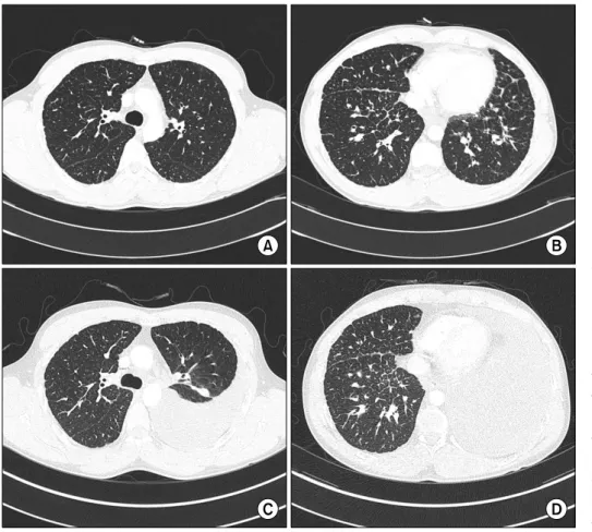
Figure 2. (A, B) Chest CT shows peripheral and axial interstitial thickening in both lungs (1st admission).
(C, D) Chest CT shows newly appeared large amount of pleural effusion and passive atelectasis in left lung. Peripheral and axial interstitial thickening in both lungs shows no significant interval changes since 1st admission (2nd admission).
Figure 1. Chest X-ray shows peripheral and axial inter- stitial thickening in both lungs with left pleural effusion.
관지 내시경을 이용한 경기관지 폐생검을 시행하였으나 출혈이 동반된 정상 폐조직 소견으로 진단을 내리지 못하 여 흉강경을 이용한 수술적 폐생검을 시행하였다. 폐조직
소견에서 간질 내 단핵구 침윤 및 폐포 내 혈철소 탐식 대식구(hemosiderin laden macrophage)가 관찰되었으나 진단이 명확히 내려지지 않아 우선 경과관찰 하기로 하고 퇴원하였다. 퇴원 후 불편감 없이 지내오다가 내원 1개월 전 호흡곤란이 발생하여 외래 방문하였고, 흉부 방사선 사진에서 좌측 흉수가 확인되어 흉수 천자 등의 검사를 위해 입원하였다.
가족력: 특이사항 없음.
개인력: 전기건설 현장에서 15년간 일해왔고, 10갑년의 흡연력이 있었으나 5개월 전 흡연을 중단하였다.
진찰 소견: 내원 시 혈압은 110/86 mm Hg, 맥박수 77 회/분, 호흡수 20회/분, 체온 36.6oC였다. 환자는 건강해 보였고, 좌측 폐하부의 호흡음은 감소되어 있었으며, 타진 시 둔탁음이 들렸다.
검사 소견: 일반 혈액 검사는 백혈구 6,800/mm3, 혈색 소 16.8 g/dL, 혈소판 235,000/mm3, 혈청 생화학 검사는 혈청 총 단백 및 알부민이 각각 6.9 g/dL와 4.3 g/dL, 이외 에 총 빌리루빈 0.7 mg/dL, AST 37 IU/L, ALT 53 IU/L, alkaline phosphatase 87 IU/L, gamma-glutamyl trans- peptidase 26 IU/L로 정상범위였다.
Figure 3. Surgical lung biopsy shows proliferation of com- plex anastomosing lymphatic channels in the interstitium
(H&E stain, ×40). Figure 5. Chest CT after lymphangiography shows multi- focal areas of residual contrast material within abnormal lymphatic spaces in bilateral lower mediastinum and left lower lobe. CT: computed tomography.
Figure 4. Lymphangiogram shows tortuous lymphatic channels and lymphatic leakage (arrow) idenitified at the medial aspect of the descending thoracic aorta.
폐기능 검사: 6개월 전 시행한 폐기능 검사에서 노력성 폐활량(forced vital capacity, FVC)은 4.33 L (정상 예측치 의 94%), 1초간 노력성호기량(forced expiratory volume in one second, FEV1)은 3.66 L (91%), FEV1/FVC은 83, 총 폐용적(total lung capacity, TLC)은 5.37 L (84%), 폐확 산능(diffusing capacity for carbon monoxide, DLCO)은 28.2 mL/min/mm Hg (99%)이었다.
방사선 소견: 단순 흉부 방사선 사진에서 좌측에 흉수 와 양측성 간질 비후가 관찰되었다(Figure 1). 흉부 CT에
서도 좌측에 다량의 흉수와 양측성 간질 비후가 관찰되었 고, 6개월 전과 비교할 때 흉수를 제외한 다른 소견은 차 이가 없었다(Figure 2).
흉수 검사 소견: 흉수 검사에서 색깔은 우윳빛이었고, 적혈구 60,000/mm3, 백혈구 2,250/mm3로 다형 백혈구는 1%, 임파구 77%, 조직구 21%였다. LDH 347 U/L, protein 5.1 g/dL였고, 중성지방은 2146 mg/dL로 증가되어 있었 다. 흉수에서 시행한 균 도말 검사와 배양 검사에서 균은 확인되지 않았고, 세포 검사에서 악성 세포는 관찰되지 않았다.
폐조직 검사 소견: 간질 내 림프관이 확장되어 있었고, 림프관의 수도 증가되어 있었으며, 림프관들이 서로 복잡 하게 문합을 이루고 있었다(Figure 3).
임상 경과: 폐림프관종증 및 동반된 유미흉으로 진단하 였고, 유미흉에 대해서 흉관 삽입 후 경구식이를 중단하고 총정맥 영양을 실시하였다. 림프관 유출부위를 찾고자 림 프관 조영술(lymphangiography)을 시행하였고, 하행 대 동맥 안쪽에서 림프관의 유출이 확인되었다(Figure 4). 이 후 시행한 흉부 CT에서도 양측 종격동 하방과 좌측 폐하 부에 확장된 림프관이 관찰되어(Figure 5) 림프관 결찰술 을 고려하였으나 환자가 수술을 원하지 않아 우선 보존적 치료를 시행하기로 하였다. 3일간 흉수는 3 L 배액되었고, 이후 더 이상 배액되지 않아 저장쇄중성 지방식이(me- dium chain trigliceride)로 경구식이를 시작하였다. 저장
쇄중성 지방식이를 시행한 뒤에도 유미흉은 관찰되지 않 았고, 재발 방지를 위해 화학적 흉막 유착술을 시행한 후 흉관을 제거하였다. 외래 추적관찰 시에도 흉수는 더 이 상 발생하지 않았고 별다른 불편감을 호소하지 않아 경과 관찰 중이다.
고 찰
폐림프관 질환은 Faul 등의 분류에 따르면 림프관종 (lymphagioma), 림프관확장증(lymphangiectasia), 림프 관종증(lymphagiomatosis), 림프관 이형성증(lymphatic dysplasia syndrome)으로 구분할 수 있고, 병리학적 구분 에 따라 다양한 임상상을 보인다1,2. 그 중 림프관종증은 림프관종이 다발성으로 존재하는 질환으로, 다른 림프계 이상을 동반하거나 여러 장기를 침범하는 경우(75%) 가 흔하다3.
폐림프관종증은 조직 소견에서 폐와 종격동 내 정상 림 프관이 문합을 이루어 증식되어 있는 양상을 보인다3. 임 상상은 본 증례에서와 같이 유미흉을 흔히 동반하고, 천명 음, 호흡곤란, 객혈, 유미성 객담, 기관지 원주(bronchial cast)가 나타날 수 있다. 그 외에도 유미성 심막염, 유미성 복수, 단백 소실성 장염, 림프구 감소증도 흔히 동반된다1. 흉부 방사선 촬영에서 양측성 간질 음영 증가, 흉수, 심낭액 등이 관찰되고1 흉부 CT에서 미만성으로 양측을 침범하는 대칭형 소엽사이막(interlobular septum) 비후와 기관지혈관 주위 간질 비후를 보인다. 소엽사이막의 비후 는 대개 매끄러운 형태를 보이나 종종 결절 형태를 보이기 도 한다. 병변은 상엽에서 호발하며 반점상 간유리음영을 보이는 경우 부종, 출혈, 혈철소를 포함한 대식구의 축적 을 시사한다4-6. 폐포 공간은 대개 침범하지 않고, 본 환자 의 영상에서와 같이 종격동과 흉막을 광범위하게 침범하 는 림프계 증식으로 인해 종격동과 흉막의 미만성 비후가 함께 관찰되는 경우가 흔하다4. 하지만 림프절 비대가 보 이는 경우는 매우 드물다5. 림프관 조영술 검사에서는 사 행성 주행을 보이는 확장된 림프관, 다발성 가슴림프관, 폐와 뼈를 침범하는 림프관종의 형태로 나타난다1,4,7. 폐림프관종증은 대개 서서히 진행하는 경과를 보이고1 재발이 흔하며 불량한 예후를 보이는 것으로 알려져 있는 데 현재까지 확립된 치료법은 없다. 주된 치료목적은 림 프액의 축적으로 발생하는 압박 증상을 감소시키는 것이 다8. 종격동 국소 병변이나 단일 폐병변의 경우 흉강경을 통한 절제술이 추천되지만, 미만성 병변의 경우 수술적
치료는 심막액과 재발성 흉수의 배액 및 흉막 유착술, 가 슴림프관 결찰술처럼 재발을 막는 고식적 수술에 국한된 다9. 광범위한 침범으로 수술적 치료가 불가능한 경우 전 신적 항암 치료와 인터페론 알파 치료를 시도할 수 있고 일부에서 효과를 거두었다는 보고가 있었다9,10. 방사선 치 료는 대개 광범위한 피부 침범을 동반한 림프종증 환자에 서 주로 시도되었으나, Kandil 등은 미만성 폐림프관종증 환자 3명에서 일차적으로 방사선 치료를 시도하였고, 각 각 20개월, 32개월, 8년의 완전관해를 보였다고 보고하였 다9.11. Doxycycline 등을 이용한 CT 유도하 경피적 경화 술이 림프관종증에서 유용하다는 보고도 있었다1,8. 본 환 자에서와 같이 저장쇄중성 지방식이와 같은 식이조절은 림프액 흐름을 감소시켜 증상의 호전을 돕고, 유미흉과 림프 부종에 대한 고식적 치료는 환자의 삶의 질을 향상 시키는데 도움이 된다12,13.
본 증례는 소아에서 호발하는 폐림프관종증이 무증상 으로 지내던 35세 남자에서 진단된 경우로 기존의 연구에 서도 폐림프관종증이 드물게 20대 이후에서도 나타나며, 0세에서 80세까지 전 연령에 걸쳐 나타날 수 있다고 보고 한 바 있다14. 기존의 국내보고에서는 반복된 유미흉으로 내원하여 폐림프관 평활근종증을 의심하였던 31세 여자 에서 조직 검사를 통해 폐림프관종증으로 진단된 예가 있 었는데, 진단 시 중중 제한성 환기장애를 보였고 흉관 삽 입과 흉막 유착술 후 호전되었으나 빈번한 재발을 보여 본 증례와 차이를 보였다15. 본 증례는 폐림프관종증의 가 장 흔한 증상 중 하나인 유미성 객담, 유미흉의 소견을 보였고, 흉부 CT에서 전형적인 간질의 비후와 림프관 조 영술에서 확장된 림프관을 보였으며, 폐조직 소견에서 림 프관의 확장, 증식, 문합 등 전형적인 폐림프관종증의 양 상을 보여 진단할 수 있었고 현재 증상 없이 외래관찰 중이다.
참 고 문 헌
1. Faul JL, Berry GJ, Colby TV, Ruoss SJ, Walter MB, Rosen GD, et al. Thoracic lymphangiomas, lym- phangiectasis, lymphangiomatosis, and lymphatic dys- plasia syndrome. Am J Respir Crit Care Med 2000;161:
1037-46.
2. Hilliard RI, McKendry JB, Phillips MJ. Congenital ab- normalities of the lymphatic system: a new clinical classification. Pediatrics 1990;86:988-94.
3. Tazelaar HD, Kerr D, Yousem SA, Saldana MJ,
Langston C, Colby TV. Diffuse pulmonary lymphan- giomatosis. Hum Pathol 1993;24:1313-22.
4. Raman SP, Pipavath SN, Raghu G, Schmidt RA, Godwin JD. Imaging of thoracic lymphatic diseases. AJR Am J Roentgenol 2009;193:1504-13.
5. Swensen SJ, Hartman TE, Mayo JR, Colby TV, Tazelaar HD, Müller NL. Diffuse pulmonary lymphangiomatosis:
CT findings. J Comput Assist Tomogr 1995;19:348-52.
6. Yekeler E, Dursun M, Yildirim A, Tunaci M. Diffuse pulmonary lymphangiomatosis: imaging findings. Dia- gn Interv Radiol 2005;11:31-4.
7. Jang HJ, Lee KS, Han J. Intravascular lymphomatosis of the lung: radiologic findings. J Comput Assist Tomogr 1998;22:427-9.
8. Molitch HI, Unger EC, Witte CL, vanSonnenberg E.
Percutaneous sclerotherapy of lymphangiomas. Radio- logy 1995;194:343-7.
9. Rostom AY. Treatment of thoracic lymphangiomatosis.
Arch Dis Child 2000;83:138-9.
10. Reinhardt MA, Nelson SC, Sencer SF, Bostrom BC,
Kurachek SC, Nesbit ME. Treatment of childhood lym- phangiomas with interferon-alpha. J Pediatr Hematol Oncol 1997;19:232-6.
11. Kandil A, Rostom AY, Mourad WA, Khafaga Y, Gershuny AR, el-Hosseiny G. Successful control of ex- tensive thoracic lymphangiomatosis by irradiation. Clin Oncol (R Coll Radiol) 1997;9:407-11.
12. Valentine VG, Raffin TA. The management of chylo- thorax. Chest 1992;102:586-91.
13. Calabrese PR, Frank HD, Taubin HL. Lymphangiomyo- matosis with chylous ascites: treatment with dietary fat restriction and medium chain triglycerides. Cancer 1977;40:895-7.
14. Kransdorf MJ. Benign soft-tissue tumors in a large re- ferral population: distribution of specific diagnoses by age, sex, and location. AJR Am J Roentgenol 1995;
164:395-402.
15. Cho YS, Yoo JH, Son SY, Cho HL, Kim SJ, Han MS, et al. Pulmonary lymphangiomatosis. Tuberc Respir Dis 1999;47:533-7.