DOI 10.3349/ymj.2008.49.1.90
Purpose: We reviewed the cases of 33 patients from our clinic and 142 patients from the literature with congenital bronchopulmonary vascular malformations (BPVM), systema- tically analyzed the bronchopulmonary airways, pulmonary arterial supplies, and pulmonary venous drainages, and classified these patients by pulmonary malinosculation (PM).
Materials and Methods: From January 1990 to January 2007, a total of 33 patients (17 men or boys and 16 women or girls), aged 1 day to 24 years (median, 2.5 months), with congenital BPVM were included in this study. Profiles of clinical mani- festations, chest radiographs, echocardiographs, esophago- graphs, computer tomography (CT), magnetic resonance imaging (MRI), magnetic resonance angiography (MRA), cardiac catheterizations with angiography, contrast broncho- graphs, bronchoscopies, chromosomal studies, surgeries, and autopsies of these patients were analyzed to confirm the diagnosis of congenital BPVM. A total of 142 cases from the literature were also reviewed and classified similarly. Results:
The malformations of our 33 patients can be classified as type A isolated bronchial PM in 13 patients, type B isolated arterial PM in three, type C isolated venous PM in two, type D mixed bronchoarterial PM in five, type F mixed arteriovenous PM in one, and type G mixed bronchoar- teriovenous PM in nine. Conclusion: Dysmorphogeneses of the primitive foregut system and the primitive aortic arch system may lead to haphazard malinosculations of the airways, arteries, and veins of the lung. A systematic classification of patients with congenital BPVM is clinically feasible by assessing the three basic bronchovascular systems of the lung independently.
Key Words: Congenital bronchopulmonary vascular malforma- tion, pulmonary malinosculation, scimitar syndrome, broncho- pulmonary sequestration, sequestration spectrum, haphazard, dysmorphogeneses, primitive foregut system, primitive aortic arch system
INTRODUCTION
Congenital bronchopulmonary vascular malfor- mations (BPVM) encompass a broad spectrum of diseases involving abnormal communication or anastomosis of one or more components of the lung.
1-3Despite early definition of the sequestration spectrum,
4-7and scimitar syndrome,
8,9reports of incomplete-, atypical-, and pseudo-sequestration /scimitar syndrome continued to prevail in the literature, which implies that a universal under- standing of the sequestration spectrum and scimitar syndrome is still lacking. It is believed that malformations of the pulmonary airways, arteries, and veins may occur independently and coin- cidentally.
10-14Clements et al. proposed a "wheel theory" to explain the serial formation of congenital BPVM, and coined "pulmonary malinosculation"
(PM) to classify these malformations. We propose a "haphazard theory" to substantiate this concept of PM.
3However, there is no category in which to classify patients with isolated scimitar vein anomalies and meandering right pulmonary veins.
3Nevertheless, isolated scimitar vein anomalies have been observed in our clinical
A Systematic Classification of the Congenital Bronchopulmonary Vascular Malformations: Dysmorphogeneses of the Primitive Foregut System and the Primitive Aortic Arch System
Meng-Luen Lee,
1Hung-Chi Lue,
2Ing-Sh Chiu,
3Han-Yao Chiu,
4Lon-Yen Tsao,
5Ching-Yuan Cheng,
6and Albert D. Yang
71
Department of Pediatrics, Division of Pediatric Cardiology,
4Department of Pediatric Pulmonology,
5Department of Pediatric Neonatology,
6Department of Surgery, Division of Chest Surgery and
7Department of Radiology, Changhua Christian Hospital, Changhua;
2Department of Pediatrics, Division of Pediatric Cardiology, Min-Sheng General Hospital, Tao-Yuan;
3Department of Surgery, Division of Cardiovascular Surgery, College of Medicine, National Taiwan University Hospital, Taipei, Taiwan.
Received April 16, 2007 Accepted July 1, 2007
Reprint address: requests to Dr. Meng-Luen Lee, Department
of Pediatrics, Division of Pediatric Cardiology, Changhua Christian
Hospital, No. 135, Nanhsiao St., Changhua 50050, Taiwan. Tel: 886-
4-7238595 (#1902) Fax: 886-4-7238847, E-mail: [email protected]
practice
15and in that of others'.
16In this article, we will make a review and classification from an embryologic perspective of our 33 patients and 142 patients reported in the literature who had scimitar syndrome, bronchopulmonary sequestra- tion, sequestration spectrum, congenital horseshoe lung, crossover lung segment, and other similar conditions.
MATERIALS AND METHODS
Between January 1990 and January 2007, 33 consecutive patients (17 men or boys and 16 women or girls), aged 1 day to 24 years (median, 2.5 months) with congenital BPVM were included in this retrospective analysis. Patients with total or partial anomalous pulmonary venous connections were excluded from this study. Study modalities included clinical features and plain chest radio- graphs (n = 33) plus at least two of the following laboratory profiles: two-dimensional echocardio- graphy with Doppler (Acuson 128XP/10c, Mountain View, CA, USA) (n = 22), cardiac catheterization with cineangiography (n = 19), cardiothoracic surgery (n = 12), bronchography (n = 10), electron- beam CT of the chest (Imatron, South San Francisco, CA, USA) (n = 11), MRI (n = 11), bronchoscopy (n
= 5), barium esophagography (n = 2), chromosomal study (n = 2), autopsy (n = 2), high resolution CT of the chest (n = 1), and MRA (n = 1).
Definitions of malinosculation, sequestration, syndrome, and scimitar syndrome
1. Stedman's Medical Dictionary (2000) defines malinosculation (Latin: mal-ill, bad, abnormal;
in-in; osculum-mouth) as "the establishment of abnormal communications by means of small openings or anastomoses".
2. The Concise Oxford Dictionary (2001) defines sequestration (from the Latin sequestrare, to separate) as something in a state of being secluded, isolated, or separated from other things.
3. The ENCARTA World English Dictionary (1999) defines syndrome (from the Latin and Greek sundrome, literally, running together) as a group of symptoms that consistently
occur together.
4. Scimitar syndrome was originally defined as a syndrome having a triad of 1) an anoma- lous pulmonary venous connection to the inferior vena cava as scimitar vein, 2) a systemic arterial supply to the right lung, and 3) bronchial anomalies of the hypoplastic right lung.
8,9The lung is mainly composed of six basic bronchovascular trees attributed to three different systems, including: 1) airways: bronchial trees, 2) artery: systemic and pulmonary arterial trees, and 3) vein: systemic and pulmonary venous trees, and lymphatic trees. Thus, these six basic bronchovascular trees can be classified into three different systems (airways, arteries, and veins).
We regard anomalies of the venous drainage of the lungs (systemic, pulmonary, and lymphatic) along with anomalies of the pulmonary airways and pulmonary arterial supply simultaneously, rather than consider them as the aftermath of malinosculations of the pulmonary airways and the pulmonary artery.
Nomenclature and definition of PM (Fig. 1)
Type A isolated bronchial PM (area A in Fig. 1)
is defined as isolated malinosculation of the
pulmonary airways into the lung (with or without
lung parenchymal abnormalities), but with a
normal pulmonary arterial supply and a normal
pulmonary venous connection. These malforma-
tions could be further subclassified into: 1)
proximal lesions of tracheal airways, e.g., tracheal
stenosis, tracheal bronchus, and tracheal cyst; 2)
distal lesions of bronchial and smaller airways,
e.g., a localized lesion of congenital bronchial
stenosis, bronchial atresia, bronchogenic cyst,
congenital cystic adenomatoid malformation, and
congenital lobar emphysema. Type B isolated
arterial PM (area B in Fig. 1) is defined as isolated
malinosculation of the pulmonary artery, but with
normal pulmonary airways and a normal
pulmonary venous connection. Patients with a
systemic arterial supply (systemic arterialization)
to the normal lung, without either broncho-
pulmonary airways sequestration or anomalous
pulmonary venous connections, have isolated
arterial PM. Type C isolated venous PM (area C
in Fig. 1) is defined as isolated malinosculation of the pulmonary vein, but with normal pulmonary airways and a normal pulmonary arterial supply.
Patients with isolated anomalies of the scimitar vein (to the inferior vena cava/right atrium) and meandering right pulmonary vein could be categorized into type C isolated venous PM. Type D mixed bronchoarterial PM (area D in Fig. 1) is defined as a combination of malinosculations of the pulmonary airways and the pulmonary artery but with a normal pulmonary venous connection.
In that regard, cases of patients with broncho- pulmonary airways sequestration with a systemic arterial supply and a normal pulmonary venous connection could be classified into type D mixed bronchoarterial PM. Type E mixed bronchovenous PM (area E in Fig. 1) is defined as a combination of malinosculations of the pulmonary airways and the pulmonary vein, but with a normal pulmonary arterial supply. Tracheal stenosis, tracheal bronchus,
tracheal cyst, bronchogenic cyst, bronchial stenosis, congenital cystic adenomatoid malformation, and congenital lobar emphysema could be found with anomalous pulmonary venous connections and a normal pulmonary arterial supply. Type F mixed arteriovenous PM (area F in Fig. 1) is defined as a combination of malinosculations of the pulmonary artery and the pulmonary vein, but with normal pulmonary airways. The cases of patients with a congenital fistula between a systemic or pulmonary artery and a pulmonary vein, and of patients with a scimitar vein (to the inferior vena cava) and a systemic arterial supply from the aorta (systemic arterialization) to a segment or a lobe of the lung, which is not secluded from the normal pulmonary airways (i.e., without broncho- pulmonary airways sequestration), could be grouped into type F mixed arteriovenous PM.
Type G mixed bronchoarteriovenous PM (area G in Fig. 1) is defined as malinosculations involving the pulmonary airways, the pulmonary artery, and the pulmonary vein. Classical scimitar syndrome with the above triads can be grouped into type G mixed bronchoarteriovenous PM. The aforementioned PM will be applied to analyze the cases of our 33 patients with congenital BPVM and those of another 142 patients from the literature with scimitar syndrome, bronchopulmonary sequestra- tion, sequestration spectrum, congenital horseshoe lung, crossover lung segment, and other similar conditions.
RESULTS
The clinical profiles of our own 33 patients with congenital BPVM are summarized in Table 1.
They were aged one day to 24 years (median, 2.5 months; range, 0.03-288 months). There were 24 infants (73%), eight children (> 1 and 18 years old; 24%), and one adult (3%). Among these 33 patients, the salient clinical picture was recurrent pneumonia in 23 patients (70%), respiratory distress in 21 (64%), wheezy respiration in 17 (52%), pulmonary hypertension in 14 (42%), cyanosis in 13 (39%), and congestive heart failure in 12 (36%). Situs solitus was noted in 28 patients, situs ambiguus with asplenia or heterotaxy syndrome in three, and situs inversus in two. Ellis-van Creveld
Fig. 1. Pulmonary malinosculation (PM) involving the pulmonary airways, the pulmonary artery, and the pulmonary vein independently. Each circle represents the occurrence of PM involving the pulmonary airways, the pulmonary artery, and the pulmonary vein within the lung parenchyma, respectively. Each single circle converges to intersect (denoted by 3 sets of reverse arrows) another two circles to form the geometric figure of a trefoil (as a mathematical Venn diagram), within which are seven exclusive areas (areas A-G) denoting seven distinct PM.
Area A represents type A isolated bronchial PM (with normal pulmonary artery and veins). Area B represents type B isolated arterial PM (with normal pulmonary airways and veins). Area C represents type C isolated venous PM (with normal pulmonary airways and artery).
Area D represents type D mixed bronchoarterial PM (with
normal pulmonary veins). Area E represents type E mixed
bronchovenous PM (with normal pulmonary artery). Area
F represents type F mixed arteriovenous PM (with normal
pulmonary airways). Area G represents type G mixed
bronchoarteriovenous PM.
syndrome was noted in one patient, Down syndrome in one, and tibial agenesis with ectrodactyly syndrome in one. Congenital syphilis, E. coli sepsis, and Klebsiella oxytocia sepsis were documented in three individual patients. Lymphedema of the arm and shoulder was noted at the sixth day of life in a patient with congenital pulmonary lymphangiec- tasia. One patient had microcephaly, cleft palate, dermal sinus tract, and hyperlactic acidemia.
Absence of a gallbladder was noted in one patient.
Among the 33 patients with congenital BPVM, 17 patients had problems involving the broncho- pulmonary system and the cardiovascular system (52%), 10 had problems of the bronchopulmonary system (30%), and six had problems of the cardiovascular system (18%). Ten patients had associated simple congenital heart disease (30%), nine of them had situs solitus (27%), and one had situs inversus (3%). In ten patients associated with simple congenital heart disease, ventricular septal defects were noted in six, patent ductus arteriosus in five, secundum atrial septal defects in five, aortic coarctation in three, sling left pulmonary artery (vascular ring) in three, and supravalvular pulmonary stenosis in one. Four patients had associated complex congenital heart disease (12%), three of them had situs ambiguus (9%), and one had situs inversus (3%). Dextroversion of the heart was found in 12 patients (36%), and dextrocardia in five (15%). Scimitar vein anomaly was found in four patients (nos. 17, 24, 32, and 33), and was silhouetted on the plain chest film as a right-sided sword in three patients (nos. 17, 24, and 32) and a left-sided sword in one (no. 33). The drainage site of the scimitar vein was the inferior vena cava in two patients (right middle and lower lobes in no. 24; right lower lobe in no. 32), the right atrium in one (a dual drainage of the right trilobes to the left atrium and the right atrium in no. 17), and the hepatic vein in one (left-sided scimitar in no. 33).
Patient 17, who had a scimitar vein draining the whole right lung both to the left atrium and to the right atrium (a dual system of right pulmonary venous drainage), did not present with desa- turation or discernible lip cyanosis. Broncho- pulmonary (airways) sequestration was found in four patients (nos. 12, 20, 32, and 33), including systemic arterial supply to the left lower lobe in two patients (nos. 20 and 33), the right lower lobe
in one (no. 32), and normal pulmonary arterial supply to the right lower lobe in one (no. 12). Of three patients with vascular ring (nos. 19, 21, and 30), an isolated left pulmonary artery sling was noted in one (no. 21), and a complex left pulmonary artery sling (associated with other rare aortic arch anomalies) was noted in the remaining two (nos. 19 and 30). Aortic arch anomalies included ductal sling, a right-sided patent ductus arteriosus, a left ascending aorta and aortic arch, and a right descending aorta in one (no. 19); an aberrant right subclavian artery from a left aortic arch, a retroesophageal aortic arch, and a descending aorta in one (no. 30).
Interventional cardiac catheterization was performed in four patients (nos. 16, 22, 23, and 30), including balloon dilatation for aortic coarctation in three (nos. 16, 22, and 23) and supravalvular pulmonary stenosis in one (no. 30), and transarterial coil embolization for the systemic pulmonary artery in one (no. 16). Surgical intervention was performed in 12 patients, including thoracic surgery in six patients, cardiovascular surgery in five, and thoracic-cardiovascular surgery in one. The overall mortality rate was 8 out of 33 (24%).
A systematic classification for the congenital BPVM was: type A isolated bronchial PM in 13 patients, type B isolated arterial PM in three, type C isolated venous PM in two, type D mixed bronchoarterial PM in five, type F mixed arterio- venous PM in one, and type G mixed bronchoar- teriovenous PM in nine (Table 1).
We also reappraised the cases of 142 patients from the literature reported to have had scimitar syndrome, bronchopulmonary sequestration, sequestration spectrum, congenital horseshoe lung, crossover lung segment, congenital pulmonary lymphangiectasia, meandering right pulmonary vein, and isolated scimitar vein anomaly, and applied this systematic PM to assess and classify them (Table 2).
DISCUSSION
Congenital BPVM can manifest with various
symptoms. One disease entity, for example,
congenital cystic adenomatoid malformation, may
have different pathology and protean features. On
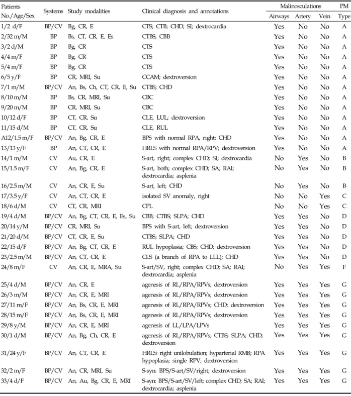
Table 1. A Systematic Approach to Our 33 Patients with Congenital BPVM and Their Classification Patients
No./Age/Sex Systems Study modalities Clinical diagnosis and annotations Malinosculations PM Airways Artery Vein Type
1/2 d/F BP/CV Bg, CR, E CTS; CTB; CHD; SI; dextrocardia Yes No No A
2/32 m/M BP Bs, CT, CR, E, Es CTBS; CBB Yes No No A
3/2 d/M BP Bg, CR CTS Yes No No A
4/4 m/F BP Bg, CR CTS Yes No No A
5/4 m/F BP Bg, CR CTS Yes No No A
6/5 y/F BP CR, MRI, Su CCAM; dextroversion Yes No No A
7/1 m/M BP/CV An, Bs, Ch, CT, CR, E, Su CTBS; CHD Yes No No A
8/10 m/M BP Bs, CR, MRI, Su CBC Yes No No A
9/20 m/M BP CR, MRI, Su CBC Yes No No A
10/12 d/F BP CT, CR, Su CLE, LUL; dextroversion Yes No No A
11/15 d/M BP CT, CR, Su CLE, RUL Yes No No A
A12/1.5 m/F BP/CV An, Bg, CR, E BPS with normal RPA, right; CHD Yes No No A
13/13 y/F BP An, CT, CR, E HRLS with normal RPA/RPV; dextroversion Yes No No A 14/1 m/M CV Au, CR, E S-art, right; complex CHD; SI; dextrocardia No Yes No B 15/1.5 m/F CV An, Bg, CR, E S-art, both; complex CHD; SA; RAI;
dextrocardia; asplenia No Yes No B
16/2.5 m/M CV An, CR, E, Su S-art, left; CHD No Yes No B
17/3.5 y/F CV An, CT, CR, E isolated SV anomaly, right No No Yes C
18/6 d/M CV CT, CR, MRI CPL No No Yes C
19/4 d/M BP/CV An, Bg, CT, CR, E, Es, Su CBB; CTBS; SLPA; CHD Yes Yes No D
20/14 y/M BP/CV CR, MRI, Su BPS with S-art, left; dextroversion Yes Yes No D
21/20 d/M BP/CV CT, CR, E, Su CTBS; SLPA; CHD Yes Yes No D
22/15 d/F BP/CV An, Bg, CT, CR, E RUL hypoplasia; CBS; CHD; dextroversion Yes Yes No D 23/2.5 m/M BP/CV An, CT, CR, E CLS (a branch of RPA to LLL); CHD Yes Yes No D 24/8 m/F CV An, CR, E, MRA, Su S-art/SV, right; complex CHD; SA; RAI;
dextrocardia; asplenia No Yes Yes F
25/4 d/M BP/CV An, CR, E agenesis of RL/RPA/RPVs; dextroversion Yes Yes Yes G 26/3 m/M BP/CV An, CR, E, MRI agenesis of RL/RPA/RPVs; dextroversion Yes Yes Yes G 27/11 m/F BP/CV An, Bs, CR, E, MRI agenesis of RL/RPA/RPVs; CHD; dextroversion Yes Yes Yes G 28/15 m/F BP/CV An, Bs, CR, E, MRI agenesis of RL/RPA/RPVs; dextroversion Yes Yes Yes G
29/8 y/M BP/CV An, CR, E, MRI agenesis of LL/LPA/LPVs Yes Yes Yes G
30/1 d/M BP/CV An, Bg, Ch, CR, E agenesis of RL/RPA/RPVs; CTBS; SLPA; CHD;
dextroversion Yes Yes Yes G
31/24 y/F BP/CV An, CT, CR, E HRLS: right unilobulation; hyparterial RMB; RPA
hypoplasia; single RPV; dextroversion Yes Yes Yes G 32/2 m/F BP/CV An, CR, MRI, Su S-syn: BPS/S-art/SV/right; dextroversion Yes Yes Yes G 33/4 d/F BP/CV An, Au, Bg, CR, E, MRI S-syn: BPS/S-art/SV/left; complex CHD; SA; RAI;
dextrocardia; asplenia Yes Yes Yes G
An, angiography; Au, autopsy; Bg, bronchography; BP, bronchopulmonary; BPS, bronchopulmonary sequestration; BPVM, bronchopulmonary vascular malformations; Bs, bronchoscopy; CBB, congenital bridging bronchus; CBC, congenital bronchogenic cyst; CBS, congenital bronchial stenosis;
CCAM, congenital cystic adenomatoid malformation; CHD, congenital heart disease; Ch, chromosomal study; CLE, congenital lobar emphysema;
CLS, crossover lung segment; CPL, congenital pulmonary lymphangiectasia; CR, chest radiography; CT, computer tomography; CTB, congenital tracheal bronchus; CTBS, congenital tracheobronchial stenosis; CTS, congenital tracheal stenosis; CV, cardiovascular; E, echocardiography; Es, esophagography; HRLS, hypogenetic right lung syndrome; LL, left lung; LLL, left lower lobe; LPA, left pulmonary artery; LPVs, left pulmonary veins; LUL, left upper lobe; MRA, magnetic resonance angiography; MRI, magnetic resonance imaging; RAI, right atrial isomerism; RL, right lung;
RLL, right lower lobe; RMB, right main bronchus; RPA, right pulmonary artery; RPVs, right pulmonary veins; RUL, right upper lobe; SA, situs ambiguus; S-art, systemic arterial supply (arterialization) to the lung; SI, situs inversus; SLPA, sling left pulmonary artery; S-syn, scimitar syndrome;
Su, surgery; SV, scimitar vein.