493 http://dx.doi.org/10.4196/kjpp.2013.17.6.493
ABBREVIATIONS: i.c.v., intracerebroventricular; i.t., intrathecal;
STZ., streptozotocin.
Received March 28, 2013, Revised September 26, 2013, Accepted October 17, 2013
Corresponding to: Hong-Won Suh, Department of Pharmacology, Institute of Natural Medicine, College of Medicine, Hallym Univer- sity, 39 Hallymdaehak-gil, Chuncheon 200-702, Korea. (Tel) 82-33- 248-2614, (Fax) 82-33-248-2612, (E-mail) [email protected]
This is an Open Access article distributed under the terms of the Creative Commons Attribution Non-Commercial License (http://
creativecommons.org/licenses/by-nc/3.0) which permits unrestricted non-commercial use, distribution, and reproduction in any medium, provided the original work is properly cited.
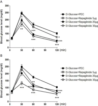
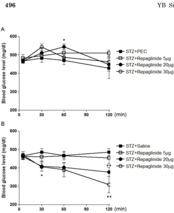
Repaglinide, but Not Nateglinide Administered Supraspinally and Spinally Exerts an Anti-Diabetic Action in D-Glucose Fed and Streptozotocin-Treated Mouse Models
Yun-Beom Sim
1, Soo-Hyun Park
1, Yu-Jung Kang
1, Sung-Su Kim
1, Chea-Ha Kim
1, Su-Jin Kim
1, Su-Min Lim
1, Jun-Sub Jung
1, Ohk-Hyun Ryu
2, Moon-Gi Choi
2, and Hong-Won Suh
11