• 검색 결과가 없습니다.

Pulmonary Embolism and Uterine Venous PlexusThrombosis in the Postpartum Period

N/A
N/A
Protected

Academic year: 2022

Share "Pulmonary Embolism and Uterine Venous PlexusThrombosis in the Postpartum Period"

Copied!
4
0
0

로드 중.... (전체 텍스트 보기)

전체 글

(1)

Introduction

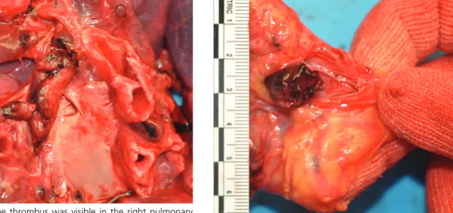
Pregnancy is associated with an increased risk of venous thromboembolism, which includes pulmonary embolism (PE) and deep vein thrombosis. The inci- dence of venous thromboembolism is estimated at 0.76 to 1.72 per 1,000 pregnancies, which is four times higher than that in non-pregnant women [1]. The inci- dence of PE in the postpartum period is much higher

than during pregnancy [2]. There are few reports on uterine vein thrombosis, consisting mostly of cases involving thrombi in external and internal iliac veins [3]. Uterine venous plexus thrombosis (UVPT) is a rare diagnosis. Sudden death linked to pulmonary thromboembolism (PTE) in the presence of UVPT without deep vein thrombosis in the lower extremities is even more uncommon. We report a case of sudden, unexpected death due to acute PTE associated with UVPT in a postpartum woman.

41

pISSN 2383-5702 eISSN 2383-5710

ⓒCopyright 2015 by the Korean Society for Legal Medicine

This is an Open Access article distributed under the terms of the Creative Commons Attribution Non-Commercial License (http://creativecommons.org/licenses/

by-nc/3.0) which permits unrestricted non-commercial use, distribution, and reproduction in any medium, provided the original work is properly cited.

Korean J Leg Med 2015;39:41-44

http://dx.doi.org/10.7580/kjlm.2015.39.2.41

Pulmonary Embolism and Uterine Venous Plexus Thrombosis in the Postpartum Period

Young Keum Kim

1

Kyung Bin Kim

2

,

Chung Hwan Kim

3

, Hongil Ha

3

1

Department of Pathology, Pusan National University Hospital, Busan, Korea,

2

Department of Pathology, Ulsan University Hospital, Ulsan, Korea,

3

Division of Forensic Medicine, Busan

Institute, National Forensic Service, Yangsan, Korea

Received: April 26, 2015 Revised: May 15, 2015 Accepted: May 26, 2015

Correspondence to Hongil Ha

Division of Forensic Medicine, Busan Institute, National Forensic Service, 50 Geumo-ro, Mulgeum-eup, Yangsan 626-742, Korea

Tel: +82-55-380-4050 Fax: +82-55-380-4060 E-mail: [email protected]

Pulmonary thromboembolism (PTE) associated with uterine venous plexus thrombo- sis is very rare. This was recently observed in a puerperal woman without significant medical history. The woman had two pregnancy-associated risk factors for venous thromboembolism, which were obesity and cesarean delivery. A day after cesarean delivery, she presented with dyspnea and cyanosis. When transferred to a larger hos- pital, no pulse was detected, and she was pronounced dead. Autopsy examination revealed that the cause of death was related to PTE, apparently due to thrombi that originated in the uterine venous plexus.

Key Words: Pulmonary embolism; Venous thrombosis; Pregnancy; Autopsy

Case Report

(2)

Case Report

A 34-year-old woman with a prior history of cesarean delivery was admitted to a local hospital at 38 weeks of gestation. Her previous pregnancy was uncompli- cated, and she had no significant medical history. A day after admission, she underwent cesarean delivery.

The newborn baby weighed 3,520 g at birth, with an Apgar score of 9/10. During surgery, no abnormal symptoms were observed. Twenty-seven hours after the surgery, the woman presented with acute-onset dyspnea and dizziness. Her vital signs indicated a slightly elevated blood pressure (150/100 mm Hg), tachypnea (respiratory rate, 27 breaths per minute), tachycardia (heart rate, 126 beats per minute), and normal temperature (36.8 C). She was transferred to a nearby larger hospital. Cyanosis was seen on her face, hands, and feet. No pulse was detected and cardiopul- monary resuscitation was consequently performed.

Two hours after the procedure, she died due to sudden cardiopulmonary arrest.

The patient autopsied was an extremely obese woman 162 cm in height and 95 kg in weight (body mass index, 36.2). External examination showed no evidence of injury, except bruises in the anterior chest

(due to cardiopulmonary resuscitation). Internal exam- ination revealed a horizontal fracture in the sternum and multiple bilateral anterior fractures from the sec- ond to the fifth ribs (due to cardiopulmonary resusci- tation). The right and left lung weighed 468 g and 642 g, respectively. The cut surface of the right lung revealed marked bloody congestion and edema with a thrombus filling the right pulmonary artery (Fig. 1).

No findings were identified in the left lung or associat- ed vessels. The uterus weighed 1,394 g. One intramur- al mass, measuring 3.5 3.0 cm in size, was identified.

The right uterine venous plexus revealed a thrombus, measuring 1.5 1.0 cm in size (Fig. 2). Findings of the remaining internal organs were unremarkable. There was no evidence of deep vein thrombosis in the lower extremities. Microscopic examination confirmed a thrombus that filled the right pulmonary artery, in addition to congestion and edema in the right lung.

The combined evidence suggests that the cause of death was PTE, possibly associated with UVPT.

Discussion

The literature supports an association between deep vein thrombosis of the lower limbs and PE. During pregnancy, the uterus increases in size and has

http://www.kjlm.or.kr http://dx.doi.org/10.7580/kjlm.2015.39.2.41

42 Korean Journal of Legal Medicine│2015;39:41-44

Fig. 1. A large thrombus was visible in the right pulmonary

artery. Fig. 2. A thrombus was identified in the uterine venous plexus.

(3)

increased blood flow [3]. These changes may impede venous outflow from the lower limbs until approxi- mately six weeks after delivery. Pregnancy is also clas- sically thought to be a hypercoagulable state [1]. This predisposes women to developing venous thromboem- bolism. Additional risk factors for venous thromboem- bolism include heart disease, diabetes, lupus, smoking, multiple pregnancy, age greater than 35 years, obesity, and cesarean delivery. Because of their high preva- lence, age greater than 35 years, obesity, and cesarean delivery are the most important factors that can increase the incidence rates of venous thromboem- bolism [4,5]. A clear relationship has been established between deep vein thrombosis and PE. Deep vein thrombosis associated with PE usually occurs in deep vessels of lower limbs, especially in those of the left leg (approximately 70% to 90% of cases). Occasionally, a large uterine leiomyoma can cause compression of the pelvic veins, leading to persistent pressure on these veins, and inducing chronic venous stasis in the pelvis and lower extremities [6]. The uterine venous plexus extends along the lateral aspect of the uterus and communicates with the ovarian and vaginal plexuses within the broad ligament. They are drained by the uterine veins arising from the lower parts of the uterine plexuses, lateral to the cervix. More common forms of pelvic venous thrombosis are uterine vein thrombosis, ovarian vein thrombosis, and cervical varices [7-10]. Because an anastomosis between the ovarian and uterine venous plexuses may obscure the venous origin, a radiologic diagnosis of UVPT may be difficult [7].

The present case had no other origins of PE except UVPT. The thrombus was found within the venous plexus located in the lateral side of the lower uterus.

The femoral vessels of both legs and deep vessels of both lower legs had no thrombi. Although an intra- mural leiomyoma was observed, it was too small to compress one of the pelvic vessels. Although PE resulting from UVPT is very rare, this patient had two risk factors for venous thromboembolism, obesity and cesarean delivery. These factors may have contributed

to the development of UVPT, finally leading to PE.

The diagnosis of proximal thrombosis involving the pelvic veins is usually made only after the thrombus extends into the veins of the lower legs [3]. UVPT may therefore be a precursor to deep vein thrombosis of the lower limbs, leading to the development of PTE.

On the other hand, it must be taken into account that deep vein thrombosis of the lower limbs may extend to proximal areas and be detected as UVPT. In both cases, it can be concluded that UVPT contributes to PTE.

PTE can occur exclusively without deep vein throm- bosis. For this reason, PTE and UVPT may be consid- ered independent events that do not affect each other.

However, since deep vein thrombosis is an established cause of PTE, the connection between UVPT and PTE cannot be excluded in a case of confirmed UVPT, even though UVPT is rare in itself and its association with PTE is uncommon. For this reason, we infer that PE and UVPT are related in this case.

In conclusion, PE concurrent with UVPT during pregnancy is very rare. To our knowledge, this case is the first in the literature to report sudden, unexpected death caused by acute PTE related to UVPT in a post- partum woman without deep vein thrombosis of the lower limbs. This case study suggests that UVPT can lead to PTE, and that careful examination of vessels surrounding the uterus may help discern the origin of PE in women whose lower limbs show no signs of deep vein thrombosis.

Conflicts of Interest

No potential conflict of interest relevant to this article was reported.

References

1. Marik PE, Plante LA. Venous thromboembolic disease and pregnancy. N Engl J Med 2008;359:2025-33.

2. Heit JA, Kobbervig CE, James AH, et al. Trends in the inci- dence of venous thromboembolism during pregnancy or postpartum: a 30-year population-based study. Ann Intern Med 2005;143:697-706.

http://dx.doi.org/10.7580/kjlm.2015.39.2.41 http://www.kjlm.or.kr

Pulmonary Embolism and Uterine Venous Plexus Thrombosis│Young Keum Kim, et al. 43

(4)

3. Mavrelos D, Cohen H, Pateman K, et al. Diagnosis of uter- ine vein thrombosis on transvaginal ultrasound.

Ultrasound Obstet Gynecol 2013;42:480-3.

4. James AH, Jamison MG, Brancazio LR, et al. Venous thromboembolism during pregnancy and the postpartum period: incidence, risk factors, and mortality. Am J Obstet Gynecol 2006;194:1311-5.

5. Larsen TB, Sorensen HT, Gislum M, et al. Maternal smok- ing, obesity, and risk of venous thromboembolism during pregnancy and the puerperium: a population-based nested case-control study. Thromb Res 2007;120:505-9.

6. Fernandes FL, Dinardo CL, Terra-Filho M. Uterine my- oma as a cause of iliac vein thrombosis and pulmonary em- bolism: common disease, rare complication. Respirol Case

Rep 2014;2:132-4.

7. Leibovitz Z, Degani S, Shapiro I, et al. Diagnosis of preg- nancy-associated uterine venous plexus thrombosis on the basis of transvaginal sonography. J Ultrasound Med 2003;22:287-93.

8. Al-toma A, Heggelman BG, Kramer MH. Postpartum ovar- ian vein thrombosis: report of a case and review of litera- ture. Neth J Med 2003;61:334-6.

9. Royo P, Alonso-Burgos A, Garcia-Manero M, et al.

Postpartum ovarian vein thrombosis after cesarean deliv- ery: a case report. J Med Case Rep 2008;2:105.

10. Sammour RN, Gonen R, Ohel G, et al. Cervical varices complicated by thrombosis in pregnancy. Ultrasound Obstet Gynecol 2011;37:614-6.

http://www.kjlm.or.kr http://dx.doi.org/10.7580/kjlm.2015.39.2.41

44 Korean Journal of Legal Medicine│2015;39:41-44

수치

Fig. 1. A large thrombus was visible in the right pulmonary

참조

관련 문서

Percutaneous polymethylmethacrylate vertebroplasty in the treatment of osteoporotic vertebral body compression fractures: technical aspects. Pulmonary embolism caused

period was prolonged unavoidably, (3) by explaining the risk factors associated with the failure to patients honestly, and subsequently performing

For example, the public’s sentiments of risk regarding nuclear power plants exceeds the risk felt by an expert in the area, and therefore the public sentiment is a negative

Undiagnosed malignancy in patients with deep vein thrombosis: incidence, risk indicators, and diagnosis.. Monreal M, Fernandez-Llamazares J, Perandreu

With these results, we can suggest the hypothesis that increased cardiac output in the patients who administered ketamine increased muscle blood flow, and this is the

When the barrier is detected in the pathways of an automatic aircraft, the MCU which is located in the inner structure of the craft decides the

It is to trace the period of their appearances and changes and also to illuminate coinage characteristics and method of architectural terminology used

1 John Owen, Justification by Faith Alone, in The Works of John Owen, ed. John Bolt, trans. Scott Clark, "Do This and Live: Christ's Active Obedience as the