Received August 12, 2019 Revised March 3, 2020 Accepted March 8, 2020
Corresponding author: Shin Hwang Department of Surgery, Asan Medical Center, University of Ulsan College of Medicine, 88 Olympic-ro 43-gil, Songpa- gu, Seoul 05505, Korea
Tel: +82-2-3010-3930 Fax: +82-2-3010-6701 E-mail: [email protected]
Prolonged hepatic inflow occlusion to reduce bleeding during recipient hepatectomy in living donor liver transplantation
Jin-Uk Choi, Shin Hwang, I-Ji Chung, Sang-Hyun Kang, Chul-Soo Ahn, Deok-Bog Moon, Tae-Yong Ha, Ki-Hun Kim, Gi-Won Song, Dong-Hwan Jung, Gil-Chun Park, Young-In Yoon, Hui-Dong Cho, Sung-Gyu Lee
Division of Hepatobiliary Surgery and Liver Transplantation, Department of Surgery, Asan Medical Center, University of Ulsan College of Medicine, Seoul, Korea
Background: Living donor liver transplantation (LDLT) causes bleeding in recipients during the careful preservation of most perihilar structures during this surgery. This case-control study aimed to analyze the effect of prolonged hepatic inflow occlusion (PHIO) when applied during recipient hepatectomy in LDLT.
Methods: The study group comprised patients who underwent PHIO with Model for End-Stage Liver Disease (MELD) scores ranging from 26 to 35 (n=20). The following two control groups were selected according to their MELD scores: the low-MELD score group (MELD scores of 15–20, n=40) and the high-MELD score group (MELD scores of 26–35, n=40). Total dissection time for hepatic mobilization and dissection and blood loss during these procedures were compared between the two groups.
Results: In the PHIO study group, mean total dissection time and mean PHIO duration were 226.3±59.4 and 68.2±19.1 minutes, respectively. Twelve patients underwent PHIO twice, and the other eight patients underwent PHIO once. The low-MELD score con- trol group and the PHIO study group showed similar dissection duration (216.0±43.9 vs. 226.3±59.4 minutes, P=0.82) and similar blood loss volume during dissection (2,112.5±1,614.9 vs. 2,350.0±951.9 mL, P=0.17). The high-MELD score control group and the PHIO study group showed similar dissection duration (241.0±41.9 vs. 226.3±59.4 minutes, P=0.71), but the PHIO group showed a significantly lower blood loss during dissection than the high-MELD score group (2,350.0±951.9 vs. 2,815.0±1,813.9 mL, P=0.002). During and after PHIO, no adverse complication was observed, except for transient splanchnic congestion.
Conclusions: Our findings suggest that PHIO is a simple effective method to reduce in- traoperative bleeding during hepatic mobilization and dissection during LDLT operation requiring difficult dissection.
Keywords: Living donor liver transplantation; Bleeding; Pringle maneuver; Portal hypertension
© The Korean Society for Transplantation This is an Open Access article distributed under the terms of the Creative Commons Attribution Non-Commercial License (http://creativecommons.org/licenses/
by-nc/4.0/) which permits unrestricted non-commercial use, distribution, and reproduction in any medium, provided the original work is properly cited.
pISSN 2671-8790 eISSN 2671-8804
INTRODUCTION
In living donor liver transplantation (LDLT), every ana- tomical structure of the recipient liver should be carefully
dissected because it can be used for graft reconstruc-
tion. If bleeding is observed during the procedure, then
anatomical dissection becomes difficult primarily due to
excessive bleeding from the dissected surfaces. Recipient
hepatectomy also becomes difficult to perform if portal hypertension exists or liver surgery was performed previ- ously [1,2].
To manage excessive intraoperative bleeding, pro- longed occlusion of the hepatoduodenal ligament can be performed [3]. This approach is the same as the Pringle maneuver. In the Pringle maneuver, temporary occlusion is repeated to prevent ischemic damage in the liver [4]. By contrast, the native liver would be sacrificed in LDLT; thus, ischemic damage is not a matter of concern, and inflow control can be prolonged over several hours. This retro- spective case-control study aimed to analyze the effect of prolonged hepatic inflow occlusion (PHIO), considering blood loss volume during bleeding, when applied during recipient hepatectomy in LDLT.
METHODS
This study protocol was approved by the Institutional Re- view Board of Asan Medical Center (IRB No. 2019-0599).
Study Groups
This study was designed to be a retrospective case-con- trol study to compare the amount of bleeding during re- cipient hepatectomy of LDLT. The amount of blood loss during bleeding was defined as the sum of the amount of the transfused blood components (packed red blood cells and fresh frozen plasma). The study group comprised patients who underwent PHIO due to anticipated difficult dissection with the Model for End-Stage Liver Disease (MELD) scores ranging from 26 to 35. The two control groups included patients who did not undergo PHIO.
These patients were selected according to their MELD scores considering that the MELD score is closely asso- ciated with extensive bleeding [5]. Patients with MELD scores of 15–20 were assigned in the low-MELD score group, while patients with MELD scores of 26–35 were
assigned in the high-MELD score group. Patients who re- quired cell saver were excluded because the assessment of bleeding is difficult in these patients. We also excluded patients who underwent PHIO aimed at preventing intra- operative metastasis caused by hepatocellular carcinoma (HCC) because most of these patients had a low MELD score or had no portal hypertension.
Patient Selection
The clinical application of PIHO in LDLT was performed since 2014, and technical refinement was completed at the end of 2016. Thus, we set the study period for 30 months, from January 2017 to June 2019. We used our institutional LDLT database to select patients meeting the inclusion criteria. Twenty patients were included in the study group. Considering a 1 to 2 matching, 40 patients were distributed into the two control groups depending on their scores. Patients’ medical records were retro- spectively reviewed. The input and output records at the anesthesia record sheet and operation nursing chart were comprehensively collected and integrated to calculate the amount of blood transfused.
Techniques for PHIO
For PHIO, we attached a single curved intestinal clamp to the hepatoduodenal ligament without dissecting the hep- atoduodenal ligament. The clamping power of the curved intestinal clamp was set at 2 or 3 jaw steps according to the thickness of the hepatoduodenal ligament (Fig. 1).
PHIO was performed according to two steps. First, we dissected the retrohepatic inferior vena cava to detach the liver under PHIO. Second, if brisk bleeding was anticipat- ed during the dissection of the hepatoduodenal ligament, then PHIO was intermittently performed at the distal part of the hepatoduodenal ligament to reduce bleeding during dissection. PHIO could be temporarily released to palpate the pulsation of the right hepatic artery.
Statistical Analysis
Dissection duration was simply calculated as the time from skin incision to completion of dissection of the native liver.
Our recipients usually underwent greater saphenous vein harvest after liver dissection; thus, the time for this extra-ab- dominal procedure was excluded when calculating the total dissection duration. The amount of bleeding was calculated as the total amount of the transfused blood components (sum of packed red blood cells and fresh frozen plasma).
All numerical data were presented as mean values with HIGHLIGHTS
• Prolonged hepatic inflow occlusion is a simple effective
method to reduce intraoperative bleeding during hepat-
ic mobilization and dissection during living donor liver
transplantation operation requiring difficult dissection.
standard deviations. Incidence variables were compared with chi-square test or Fisher’s exact test. Continuous vari- ables were compared with Student t-test. Statistical anal- yses were performed using IBM SPSS ver. 22.0 (IBM Corp., Armonk, NY, USA).
RESULTS
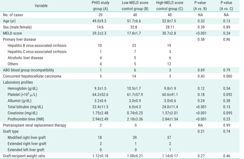
The clinical profiles of patients in the study group and the two control groups are summarized in Table 1. These profiles were relatively similar in all groups except for the
A B
Fig. 1.
Concept of prolonged hepatic inflow occlusion. (A) Interruption of the main portal flow and hepatic arterial flow in a patient with liver cirrhosis, and portal hypertension does not induce significant splanchnic congestion because of portal bypass through venous collaterals. Adapted from Choi et al. Ann Hepatobiliary Pancreat Surg 2019;23:61-4 [3]. (B) A curved intesti- nal clamp is attached to the hepatoduodenal ligament for right liver mobilization.
Table 1. Clinical profiles of the PHIO study group and two control groups
Variable PHIO study
group (A)
Low-MELD score control group (B)
High-MELD score control group (C)
P-value (A vs. B)
P-value (A vs. C)
No. of cases 20 40 40 NA NA
Age (yr) 49.0±9.2 51.7±6.6 52.8±7.5 0.32 0.13
Sex (male:female) 14:6 32:8 28:11 0.39 0.89
MELD score 29.2±2.3 17.8±1.7 30.7±2.8 <0.001 0.24
Primary liver disease 0.58 0.86
Hepatitis B virus-associated cirrhosis 10 23 19
Hepatitis C virus-associated cirrhosis 1 7 3
Alcoholic liver disease 4 5 6
Others 4 5 12
ABO blood group incompatibility 1 6 3 0.69 0.79
Concurrent hepatocellular carcinoma 5 14 3 0.43 0.060
Laboratory profiles
Hemoglobin (g/dL) 9.3±1.5 10.5±1.7 9.8±1.9 0.12 0.54
Platelet (×10
3µ/L) 64.2±52.6 61.7±57.9 60.6±41.1 0.18 0.092
Albumin (g/dL) 3.2±0.6 3.0±0.9 3.0±0.6 0.24 0.38
Total bilirubin (mg/dL) 22.4±11.5 6.0±4.3 24.0±11.4 <0.001 0.13
Creatinine (mg/dL) 1.75±2.48 0.74±0.25 1.57±2.01 <0.001 0.095
Prothrombin time (INR) 2.94±2.49 2.18±3.36 2.84±1.34 <0.001 0.23
Pretransplant renal replacement therapy 2 0 4 NA 0.99
Graft type 0.21 0.74
Modified right liver graft 18 39 37
Extended right liver graft 2 1 2
Extended left liver graft 0 0 1
Graft-recipient weight ratio 1.12±0.18 1.08±0.21 1.14±0.17 0.27 0.46
Values are presented as mean±standard deviation.
PHIO, prolonged hepatic inflow occlusion; MELD, Model for End-Stage Liver Disease; NA, not applicable.
MELD score and its three components. In the PHIO study group, mean total dissection duration and mean PHIO duration were 226.3±59.4 and 68.2±19.1 minutes, respec- tively. Twelve patients (60%) underwent PHIO twice: one for liver mobilization and detachment from the retrohe- patic inferior vena cava and the other for the dissection of the hepatoduodenal ligament. Their mean total dissection duration and mean total PHIO time were 243.5±55.3 and 78.7±18.5 minutes, respectively. The other eight patients underwent PHIO once for liver mobilization and detach- ment from the retrohepatic vena cava, and their mean total dissection duration and mean total PHIO duration were 201.5±65.3 and 51.9±24.3 minutes, respectively.
The mean amount of blood loss in all 20 patients was 2,350.0±951.9 mL.
In the low-MELD score control group, mean total dissection duration and mean amount of blood loss were 216.0±43.9 minutes and 2,112.5±1,614.9 mL, re- spectively. In the high-MELD score control group, mean total dissection duration and mean amount of blood loss were 241.0±41.9 minutes and 2,815±1,813.9 mL, respectively. The PHIO study group and the low-MELD score control group showed similar total dissection du- ration (226.3±59.4 vs. 216.0±43.9 minutes, P=0.82) and similar blood loss during dissection (2,350.0±951.9 vs.
2,112.5±1,614.9 mL, P=0.17). The PHIO study group and the high-MELD score control group showed similar total dissection duration (226.3±59.4 vs. 241.0±41.9 minutes, P=0.71), but the PHIO group showed a significantly lower blood loss during dissection than the high-MELD score group (2,350.0±951.9 vs. 2,815.0±1,813.9 mL, P=0.002) (Figs. 2 and 3).
During LDLT operation using PHIO, major serosal peri- toneal tearing-associated bleeding and hepatic artery dissection did not develop in all patients. Six of the 20 patients (30%) showed noticeable edematous change af- ter PHIO for more than 1 hour, but this was immediately resolved after releasing the intestinal clamp. The other 14 patients (70%) did not show noticeable signs of splanch- nic congestion such as bowel edema or mesenteric dis- coloration. None of the patients experienced posttrans- plant acute pancreatitis.
DISCUSSION
Excessive bleeding is considered a serious complication
of LDLT operation because of difficult dissection, resulting in bleeding tendency. Thus far, we have performed more than 5,000 LDLT operations, and a non-negligible number of patients required massive transfusion due to excessive bleeding during LDLT operation. Intraoperative bleeding is common during LDLT when compared to that during deceased donor liver transplantation because the whole retrohepatic inferior vena cava should be preserved and all perihilar structures should be meticulously dissected to preserve the small hepatic artery branches and hilar bile duct openings [1,5,6]. Excessive bleeding and massive transfusion can cause several adverse effects on intraop- erative management and posttransplant recovery [7]. Thus, intraoperative blood loss should be reduced as much as
PHIO study group 300
250
200
150
100
Dissectionduration(min) 50
Low-MELD score control group 0
High-MELD score control group
Fig. 2. Comparison of the total liver dissection duration in the prolonged
hepatic inflow occlusion (PHIO) study group and two Model for End-Stage Liver Disease (MELD) score control groups.
PHIO study group 5,000
4,000
3,000
2,000
1,000
Bloodloss(mL)
Low-MELD score control group 0
High-MELD score control group Fig. 3. Comparison of amount of blood loss during liver dissection in the