© 2017 The Korean Ophthalmological Society
This is an Open Access article distributed under the terms of the Creative Commons Attribution Non-Commercial License (http://creativecommons.org/licenses /by-nc/3.0/) which permits unrestricted non-commercial use, distribution, and reproduction in any medium, provided the original work is properly cited.
Original Article
A Valid Indication and the Effect of Bilateral Inferior Oblique Transposition on Recurrent or Consecutive Horizontal Deviation in
Infantile Strabismus
Suk-Gyu Ha, Gun-Hoo Na, Seung-Hyun Kim
Department of Ophthalmology, Korea University College of Medicine, Seoul, Korea
Purpose: To evaluate the effects of bilateral inferior oblique transposition (BIOT) on horizontal deviation from primary position among patients with bilateral dissociated vertical deviation (DVD) associated with inferior oblique overaction (IOOA) in infantile strabismus.
Methods: Retrospective chart review was conducted among 19 patients with infantile strabismus. All patients had DVD and IOOA with consecutive or recurrent horizontal deviation and underwent modified BIOT surgery.
Patients were divided into three subgroups: patients who underwent BIOT (BIOT group, n = 9) alone, BIOT with medial rectus recession or lateral rectus resection simultaneously (ET BIOT group, n = 6), or BIOT with lateral rectus recession or medial rectus resection simultaneously (XT BIOT group, n = 4). Postoperative angle of horizontal deviation (prism diopter, PD) and corrected magnitude of horizontal deviation (PD) at final visit af- ter surgery were analyzed in each group.
Results: The mean age was 55.11 ± 21.05 months (range, 32 to 115). The mean follow-up period was 8.68 ± 2.87 months (range, 6 to 18). Preoperative horizontal deviation was 4.23 ± 5.99 PD (range, 0 to 16) in BIOT, –17.33 ± 6.76 PD (range, –30 to –10) in ET BIOT, and 17.50 ± 2.52 PD (range, 14 to 20) in XT BIOT. Esodeviation is rep- resented by negative values. DVD and IOOA were reduced less than +1 in all patients. The corrected amount of horizontal deviation was 3.56 ± 5.18 PD (range, 0 to 16) in BIOT surgery alone and larger in XT BIOT (18.50
± 3.41 PD) than in ET BIOT (12.33 ± 5.57 PD, p = 0.004).
Conclusions: Minimal exodeviation was corrected by BIOT alone. In addition, secondary eso- or exodeviation at great magnitudes should be corrected with proper horizontal muscle surgery along with BIOT.
Key Words: Infantile, Inferior oblique muscle, Strabismus, Surgery, Transposition
Elevation in adduction and dissociated vertical deviation (DVD) are common components of infantile strabismus.
The reported prevalence of inferior oblique overaction (IOOA) and DVD ranges from 76% to 88% in infantile es- otropia and 50% in infantile exotropia [1,2]. It was previ- ously believed that procedures that weaken the inferior oblique muscle do not affect the DVD. In addition, it was thought that IOOA associated with DVD should be cor- rected with procedures of the anterior or modified anterior transposition of the inferior oblique muscle in order to
Received: November 23, 2015 Accepted: March 14, 2016
Corresponding Author: Seung-Hyun Kim, MD, PhD. Department of Ophthalmology, Korea University Anam Hospital, #73 Inchon-ro, Seong- buk-gu, Seoul 02841, Korea. Tel: 82-2-920-5520, Fax: 82-2-924-6820, E-mail: [email protected]
maintain DVD in the primary position. Furthermore, it is thought reasonable to use this procedure in children with bilateral IOOA associated with DVD as part of the infan- tile strabismus complex [3,4].
Generally, among patients with IOOA and DVD, indica- tion for surgery is variable. Typically, DVD appears in pre- school-age and school-age children who have had horizon- tal muscle surgery earlier in life to correct horizontal strabismus. Currently, recommended indications for sur- gery include increasing frequency of manifest large DVD, associated anomalous head posture, and significant IOOA [1,2].
In patients with IOOA associated with DVD, the brain circuit is likely insufficient to permanently lock the eyes;
hence, the average child with infantile strabismus requires at least two surgeries [1,2]. Therefore, if IOOA associated with DVD develops in patients with previously corrected infantile strabismus, the patient may also have poorly con- trolled horizontal deviation. Eso- or exodeviation may re- develop in these patients; in addition, there is a possibility that inferior oblique muscle weakening or transposition it- self may affect horizontal alignment in the primary posi- tion.
This study was conducted to determine whether recur- rent or consecutive horizontal deviation is a valid indica- tion for surgery in IOOA associated with DVD and to evaluate the effects of modified bilateral inferior oblique transposition (BIOT) on horizontal deviation in the prima- ry position.
Materials and Methods
This study followed the tenets of the Declaration of Hel- sinki and was approved by the institutional review board at Korea University Medical Center. A retrospective chart review was conducted on all patients with BIOT performed at Korea University Medical center from 2008 August to 2014 October. The minimum follow-up period after BIOT was more than 6 months. All patients who had prior bilat- eral lateral rectus (LR) or bilateral medial rectus (MR) re- cession to correct infantile exotropia or esotropia had re- current or consecutive exo- and esodeviation ranging from 30 prism diopter (PD) (esotropia) to 20 PD (exotropia), and were associated with both IOOA and DVD.
Patients were excluded if they had paretic or restrictive
strabismus, prior oblique muscle or vertical rectus muscle surgery, or any systemic, neurologic disorder that could af- fect extraocular muscle alignment.
DVD was measured using the prism and alternate cover test, with the eyes in the primary position and fixating on an accommodative target at 6 m with full refractive cor- rection. Any concurrent horizontal deviation was initially neutralized with a horizontal prism over the contralateral eye and DVD was measured in the ipsilateral eye. The de- gree of IOOA was estimated from +1 to +4, according to the method described previously [5].
One surgeon (SHK) performed all of the surgeries. As previously reported, the surgical technique of BIOT is de- scribed. The inferior oblique muscle was approached using an inferior-temporal fornix incision through the conjuncti-
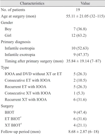
Table 1. Demographic characteristics
Characteristics Value
No. of patients 19
Age at surgery (mon) 55.11 ± 21.05 (32–115) Gender
Boy 7 (36.8)
Girl 12 (63.2)
Primary diagnosis
Infantile esotropia 10 (52.63)
Infantile exotropia 9 (47.37)
Timing after primary surgery (mon) 35.84 ± 19.14 (7–87) Type
IOOA and DVD without XT or ET 5 (26.3) Consecutive ET with IOOA 2 (10.5)
Recurrent ET with IOOA 5 (26.3)
Consecutive XT with IOOA 1 (5.3)
Recurrent XT with IOOA 6 (31.6)
Surgery
BIOT 9 (47.4)
ET BIOT* 6 (31.6)
XT BIOT† 4 (21.1)
Follow-up period (mon) 8.68 ± 2.87 (6–18) Values are presented as mean ± standard deviation (range) or number (%).
IOOA = inferior oblique overaction; DVD = dissociated vertical deviation; XT = exodeviation; ET = esodeviation; BIOT = bilater- al inferior oblique transposition.
*Modified BIOT with bilateral medial rectus muscle recession or lateral rectus muscle resection; †Modified BIOT with unilateral lateral rectus muscle recession or unilateral medial rectus muscle resection.
va and Tenon’s capsule. Following isolation of the inferior oblique muscle, posterior fibers of the inferior oblique muscle were bunched using one suture toward the main muscle belly. Next, the isolated inferior oblique muscle was placed onto the equator (i.e., 1 mm anterior to the equator) and was dependent on severity of DVD and IOOA such that the distal end of the inferior oblique muscle was positioned parallel to the proximal end and on a line per- pendicular to the lateral corneal limbus, 0.5 mm temporal to the lateral border of the inferior rectus muscle [6].
Correction of concurrent horizontal deviation was not performed if the horizontal deviation was less than 8 PD;
however, only one female patient underwent BIOT alone even though she had 16 PD of exodeviation. For most cas- es, if the angle was greater than that of esodeviation or any magnitude of exodeviation, it was simultaneously correct- ed with MR recession or LR resection for consecutive or recurrent esotropia, LR recession or MR resection for re- current or consecutive exotropia. The surgical magnitude of horizontal muscle was determined by the patient’s angle of deviation according to standardized values [7]. The de- tailed surgical method of horizontal muscle combined with BIOT is shown in Table 1.
We divided patients into three groups: patients who had undergone BIOT alone (BIOT group), BIOT with MR re-
cession or LR resection simultaneously (ET BIOT group), or BIOT with LR recession or MR resection simultaneous- ly (XT BIOT group).
After surgery, all patients were observed at postopera- tive 1 day, 3 months, 6 months, and final visit. Preoperative horizontal deviation, postoperative horizontal deviation, degree of IOOA associated with DVD, and corrected mag- nitude of horizontal deviation were evaluated in each group. The corrected magnitude of horizontal deviation was calculated as the difference between preoperative hor- izontal deviation and horizontal deviation at the final visit.
Statistical analyses were performed using the SPSS soft- ware ver. 21.0 (IBM Corp., Armonk, NY, USA). Non-con- tinuous variables were analyzed using the chi-square test.
Kruskal-Wallis was used for the analysis of preoperative and postoperative data according to the three groups. Sta- tistical significance was approved for p < 0.05.
Results
Nineteen patients were enrolled and assigned to three groups, following BIOT. The mean age at the time of sur- gery was 55.11 ± 21.05 months (range, 32 to 115). There were seven boys (36.8%) and 12 girls (63.2%). Mean timing
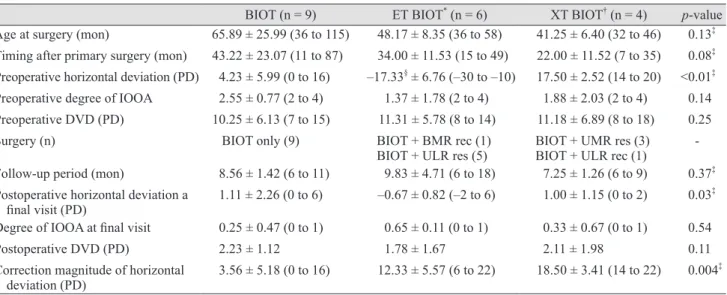
Table 2. Postoperative changes in horizontal deviation, IOOA, and DVD according to surgery
BIOT (n = 9) ET BIOT* (n = 6) XT BIOT† (n = 4) p-value Age at surgery (mon) 65.89 ± 25.99 (36 to 115) 48.17 ± 8.35 (36 to 58) 41.25 ± 6.40 (32 to 46) 0.13‡ Timing after primary surgery (mon) 43.22 ± 23.07 (11 to 87) 34.00 ± 11.53 (15 to 49) 22.00 ± 11.52 (7 to 35) 0.08‡ Preoperative horizontal deviation (PD) 4.23 ± 5.99 (0 to 16) –17.33§ ± 6.76 (–30 to –10) 17.50 ± 2.52 (14 to 20) <0.01‡ Preoperative degree of IOOA 2.55 ± 0.77 (2 to 4) 1.37 ± 1.78 (2 to 4) 1.88 ± 2.03 (2 to 4) 0.14 Preoperative DVD (PD) 10.25 ± 6.13 (7 to 15) 11.31 ± 5.78 (8 to 14) 11.18 ± 6.89 (8 to 18) 0.25
Surgery (n) BIOT only (9) BIOT + BMR rec (1)
BIOT + ULR res (5) BIOT + UMR res (3) BIOT + ULR rec (1) - Follow-up period (mon) 8.56 ± 1.42 (6 to 11) 9.83 ± 4.71 (6 to 18) 7.25 ± 1.26 (6 to 9) 0.37‡ Postoperative horizontal deviation a
final visit (PD) 1.11 ± 2.26 (0 to 6) –0.67 ± 0.82 (–2 to 6) 1.00 ± 1.15 (0 to 2) 0.03‡ Degree of IOOA at final visit 0.25 ± 0.47 (0 to 1) 0.65 ± 0.11 (0 to 1) 0.33 ± 0.67 (0 to 1) 0.54
Postoperative DVD (PD) 2.23 ± 1.12 1.78 ± 1.67 2.11 ± 1.98 0.11
Correction magnitude of horizontal
deviation (PD) 3.56 ± 5.18 (0 to 16) 12.33 ± 5.57 (6 to 22) 18.50 ± 3.41 (14 to 22) 0.004‡ Values are presented as mean ± standard deviation (range) or mean ± standard deviation.
IOOA = inferior oblique overaction; DVD = dissociated vertical deviation; BIOT = bilateral inferior oblique transposition; PD = prism diopter; BMR rec = bilateral medial rectus muscle recession; UMR res = unilateral medial rectus muscle resection; ULR res = unilateral lateral rectus muscle resection; ULR rec = unilateral rectus muscle recession.
*Modified BIOT with bilateral medial rectus muscle recession or lateral rectus muscle resection; †Modified BIOT with unilateral lateral rec- tus muscle recession or unilateral medial rectus muscle resection; ‡Kruskal-Wallis test; §Esodeviation is expressed with negative values.
after primary surgery was 35.84 ± 19.14 months (range, 7 to 87). Five patients (26.3%) had only IOOA associated with DVD without horizontal deviation. Recurrent exode- viation with IOOA associated with DVD was the most prevalent (31.6%). There were nine patients (47.4%) in the BIOT group, six patients (31.6%) in ET BIOT, and four pa- tients (21.1%) in XT BIOT. The mean follow-up period af- ter surgery was 8.68 ± 2.87 months (range, 6 to 18). Table 1 summarizes the demographic characteristics.
The analysis of surgical outcomes is described in Table 2.
The mean age at surgery was older (65.89 ± 25.99 months) in the BIOT group compared to other groups (48.17 ± 8.35 in ET BIOT and 41.25 ± 6.40 in XT BIOT) without statisti- cal significance (p = 0.13). Preoperative horizontal devia- tion in the BIOT, ET BIOT, and XT BIOT groups were 4.23
± 5.99 PD (range, 0 to 16), –17.33 ± 6.76 PD (range, –30 to –10), and 17.50 ± 2.52 PD (range, 14 to 20), respectively.
Esodeviation is expressed by negative values. Preoperative DVD was 10.25 ± 6.13 PD (range, 7 to 15), 11.31 ± 5.78 PD (range, 8 to 14), and 11.18 ± 6.89 PD (range, 8 to 18) in the BIOT, ET BIOT, and XT BIOT groups, respectively. Post- operative horizontal deviation at final visit was 1.11 ± 2.26 PD (range, 0 to 6) in the BIOT, –0.67 ± 0.82 PD (range, –2 to 6) in the ET BIOT, and 1.00 ± 1.15 PD (range, 0 to 2) in the XT BIOT groups. The postoperative degree of IOOA at the final visit was less than +1 in all patients. Postoperative DVD decreased to less than 5 PD in all groups (Table 2).
Magnitude of correction in exodeviation was 3.56 ± 5.18 PD (range, 0 to 16) in BIOT and 18.50 ± 3.41 PD (range, 14 to 22) in XT BIOT groups, respectively. The corrected magnitude of esodeviation was 12.33 ± 5.57 PD (range, 6 to 22) in ET BIOT. The total horizontal deviation corrected in the XT BIOT group was significantly larger than in the ET BIOT group (p = 0.004).
Discussion
More than one surgery is often needed to achieve binoc- ular vision and stability in infantile strabismus. Reopera- tion is necessary to correct residual or consecutive hori- zontal strabismus, oblique muscle dysfunction, DVD, or a combination of these in infantile strabismus [8,9]. In par- ticular, IOOA and DVD are frequently associated with in- fantile esotropia after surgery [10].
Monofixation syndrome is an eye condition defined by a
small angle deviation within 8 PD and is often considered to be a stable condition in infantile strabismus. The condi- tion of some patients will deteriorate over time with an in- crease in ocular deviation and loss of fusion [11].Uncorrect- ed IOOA and DVD may be disruptive to the maintenance of the primary stable eye position, despite primary surgery in the early period [12].
Whether procedures weakening the single inferior oblique muscle affect horizontal deviation has been con- troversial. Stager and Parks [13] reported that in 50 patients with primary IOOA, no significant horizontal change oc- curred in 84% of the patients, and in 16% of those who manifested a change, it was never greater than 8 PD after bilateral inferior oblique muscle weakening alone.Wright [5] also described that the weakening inferior oblique mus- cle did not significantly alter the horizontal alignment in the primary position. He suggested that when planning si- multaneous horizontal and inferior oblique surgery, the magnitude of horizontal surgery should be based on mea- surement in the primary position, independent of inferior oblique muscle surgery. In other studies, it has been re- ported that the weakening of inferior oblique and superior oblique muscles resulted in esodeviation of approximately 5 to 10 PD and esodeviation of approximately 10 to 15 PD [14]. Recently, it was reported that the median magnitude of correction of horizontal deviations with inferior oblique muscle weakening was 4 PD; however, this was variable and ranged from 0 to 20. These results are in accordance with the results from our study [15]. Our study revealed that exodeviation (3.56 ± 5.18 PD) on average was correct- ed by BIOT alone. Theoretically, minimal esoshift after BIOT is due to decreasing tertiary action of inferior oblique muscles that is abducted from the primary position [16]. In the case of prior horizontal muscle surgery, the binocular status of the patients may have an effect that re- quires future study. In this study, in patients with minimal exodeviation (3.56 ± 5.18 PD) associated with IOOA and DVD in infantile strabismus, BIOT alone corrected exode- viation associated with IOOA and DVD.
However, our study showed that in patients with recur- rent or consecutive esotropia associated with IOOA and DVD, it is possible to obtain favorable surgical results by additional esotropia surgery such as MR recession or LR resection, as BIOT alone might aggravate esodeviation.
The results of our study correspond with a recent report by Isaac and Chalita [17] that showed patients who had under-
gone oblique weakening surgery in combination with MR recession might require larger MR recession to correct the primary esodeviation. On the other hand, in patients with recurrent or consecutive exodeviation associated with IOOA and DVD, BIOT alone or exotropia surgery (such as MR resection or LR recession) should be performed as ap- propriate, according to the magnitude of exodeviation.
Our study has limitations. First, this study was retro- spective in design. Second, a small number of patients were recruited. However, currently, infantile strabismus (eso- and exo-) associated with DVD or IOOA is not a common disease. In addition, among these patients, recur- rent or overcorrected esotropia is relatively rare. Further studies conducted among a large number of patients are needed. Third, mean follow-up periods after surgery were relatively short, so further long-term follow-up is needed to determine postoperative results.
In conclusion, uncorrected IOOA associated with DVD in infantile strabismus may be disruptive to the mainte- nance of the stable alignment after primary surgery. This condition was a valid surgical indication for BIOT. Mini- mal exodeviation was corrected by BIOT alone. In addi- tion, secondary eso- or exodeviation at greater magnitudes should be corrected with proper horizontal muscle surgery along with BIOT.
Conflict of Interest
No potential conflict of interest relevant to this article was reported.
References
1. Kraft SP. Infantile exotropia. In: Rosenbaum AL, Santiago AP, editors. Clinical strabismus management: principles and surgical techniques. Philadelphia: W.B. Saunders;
1999. p. 176-81.
2. Tychsen L. Infantile esotropia: current neurophysiologic concepts. In: Rosenbaum AL, Santiago AP, editors. Clini- cal strabismus management: principles and surgical tech- niques. Philadelphia: W.B. Saunders; 1999. p. 117-38.
3. Kushner BJ. Restriction of elevation in abduction after in- ferior oblique anteriorization. J AAPOS 1997;1:55-62.
4. Mims JL 3rd, Wood RC. Antielevation syndrome after bi- lateral anterior transposition of the inferior oblique mus- cles: incidence and prevention. J AAPOS 1999;3:333-6.
5. Wright KW, editor. Color atlas of strabismus surgery:
strategies and techniques. 3rd ed. New York: Springer;
2007. p. 136.
6. Kim SH, Na JH, Cho YA. Inferior oblique transposition onto the equator: the role of the equator in development of contralateral inferior oblique overaction. J Pediatr Oph- thalmol Strabismus 2012;49:98-102.
7. Wright KW, editor. Color atlas of strabismus surgery:
strategies and techniques. 3rd ed. New York: Springer;
2007. p. 191.
8. Hiles DA, Biglan AW. Early surgery of infantile exotropia.
Trans Pa Acad Ophthalmol Otolaryngol 1983;36:161-8.
9. Rubin SE, Nelson LB, Wagner RS, et al. Infantile exotropia in healthy children. Ophthalmic Surg 1988;19:792-4.
10. Yoo EJ, Kim SH. Modified inferior oblique transposition considering the equator for primary inferior oblique over- action (IOOA) associated with dissociated vertical devia- tion (DVD). Strabismus 2014;22:13-7.
11. Parks MM. The monofixation syndrome. Trans Am Oph- thalmol Soc 1969;67:609-57.
12. Tychsen L. Why do humans develop strabismus? In: Hoyt CS, Taylor D, editors. Pediatric ophthalmology and strabis- mus. 4th ed. New York: Elsevier Saunders; 2013. p. 756-63.
13. Stager DR, Parks MM. Inferior oblique weakening proce- dures: effect on primary position horizontal alignment.
Arch Ophthalmol 1973;90:15-6.
14. Diamond GR, Parks MM. The effect of superior oblique weakening procedures on primary position horizontal alignment. J Pediatr Ophthalmol Strabismus 1981;18:35-8.
15. Taylan Sekeroglu H, Dikmetas O, Sanac AS, et al. Inferior oblique muscle weakening: is it possible to quantify its effects on horizontal deviations? J Ophthalmol 2012;2012:813085.
16. Von Noorden GK, Campos EC, editors. Binocular vision and ocular motility: theory and management of strabis- mus. 4th ed. St. Louis: Mosby; 1990. p. 55.
17. Isaac CR, Chalita MR. Effect of combining oblique muscle weakening procedures with bimedial rectus recessions on the surgical correction of esotropia. J AAPOS 2015;19:54-6.