대한외과학회지:제 68 권 제 4 호
□ 증 례 □
Vol. 68, No. 4, April, 2005
339
서 론
장간막 가성낭종은 드문 질환으로 병리적으로 내벽을 구 성하는 세포 없이 섬유성 벽으로 이루어져 있어 내피세포 로 구성되는 진성낭종과 구별된다. 원인은 명확하지 않지 만 염증 반응이나 외상에 의하여 발생하는 것으로 알려져 있다.(1) 증상이 없는 경우가 많아 대부분 수술시 진단되는 경우가 많고 드물게 출혈이나 낭종의 파열, 감염 등 합병증 으로 증상이 발생할 수 있다.(2) 저자들은 회장의 장간막 가 성낭종이 장폐색을 일으킨 예를 경험하였기에 문헌고찰과 함께 보고하는 바이다.
증 례
66세의 남성이 내원 3일 전부터 복부 통증을 주소로 내원 하였다. 통증은 복부 전반에 쥐어짜는 듯한 양상이었다. 과 거력상 40년 전에 급성 충수돌기염으로 충수절제술을 시행 받았으며, 수술 후 3개월부터 좌하복부에 종괴가 만져지기 시작하여 지속적으로 크기가 증가하였다고 하였다. 당시 발열이나 통증 등의 동반증상은 없었다. 내원 시 시행한 이 학적 검사에서 체온은 37.1oC, 맥박수 76회/분, 호흡수 20회/
분, 혈압은 130/90 mmHg이었다. 복부 진찰상 전반적으로 팽만되어 있었고 장음이 증가되어 있었다. 전반적인 압통 이 있었으나 반발통은 없었다. 좌하복부에 10 cm 크기의 종 괴가 만져졌으나 종괴 부위에 압통은 없었다.
단순복부촬영상 소장의 기계적 폐색소견을 보였으며 여 러 곳의 석회화된 림프절이 관찰되었다(Fig. 1). 전산화 단 층촬영상 전체적인 장벽의 확장 소견을 보였으며 좌하복부 에 크기 10×9 cm 크기의 낭성 종괴가 관찰되었으며 일부 낭성 종괴 벽에 조영증강되는 소견이 있었다(Fig. 2). 환자 는 2일간의 보존적 치료에 증상의 호전 없어 복부 종괴와 책임저자:장명철, 충남 천안시 안서동 산 16-5
ꂕ 330-714, 단국대학교병원 외과 Tel: 041-550-3930, Fax: 041-556-3878 E-mail: [email protected]
접수일:2004년 10월 16일, 게재승인일:2004년 12월 14일 본 논문의 요지는 2004년 대한외과학회 추계학술대회에서 포스터 전시되었음.
Key Words: Mesenteric pseudocyst, Intestinal obstruction
중심 단어: 장간막 가성낭종, 장폐색ꠏꠏꠏꠏꠏꠏꠏꠏꠏꠏꠏꠏꠏꠏꠏꠏꠏꠏꠏꠏꠏꠏꠏꠏꠏꠏꠏꠏꠏꠏꠏꠏꠏꠏꠏꠏꠏꠏꠏꠏꠏꠏꠏꠏꠏꠏꠏꠏꠏꠏꠏꠏꠏ
Departments of Surgery and 1Pathology, Dankook University College of Medicine, Seoul, KoreaMesenteric Pseudocyst Causing Intestinal Ob- struction
Jun Won Min, M.D., Myung Chul Chang, M.D., Youn Chan Park, M.D. and Jai Hyang Go, M.D.
1Mesenteric pseudocyst has a fibrous cystic wall without an endothelial lining. It can develop from an inflammatory reaction or from trauma. This lesion is difficult to diagnose preoperatively, and it is rarely symptomatic except when it is complicated by bleeding, rupture or infection. A 66-year old male presented with generalized abdominal pain for 3 days. In his past medical history, an appendectomy was done forty years ago. Three months after the operation, a mass was palpated in the left lower quadrant and size of the mass had gradually increased. Physical examination revealed a distended abdomen with diffuse tenderness. The non-tender mass, which was about 10 cm in size, was palpated in the left lower abdomen. A simple abdominal x-ray showed a mechanical obstruction of the small bowel.
A CT scan showed a 10×9 cm sized cystic mass with a partially enhancing cystic wall. Surgical exploration revealed the 13 cm sized mass in the distal ileum about 40 cm proximal from the ileocecal valve, and the adjacent ileum was obstructed by this mass. The mass was a thick walled cyst that contained a non-clotting bloody material. Histopa- thological examination indicated that the cyst wall was composed of fibrosis with neutrophil infiltration, but there was no specific endothelial lining. The final pathological diagnosis was a mesenteric pseudocyst. Mesenteric pseudocyst with obstruction is rare and difficult to diagnosis, but it should be included in the differential diagnosis in the case of intestinal obstruction with mass. (J Korean Surg Soc 2005;
68:339-341)
장간막 가성낭종에 의한 장폐색
단국대학교 의과대학 외과학교실, 1병리학교실
민준원․장명철․박윤찬․고재향1
340
대한외과학회지:제 68 권 제 4 호 2005ꠏꠏꠏꠏꠏꠏꠏꠏꠏꠏꠏꠏꠏꠏꠏꠏꠏꠏꠏꠏꠏꠏꠏꠏꠏꠏꠏꠏꠏꠏꠏꠏꠏꠏꠏꠏꠏꠏꠏꠏꠏꠏꠏꠏꠏꠏꠏꠏꠏꠏꠏꠏꠏꠏꠏꠏꠏꠏꠏꠏꠏꠏꠏꠏꠏꠏꠏꠏꠏꠏꠏꠏꠏꠏꠏꠏꠏꠏꠏꠏꠏꠏꠏꠏꠏꠏꠏꠏꠏꠏꠏꠏꠏꠏꠏꠏꠏꠏꠏꠏꠏꠏꠏꠏꠏꠏꠏꠏꠏꠏꠏꠏꠏꠏꠏ
기계적 장폐색으로 개복술을 시행하였다.
수술소견상 약간의 혈장성 복수가 관찰되었으며, 회장 직경은 최대 5 cm으로 소장 전반이 늘어나 있었으나 대장 은 정상 소견이었다. 복막 및 장막에 여러 개의 석회화된 림프절이 관찰되었으며 동결절편 검사상 결핵이나 종양의 증거는 관찰되지 않았다. 회맹장 이행부 상방 40 cm의 장간 막에 13 cm 크기의 종괴가 있었으며 섬유화로 인하여 딱딱 하였고 이로 인하여 회장이 눌린 소견을 보였다. 근위부 소
장에 다른 이상소견은 관찰되지 않았다. 장간막 동맥과 인 접하고 종괴의 악성 가능성으로 회장을 포함한 장간막 종 괴를 절제하였다.
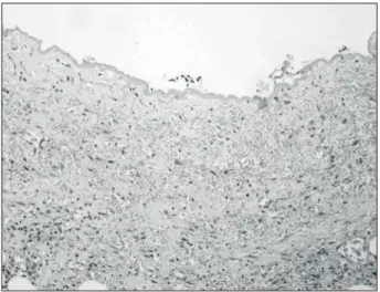
종괴는 육안적으로 회장과 연하여 있었으며 절단하였을 때 5 mm의 두꺼운 낭벽내에 응고되지 않는 오래된 혈액성 내용물을 함유하였다(Fig. 3). 병리조직학적 검사상 낭벽을 이루는 내피세포는 관찰되지 않았고 섬유성 막과 함께 염 증성 세포의 침윤이 관찰되어 장간막 가성낭종으로 진단되 었다(Fig. 4). 환자는 수술 후 합병증 없이 퇴원하였고 이후 현재까지 특별한 증상 없이 추적관찰 중이다.
고 찰
장간막 낭종은 크게 내피세포로 구성된 진성낭종과 염증 과 섬유성 낭벽으로 구성된 가성낭종으로 나눌수 있다.
Fig. 1. Plain abdominal x-ray shows multiple air-fluid levels and calcified lymph nodes.
Fig. 2. CT shows 10×9 cm sized cystic mass with a partially enhancing cystic wall (arrow).
Fig. 3. Resected small bowel shows thick walled cyst which contained non-clotting bloody material.
Fig. 4. The cyst wall was composed of fibrosis with neutrophil
infiltration without endothelial lining (H&E stain × 200).
장명철 외:장간막 가성낭종에 의한 장폐색 341
ꠏꠏꠏꠏꠏꠏꠏꠏꠏꠏꠏꠏꠏꠏꠏꠏꠏꠏꠏꠏꠏꠏꠏꠏꠏꠏꠏꠏꠏꠏꠏꠏꠏꠏꠏꠏꠏꠏꠏꠏꠏꠏꠏꠏꠏꠏꠏꠏꠏꠏꠏꠏꠏꠏꠏꠏꠏꠏꠏꠏꠏꠏꠏꠏꠏꠏꠏꠏꠏꠏꠏꠏꠏꠏꠏꠏꠏꠏꠏꠏꠏꠏꠏꠏꠏꠏꠏꠏꠏꠏꠏꠏꠏꠏꠏꠏꠏꠏꠏꠏꠏꠏꠏꠏꠏꠏꠏꠏꠏꠏꠏꠏꠏꠏꠏ
Beahrs 등(3)은 장간막 낭종을 원인 및 조직학적 특징에 따라 태생 및 발생학적 낭종(embryonic and developmental cyst), 외상성 후천성 낭종(traumatic acquired cyst), 종양성 낭 종(neoplastic cyst), 감염 및 퇴행성 낭종(infective and degen- erative cyst)의 4가지로 나누었다. 이중 태생 및 발생학적 낭 종과 종양성 낭종은 진성낭종이며, 외상성 낭종과 감염 및 퇴행성 낭종은 가성낭종에 속한다. Ros 등(1)은 조직학적 소견과 방사선학적 소견에 따라 림프관종(lymphangioma), 장관 중복낭(enteric duplication cyst), 장관 낭(enteric cyst), 중 피낭(mesothelial cyst) 및 비췌장성 가성낭종(nonpancreatic pseudocyst)의 5가지로 나누었다. Ros의 분류에 의하면 본 예는 비췌장성 가성낭종에 속한다.
가성낭종의 원인으로는 염증, 외상, 기생충, 이물반응, 지 방괴사 등이 알려져 있다.(4) 본 예에서는 진균이나 결핵균 의 증거가 없고 특별한 외상의 과거력이 없었으나 충수염 에 의한 충수돌기 절제술의 과거력이 있었으며 이후 천천 히 자라는 종괴였기 때문에 수술이 외상으로 작용하였으리 라 생각된다.
진성낭종이 젊은 나이에 호발하는 데 비하여 장간막 가 성낭종은 장년층에 많은데 이는 외상 등의 가성낭종의 발 생원인과 연관이 있으리라 생각된다. 일본의 Iida 등(5)이 보고한 15예를 정리하면 평균 연령이 42.7세이며, 흔한 증 상으로는 복통이 44.4%로 가장 많고, 종괴가 27.8%, 복부 팽만이 11.1%였다. 복부외상의 과거력이 있는 경우가 30%, 급성 충수염의 과거력이 있는 경우가 20%로 보고되고 있 다. 검사실 소견상 백혈구 증가가 13.3%에서 있었으며, CA- 125의 상승이 13.3%에서 관찰되었다. 수술 전 장간막 낭종 으로 진단된 경우는 15예 중 3예(20%)였다. 수술소견상 6예 에서 공장, 1예에서 회장의 장간막에 위치하였고, 6예에서 횡행결장, 2예에서 S상 결장에 위치하였다. 크기는 3 cm에 서 40 cm까지로 평균 9.9 cm였다. 11예에서 단방성이었고, 3예에서 다방성이었다. 낭종의 내용물은 진흙같은 경우가 5예, 장액성이 3예, 유미성이 3예, 아교질성이 2예, 혈액성 이 2예였다. 15예 중 11예에서 장 절제 없이 낭종 제거가 가능하였고 4예에서는 장 절제를 동반하였다고 보고하였 다. 본 예에서는 회장의 장간막에 위치하였고 단방성이었 으며 혈액성의 내용물이었다.
수술 전 진단은 어려우나 초음파에서 낭종과 함께 내부 에 많은 찌꺼기가 관찰되고, 전산화 단층촬영상 조영증강
되는 두꺼운 벽을 갖는 낭종으로 관찰된다.(1) 감별진단으 로는 복부 대동맥류의 대동맥 박리, 췌장염에 의한 가성낭 종, 대망 낭종, 장간막 지방종 등이 있다. 주위 장을 포함하 여 장간막을 절제하는 수술이 시행되나 만약 낭종의 크기 가 커서 절제부위가 넓은 경우는 단장 증후군(short bowel syndrome)을 방지하기 위하여 낭종의 일부 절제나 주머니 형성술(marsupialization)을 시행할 수 있다.
장간막 가성낭종은 매우 드문 질환으로 국내에서는 소아 에서 복부 외상 후 발생한 출혈성 장간막 가성낭종에 대한 보고가 있으며,(4) 일본에서는 15예의 보고(5)가 있다. 김 등 (6)은 31예의 장간막 낭종중 5예의 가성낭종을 보고하였고, 최 등(7)은 29예의 비교적 드문 소장 폐쇄의 원인중 2예의 장간막 낭종을 보고하였으나 가성낭종에 의한 장폐색은 확 인되지 않았다. 장폐색을 유발한 장간막 가성낭종은 매우 드문 질환으로 비록 수술 전 진단은 어렵지만 장폐색의 감 별진단으로 고려해야 할 것이다.