Bronchogenic cysts are congenital anomalies that re- sult from aberrant budding from the ventral diverticu- lum, and are usually discovered in the posterior part of the mediastinum, along the right paratracheal wall (1, 2). Cysts that develop below the diaphragm, especially in the retroperitoneal region, are extremely rare (3, 4), and fewer than 50 cases have been reported in English language clinical literature to date. To the best of our knowledge, there has been no report in a Korean radio- logical journal.
We have recently experienced a case of a retroperi- toneal bronchogenic cyst that was located between the stomach and pancreas. We report the case with a review of the literature.
Case Report
A 32-year-old woman was referred to our hospital
with a mass incidentally detected by ultrasonography, which showed a homogeneous hypoechoic mass in the peripancreatic region. The patiwent had symptoms of cholecystitis, but there was no symptom related to the peripancreatic mass.
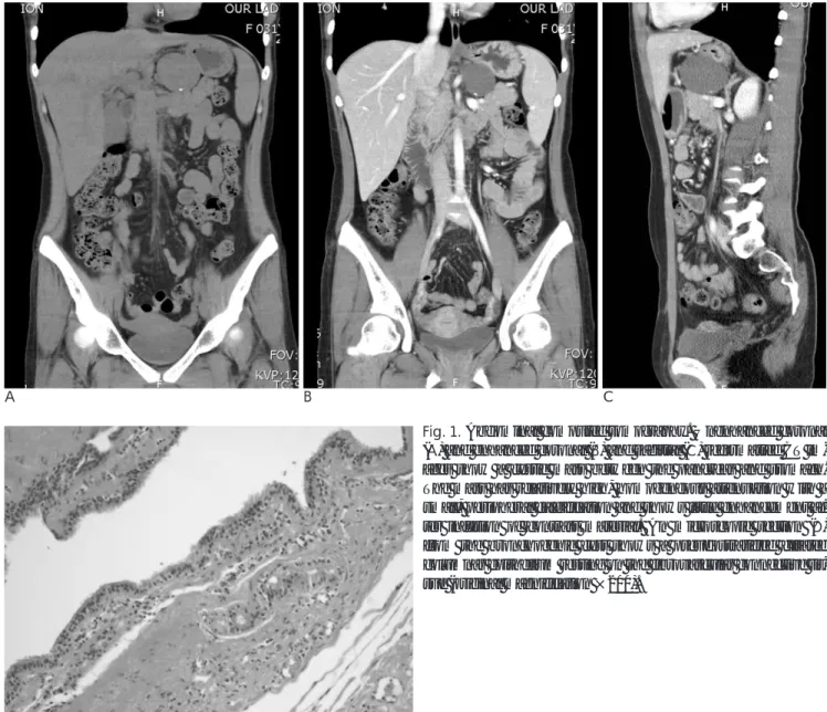
Computed tomography (CT) was performed for the further evaluation of the peripancreatic mass at our hos- pital. An unenhanced CT scan revealed a well-defined, 6 × 5 × 5 cm mass between the stomach and pancreas body. The mass had homogeneous attenuation of 45 Hounsfield units (HU) on the unenhanced CT scan and had small peripheral calcification. There was little en- hancement after infusion of contrast material. These findings suggested a highly attenuating cystic mass filled with protein-rich fluid or hemorrhage (Fig. 1A-C).
We presumed a pseudocyst,a lymphangioma, a cystic teratoma, a mucin-producing tumor of pancreatic origin, and a cystic neurogenic tumor for the possible differential diagnosis. There was no evidence of a gallstone or gall bladder wall thickening suggesting acute cholecystitis seen on the CT scan. In addition, there was no remark- able laboratory finding including amylase level, complete blood count, electrolyte levels, and blood chemistry.
Surgical resection of the mass was performed. The mass was in the retroperitoneal space between the stomach and pancreas, and was separated from the
J Korean Radiol Soc 2007;57:451-453
─ 451 ─
Retroperitoneal Bronchogenic Cyst: A Case Report1
Kyung-Myung Sohn, M.D., Ki-Jun Kim, M.D., E-So Maeng, M.D.2
1Department of Radiology, Our Lady of Mercy Hospital, The Catholic University of Korea, College of Medicine
2Department of Clinical Pathology, Our Lady of Mercy Hospital, The Catholic University of Korea, College of Medicine
Received July 28, 2007 ; Accepted September 21, 2007
Address reprint requests to : Kyung-Myung Sohn, M.D., Department of Radiology, Our Lady of Mercy Hospital, 665 Bupyeong 6-dong, Buk-gu, Incheon 403-720, Republic of Korea
Tel. 82-32-510-5531 Fax. 82-32-529-0964 E-mail: [email protected]
An retroperitoneal bronchogenic cyst is extremely rare and often mimics other cystic diseases such as a lymphangioma, pseudocyst, or cystic tumor of the pancreas. We have recently experienced a case of a peripancreatic bronchogenic cyst in 32-year-old woman.
We report this case with a description of the CT findings and a review of the literature.
Index words :Retroperitoneal space Bronchogenic cyst Tomography
stomach, pancreas, left adrenal gland and left kidney.
The lesion was a well-encapsulated cystic mass that con- tained thick, yellowish mucoid fluid. A subsequent mi- crobiological examination yielded no abnormality.
Histologically, the cyst wall was composed of fi- brovascular connective tissue containing thin bundles of smooth muscle cells and glands resembling the bronchial wall. The inner surface of the cyst was lined with ciliated pseudostratified or tall columnar epitheli- um (Fig. 1D). These findings indicated the presence of a bronchogenic cyst in the retroperitoneal region. The postoperative course was uneventful.
Discussion
Bronchogenic cysts are developmental anomalies of
the primitive foregut that are usually found above the diagphragm, especially in the mediastinum and particu- larly posterior to the carina. Rarely, they can occur in a subdiaphragmatic location, and a retroperitoneal posi- tion is exceptionally unusual (3-5).
Bronchogenic cysts arise from an abnormal budding of the tracheobronchial anlage of the primitive foregut during the third to seventh week of development. When attachment to the primitive foregut persists, the cyst is usually associated with the tracheobronchial tree or the esophagus. If complete separation occurs, the cyst may form in other unusual locations, presumably by migra- tion. A retroperitoneal location is exceptionally unusual.
Although the exact mechanism of cyst formation is un- known, Sumiyoshi et al. (3) proposed the following sce- nario: In early embryonic life, the thoracic and abdomi-
Kyung-Myung Sohn, et al: Retroperitoneal Bronchogenic Cyst
─ 452 ─
A B C
D
Fig. 1. Abdominal computed tomography. Unenhanced coronal (A) and enhanced coronal (B) and sagittal (C) reformatted CT im- ages show a cystic mass between the pancreas and stomach.
The mass has relatively high, homogeneous attenuation with a small, peripheral calcification and shows little enhancement af- ter infusion of contrast material. An microscopic section (D) from the bronchogenic cyst shows a pseudostratified ciliated columnar epithelium resting on the fibrovascular connective tis- sue (original magnification ×200).
nal cavities are linked via the pericardio-peritoneal canal. When fusion of the pleuroperitoneal membranes (forming the future diaphragm) divide the canal, a por- tion of the tracheobronchial tree could be pinched off and could migrate, resulting in a retroperitoneal bron- chogenic cyst. Most retroperitoneal bronchogenic cysts have been found in the region of the left adrenal gland or the superior body of the pancreas. In most cases, the cysts are smaller than 5 cm in diameter, and the cysts are discovered incidentally with no symptoms unless they become secondarily infected, perforated or are suf- ficiently large. Larger cysts cause various symptoms around the region due to the compression of neighbor- ing organs (6, 7). The cysts have been reported to occur in both sexes in equal ratio, and in a wide age range.
Radiologically, bronchogenic cysts often appear as a highly attenuated mass on CT (30 to 100 HU) as in the present case (8-10). As the fluid within the bron- chogenic cyst usually contains proteinaceous mucus and calcium oxalate crystals, it shows a high signal on T1- weighted images by MRI. The presence of calcification in the cystic wall is relatively frequent and milk of calci- um is rarely reported (6). The mechanism of calcium ac- cumulation in a bronchogenic cyst is unclear, but the epithelial lining of the cyst wall may secrete a mucoid fluid that contains calcium.
A radiological diagnosis of a retroperitoneal bron- chogenic cyst is difficult because of its rarity. In addi- tion, hemorrhage in the cystic mass as well as protein- rich fluid could be a cause of high attenuation. The dif- ferential diagnosis includes masses including pancreatic cysts, adrenal tumors or cysts, neurogenic tumors, lym-
phangiomas, and pseudocysts.
Although rare, a retroperitoneal bronchogenic cyst should be included in the differential diagnosis of a retroperitoneal cystic mass with high CT attenuation, in addition to the other relatively common cystic masses.
References
1. Haddon MJ, Bowen A. Bronchopulmonary and neuroenteric forms of foregut anomalies: imaging for diagnosis and manage- ment. Radiol Clin North Am 1991;29:241-254
2. Leithiser RE Jr, Capitanio MA, Macpherson RI, Wood BP. “Com- municating” bronchopulmonary foregut malformations. AJR Am J Roentgenol 1986;146:227-231
3. Sumiyoshi K, Shimizu S, Enjoji M, Iwashita A, Kawakami K.
Bronchogenic cyst in the abdomen. Virchows Arch A Pathol Anat Histopathol 1985;408:93-98
4. Itoh H, Shitamura T, Kataoka H, Ide H, Akiyama Y, Hamasuna R, et al. Retroperitoneal bronchogenic cyst. report of a case and litera- ture review. Pathol Int 1999;49:152-155
5. Coselli MP, de Ipolyi P, Bloss RS, Diaz RF, Fitzgerald JB.
Bronchogenic cysts above and below the diaphragm: report of eight cases. Ann Thorac Surg 1987;44:491-494
6. Murakami R, Machida M, Kobayashi Y, Ogura J, Ichikawa T, Kumazaki T. Retroperitoneal bronchogenic cyst: CT and MR imag- ing. Abdom Imaging 2000;25:444-447
7. Forester HM, Sengupta EE, Montag AG, Kaplan EL.
Retroperitoneal bronchogenic cyst presenting as an adrenal mass.
Arch Patho Lab Med 1991;115:1057-1059
8. Nakata H. Sato Y, Nakayama T, Yoshimatsu H, Kobayashi T.
Bronchogenic cyst with high CT number. analysis of contents. J Comput Assist Tomogr 1986;10:360
9. Mendelson DS, Rose JS, Efremidis SC, Kirschner PA, Cohen BA.
Bronchogenic cysts with high CT numbers. AJR Am J Roentgenol 1983;140:463-465
10. Yernault JC, Kuhn G, Dumortier P, Rocmans P, Ketelbant P, De Vuyst P. “Solid” mediastinal bronchogenic cyst: mineralogic analy- sis. AJR Am J Roentgenol 1986;146:73-74
J Korean Radiol Soc 2007;57:451-453
─ 453 ─
대한영상의학회지 2007;57:451-453
후복강에 발생한 기관지원성낭종: 증례 보고1
1가톨릭대학교 성모자애병원 영상의학과
2가톨릭대학교 성모자애병원 병리학과
손경명・김기준・맹이소2
후복막 기관지원성낭종은 극히 드문 질환으로 흔히 림프관종, 거짓낭 또는 췌장의 낭종으로 오인된다. 저자들은 32세 여자 환자에서 췌장주위에 발생한 기관지원성낭종을 경험하였기에 문헌고찰과 함께 컴퓨터 단층 촬영소견을 보고한다.