• 검색 결과가 없습니다.

Influence of Thromboxane A2

N/A
N/A
Protected

Academic year: 2022

Share "Influence of Thromboxane A2"

Copied!
7
0
0

로드 중.... (전체 텍스트 보기)

전체 글

(1)

Korean Circulation Journal

Introduction

The patch clamp experiment is a technique in electrophysiology that allows the study of single or multiple ion channels in cells;

it was developed in the late 1970s and early 1980s by Neher and Sakmann.
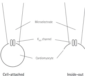
1)

Several configurations of this technique have been introduced, including cell-attached, excised inside-out, and whole-

cell patch configuration (Fig.1). In the ‘cell-attached’ mode, a tight seal is formed between the micropipette and the cell membrane, and the pipette captures the ion channel current flow. Although this configuration does not disturb the intracellular contents, it is difficult to accurately measure the membrane potential and to perfuse into the intracellular space. In the ‘excised inside-out’

mode, the micropipette is pulled away from the main body of the cell, leaving the formerly intracellular membrane surface exposed to the bath. Even though the cell body is broken in the excised patch, this technique is more likely to regulate the intracellular environment. Cell-attached and excised patch techniques are used to study the behavior of single ion channels in the section of membrane attached to the electrode. However, ‘whole-cell’ patches allow researchers to study the electrical behavior of the entire cell, instead of single channel currents.

2)

Potassium channels (K

+

channels) play a crucial role in regulating the action potential of cardiomyocytes. Among K

+

channels in the cardiovascular system, the adenosine triphosphate (ATP)-sensitive potassium channels (K

ATP

channels), the first to be discovered in cardiomyocytes,

3)

have a structure analogous to the inwardly

Print ISSN 1738-5520 • On-line ISSN 1738-5555

Influence of Thromboxane A 2 on the Regulation of Adenosine

Triphosphate-Sensitive Potassium Channels in Mouse Ventricular Myocytes

In Seok Jeong, MD 1 , Hwa Jin Cho, MD 2 , Jeong Gwan Cho, MD 3 , Sang Hyung Kim, MD 1 , Kook Joo Na, MD 1 , and Jong-Keun Kim, MD 4

1

Department of Thoracic and Cardiovascular Surgery,

2

Department of Pediatrics,

3

Department of Internal Medicine,

4

Department of Pharmacology, Chonnam National University Medical School, Gwangju, Korea

Background and Objectives: Adenosine triphosphate (ATP)-sensitive potassium (K

ATP

) channels play an important role in myocardial protection. We examined the effects of thromboxane A

2

on the regulation of K

ATP

channel activity in single ventricular myocytes.

Materials and Methods: Single ventricular myocytes were isolated from the hearts of adult Institute of Cancer Research (ICR) mice by enzymatic digestion. Single channel activity was recorded by excised inside-out and cell-attached patch clamp configurations at -60 mV holding potential during the perfusion of an ATP-free K-5 solution.

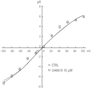
Results: In the excised inside-out patches, the thromboxane A

2

analog, U46619, decreased the K

ATP

channel activity in a dose-dependent manner; however, the thromboxane A
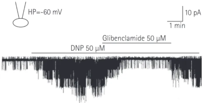
2

receptor antagonist, SQ29548, did not significantly attenuate the inhibitory effect of U46619. In the cell-attached patches, U46619 inhibited dinitrophenol (DNP)-induced K

ATP

channel activity in a dose-dependent manner, and SQ29548 attenuated the inhibitory effects of U46619 on DNP-induced K

ATP

channel activity.

Conclusion: Thromboxane A

2

may inhibit K

ATP

channel activity, and may have a harmful effect on ischemic myocardium. (Korean Circ J 2016;46(4):562-568)

KEY WORDS: KATP channels; Thromboxane A

2

; Myocytes; Cardiac.

Received: March 20, 2015 Revision Received: June 17, 2015 Accepted: November 5, 2015

Correspondence: Jong-Keun Kim, MD, Department of Pharmacology, Chonnam National University Medical School, 160 Baekseo-ro, Dong-gu, Gwangju 61469, Korea

Tel: 82-62-220-6546, Fax: 82-62-227-1636 E-mail: [email protected]

• The authors have no financial conflicts of interest.

This is an Open Access article distributed under the terms of the Creative Commons Attribution Non-Commercial License (http://creativecommons.

org/licenses/by-nc/3.0) which permits unrestricted non-commercial use,

distribution, and reproduction in any medium, provided the original work

is properly cited.

(2)

rectifying potassium channel superfamily, and their activity is regulated by the concentration of intracellular ATP metabolites.

4)

The activity of K

ATP

channels is regulated by the ratio of ATP/

Adenosine Driphosphate or ATP concentration, which is an indicator of intracellular metabolism. Intracellular K

+

loss and extracellular K

+

accumulation occur within a few minutes of the onset of myocardial ischemia. This is due to the K

+

efflux that occurs as K

ATP

channels open when intracellular ATP decreases during myocardial ischemia.

5)6)

K

ATP

channel activity simultaneously has a protective effect during ischemia, through vasodilation and the reduction of myocardial contractility, and a negative arrhythmogenic effect caused by the depolarization of the membrane potential.

7)8)

Due to this, K

ATP

channels are considered to be one of the more interesting ion channels, and research on the substances that regulate the activity of this channel has been increasing.

Thromboxane A

2

, a member of the eicosanoid family, is a typical vasoconstrictor. Because its effect is generally the opposite of prostacyclin, the balance of these two substances has major implications for the regulation of cardiovascular tension. In particular, a marked increase in thromboxane A

2

synthesis during myocardial ischemia-reperfusion has been observed, and it appears to be related to the regulation of cardiac function during myocardial ischemia. If thromboxane A

2

is involved in the regulation of K

ATP

channel activity, then, working in opposition to prostacyclin, it decreases the channel activity, increases cardiovascular tension, and likely has an overall negative impact on myocardial ischemia.

We used excised the inside-out and cell-attached patch clamp electrophysiological techniques to investigate the effects of thromboxane A

2

on the regulation of K

ATP

channels.

Materials and Methods

All experiments were performed according to the Guide for the Care and Use of Laboratory Animals published by the US National Institutes of Health (NIH Publication No. 85-23, revised 1996). The Ethics Committee of Chonnam National University Medical School approved all experimental protocols.

Isolation of single ventricular myocytes

Single ventricular myocytes were obtained from ICR mice (25-35 g).

After induction of unconsciousness through cervical dislocation, the thoracic cavity was opened and the heart was extracted.

Using a dissecting microscope at 20× magnification, adipose and connective tissues were removed from the extracted heart in a 4°C, 100% oxygen saturated Tyrode solution (composition:

137 mM NaCl, 5.4 mM KCl, 1 mM MgCl

2

, 1 mM CaCl

2

, 0.33 mM NaH

2

PO

4

, 10 mM HEPES, 10 mM dextrose, titrated to pH 7.4 with NaOH). After inserting a catheter into the aorta, the aorta was ligated and suspended in a Langendorff device where the coronary arteries were perfused for 5 minutes in a 37°C Tyrode solution at 1.5 mL/min.

Next, the extracted heart was perfused with a Ca

2+

-free Tyrode solution until the pulse stopped. With the heart completely relaxed, a Ca

2+

-free Tyrode solution containing 0.6 mg/mL collagenase (CLS2, Worthington Biochemical Co. Lakewood, NJ, USA) and 0.15 mg/mL protease (type XIV, Sigma-Aldrich Co. St. Louis, MO, USA) was perfused for around 25 minutes; the heart was then perfused for 5 minutes with a high K

+

, low Cl

-

solution (composition: 20 mM taurine, 70 mM glutamic acid, 25 mM KCl, 10 mM KH

2

PO

4

, 3 mM MgCl

2

, 0.5 mM EGTA, 10 mM HEPES, 10 mM dextrose, titrated to pH 7.35 with KOH) to remove the remaining enzymes from the heart.

We detached the ventricles from the digested heart, and placed them in a high K

+

, low Cl

-

solution. After cutting them into small fragments, they were separated into single ventricular myocytes using light mechanical stimulation with a pipette. After storing the isolated cells in a high K

+

, low Cl

-

solution, we examined the inverted microscope (American Optical Co. Buffalo, NY, USA) images, and selected the cells exhibiting no motion and having a distinct rod- shaped outline; these cells were used for all further experiments.

Fabrication of microelectrodes

The microelectrodes used in the patch clamp technique were given a resistance of 5–7 MΩ, using 1.5 mm bore diameter borosilicate glass tubing (PG150T-7.5, Clark Electromedical Instruments Co., Edenbridge, Kent, UK) and a micropipette puller (P-97, Flaming/

Brown Micropipette Puller, Sutter Instrument Co., Novato, CA, USA).

Using a stereozoom microscope (SMZ-2B, Nikon Co., Tokyo, Japan),

Cell-attached Inside-out

Microelectrode

K

ATP

channel

Cardiomyocyte

Fig. 1. Cell-attached (left) and excised inside-out (right) patch clamp

configurations.

(3)

they were then coated with Sylgard (Dow Corning Co., Midland, MI, USA) up to the area around the microelectrode’s terminal, and dried with a laboratory-made coiled heater. Once the terminal endpoints of these microelectrodes had heat reapplied to them and were polished (under an optical microscope [MF-83, Microforge, Narishige Scientific Instrument. Tokyo, Japan]), microelectrodes with a resistance of 3–5 MΩ were used in the experiment.

Single-channel current measurement and data analysis Single-channel current was recorded using the excised inside- out and cell-attached membrane patch techniques (Fig. 1).

9)

The electrical signal (at a 2 kHz cut-off frequency) measured with a patch clamp amplifier (Axopatch 200A, Axon Instruments Inc., Union City, CA, USA) was recorded onto a VCR tape with a digital data recorder (VR-10B, Instrutech Co., Longmont, CO, USA).

To analyze single-channel currents, the VCR tape was played, saved to a computer by connecting it to an A/D converter (Digidata 1200 interface, Axon Instruments Inc., Union City, CA, USA), and analyzed using the pClamp program (Version 9, Axon Instruments Inc., Union City, CA, USA). The half-maximum single-unit amplitude threshold method was used to measure the opening and closing times of a single channel.

Solutions and drugs used in the experiment

To measure single-channel currents, an ATP-free K-5 solution was used as an internal, pipette, and bath solution (composition: 140 mM KCl, 2 mM MgCl

2

, 5 mM EGTA and 10 mM HEPES, titrated to pH 7.2 with HCl). The thromboxane A

2

analog, U46619 (9, 11- dideoxy- 9α, 11α- methanoepoxy- prosta- 5Z, 13E- dien- 1- oic acid), and thromboxane A

2

receptor antagonist, SQ29548 ([1S-α,α(Z),α,α]]- phenylamino) carbonyl]hydrazino]methyl]-oxabicyclo[2.2.1]hept-yl]- heptenoic acid), used in the experiment were purchased from Cayman

Chemical Co. (Ann Arbor, MI, USA). The ATP, Glibenclamide (K

ATP

channel blocker), 2,4-Dinitrophenol (DNP, metabolite restrainer), and other reagents related to the creation of experimental solutions were purchased from Sigma-Aldrich Co. (St. Louis, MO, USA). Following manufacturers’ guidelines, each drug was highly diluted before use in the final experimental solutions.

Statistical analysis

Using a paired Student’s t-test, we interpreted p<0.05 as a significant difference between the data of the two experimental groups.

Results

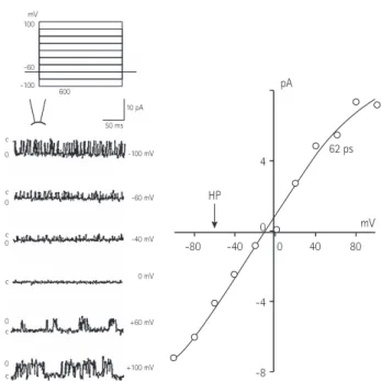
Excised inside-out patch experiment

An excised inside-out patch was prepared under a perfusion of ATP-free K-5 solution. When a holding potential of -60 mV was applied, inverted current channel activity appeared. When 1 mM ATP was added to the intracellular perfusate, the channel activity gradually decreased, and within about a minute no activity could be detected.

When the intracellular perfusate was replaced with an ATP-free K-5 solution, channel activity reappeared. Within about 3 minutes,

ATP 1 mM HP=-60 mV

1 min 10 pA Glibenclamide 50 μM

Fig. 2. Typical ATP-sensitive K

+

(K

ATP

) channel activity in an excised inside- out patch (HP=-60 mV). Channel activity appeared immediately after making an excised inside-out patch, and ATP (1 mM) almost completely inhibited the channel activity. Channel activity reappeared when the ATP was washed out with the bath solution, and the K

ATP

channel inhibitor, glibenclamide (50 μM) inhibited the channel activity. ATP: adenosine triphosphate.

pA

100

-60 -100 600

c 0

c 0

0 c

c 0

mV

HP

mV 62 ps

-80 -40 0 40 80

4

0

-4

-8

50 ms 10 pA

-100 mV

-60 mV

-40 mV

+60 mV

+100 mV 0 mV

c

0 c

Fig. 3. Current-voltage relationships of the ATP-sensitive K

+

(K

ATP

) channel

in the excised inside-out patch at different clamp potentials ranging from

-100 to +100 mV (left panel). The current-voltage relationship (right panel)

was plotted with the single channel currents in the left panel. HP: holding

potential, ATP: adenosine triphosphate.

(4)

the channel activity reached similar levels to those seen just after the excised inside-out patch procedure. The addition of glibenclamide (50 μM) caused a gradual weakening of the channel activity, and again, almost no channel activity was seen after about 1 minute.

These results suggest that the single-channel activity in the excised inside-out patch was K

ATP

channel activity (Fig. 2).

The current-voltage relationship curve showed inward rectification

with a channel conductance of 62±1.3 pS (n=5). The mean single channel current was -3.7±0.26 pA at a -60 mV holding potential (n=5), and there was an inverse relationship between the dwell time of channel opening and the degree of channel activity in a single-channel. The excised inside-out patch experiment targeted the inward current occurring at a -60 mV holding potential (Fig. 3).

Injection of U46619 (0.1–10 μM) into the intracellular perfusate decreased the channel activity in the excised inside-out patch.

Inhibition of K

ATP

channel activity by U46619 markedly decreased the frequency of channel openings and mean opening time, but did not affect the size of the single-channel current (Fig. 4).

Furthermore, based on the current-voltage relationship curves of the control and U46619 groups (injection of U46619, 10 μmol/L into the intracellular perfusate), the channel conductance values were 62±1.9 and 63±1.7 pS, respectively (n=5). There were no significant U46619 (μM)

[a]

[b]

[b]

[a]

1 min 10 pA

1 sec 10 pA

0.1 1.0 10

HP=-60 mV

Fig. 4. Effects of thromboxane A

2

analog, U46619 (range 1–10 μM) on K

ATP

channel activity in the excised inside-out patch (HP=-60 mV). U46619 inhibited K

ATP

channel activity in a dose dependent manner (upper trace).

Both the opening frequencies and the mean open-burst durations were markedly decreased at the times marked (a) and (b) on the expanded scale, but single-channel current amplitudes were not affected (lower two traces).

CTRL U46619 10 μM -100

8

6

4

2

0

-2

-4

-6

-8

-80 -60 -40 -20 0 20 40 60 80 100 mV

pA

Fig. 5. Influence of thromboxane A

2

analog, U46619, on current-voltage, K

ATP

channel relationships in the excised inside-out patch at different clamp potentials. U46619 did not significantly change the channel conductance. pA: pico ampere, CTRL: control.

U46619 (μM) SQ29548 (0.1 μM)

1 min 10 pA

0.1 1.0 10

HP=-60 mV

Fig. 6. Influence of thromboxane A

2

receptor antagonist, SQ29548 (0.1 μM), on the inhibitory effects of a thromboxane A

2

analog, U46619 in an excised inside-out patch (HP=-60 mV). SQ29548 did not influence the inhibitory effects of U46619.

U46619 (μM) 0

1.0

0.8

0.5

0.3

0.0

U46619

SQ29548 (0.1 μM) + U46619

0.1 1.0 10

Relative activity

Fig. 7. Typical ATP-sensitive K

+

(K

ATP

) channel activity in a cell-attached patch (HP=-60 mV). The K

ATP

channel inhibitor, glibenclamide, inhibited the DNP-induced channel activity. ATP: adenosine triphosphate, DNP:

dinitrophenol.

(5)

differences between the two groups, and U46619 had no effect on channel conductance (Fig. 5). SQ29548 (0.1 μM) did not attenuate the inhibitory effects of U46619 on K

ATP

channel activity (Fig. 6).

Cell-attached patch experiment

A cell-attached patch in a perfusate of ATP-free K-5 solution was prepared. When a -60 mV holding potential was applied, unlike in the excised inside-out patch, channel activity was weak. When DNP (50 μM) was added, channel activity gradually increased, reaching its peak within 3-5 minutes. When glibenclamide (50 μM) was added, the channel activity gradually weakened. Activity gradually increased when perfused in an ATP-free K-5 solution again. Such channel properties suggest that the single-channel activity within this cell-attached patch experiment was indeed K

ATP

channel activity (Fig. 7).

The mean single channel current was -3.9±0.15 pA at a -60 mV holding potential (n=5), which was similar to the inside-out patch

experiment. There was an inverse relationship between the dwell time of channel opening and the degree of channel activity in a single channel. The cell-attached patch experiment targeted the inward current appearing at a -60 mV holding potential.

K

ATP

channel activity was induced by injecting DNP (50 μM) into a bath solution in a cell-attached patch experiment. U46619 was then added (1 and 10 μM) to the perfusate to investigate its effects on channel activity. Injection of DNP caused channel activity to increase, and the activity peaked between 3 and 5 minutes after injection. At the peak of DNP activity, U46619 was added (1 and 10 μM) to the perfusate to investigate its effects on channel activity.

This injection resulted in a dose-dependent, gradual decrease in DNP-induced K

ATP

channel activity (Fig. 8). Moreover, the addition of SQ29548 (0.1 μM) attenuated the inhibitory effects of U46619 on DNP-induced K

ATP

channel activity (Fig. 9).

Discussion

This study was the first to use the patch clamp technique to directly measure the effects of a thromboxane A2 analog on the regulation of K

ATP

channel activity in an isolated single ventricular myocyte.

Known regulators of K

ATP

channel activity include endogenous adenosine, calcitonin gene-regulated peptide, vasoactive intestinal peptide, and endothelium-derived hyperpolarizing factor, as well as the prostaglandin group.

10-13)

Recently, nitrous oxide, part of endothelium-derived relaxing factor, was also identified as a regulator of K

ATP

channel activity.

13)14)

Several drugs that regulate K

ATP

channel activity have been identified: the first drug was nicorandil, used clinically for the treatment of angina,

15)

while cromakalim, developed as a coronary relaxant, was the first synthetic potassium channel opener. After that came the development of minoxidil, diazoxide, and others.

16)

Glibenclamide, a member of the sulfonylurea drug family, is the best known K

ATP

channel blocker, and is also used in the laboratory as a tool for verifying whether K

ATP

channels are responsible for the effects of drugs or endogenous substances.

17)

Prostaglandin, an arachidonic acid metabolite that relaxes cardiovascular smooth muscles, increases the K

ATP

channel activity and protects the myocardium. Using the patch clamp technique, Bouchard et al.

18)

reported that prostaglandin D

2

, E

2

, and I

2

activate K

ATP

channels in single ventricular myocytes. Jackson et al.

19)

reported that prostaglandin I

2

-induced vasodilation in rabbit hearts was partially suppressed by glibenclamide. Additionally, Ju et al.

13)

reported that prostacyclin increased K

ATP

channel activity in excised inside-out and cell-attached patch experiments in the hearts of U46619 (μM)

1 min 10 pA ATP (100 μM)

1.0 10

HP=-60 mV

Fig. 8. Effects of the thromboxane A

2

analog, U46619 (1 and 10 μM), on the DNP-induced K

ATP

channel activity in a cell-attached patch (HP=-60 mV). U46619 decreased the DNP-induced channel activity in a dose- dependent manner. DNP: dinitrophenol, ATP: adenosine triphosphate.

1 min 10 pA HP=-60 mV

Glibenclamide 50 μM DNP 50 μM

Fig. 9. Influence of the thromboxane A

2

receptor antagonist, SQ29548 (0.1 μM), on DNP-induced K

ATP

channel activity in a cell-attached patch (HP=- 60 mV). SQ29548 attenuated the inhibitory effects of the thromboxane A

2

analog, U46619, on DNP-induced K

ATP

channel activity. DNP: dinitrophenol.

(6)

white rats. These studies show that prostacyclin plays a role in increasing the K

ATP

channel activity.

12)19)

Although thromboxane A

2

, like prostacyclin, is produced by arachidonic acid via the cyclooxygenase pathway, its role in the cardiovascular system is opposite to that of prostacyclin.

Accordingly, if thromboxane A

2

is involved in the regulation of K

ATP

channel activity, then having the opposite effects of prostacyclin, it should decrease channel activity, increase cardiovascular tension, and likely have a negative impact on myocardial ischemia. Our results, in particular the finding that a thromboxane A

2

analog (U46619) inhibited K

ATP

channel activity, supports this idea.

The results of a previous study showed that thromboxane A

2

causes depolarization and suppresses voltage-gated K

+

channels in mouse pulmonary arteries,

20)

and other studies showed that U46619 suppressed the activity of Ca-activated K

+

channels in bronchial and coronary smooth muscle,

21)22)

suggesting the involvement of thromboxane A

2

in the regulation of K

+

channel activity. However, no study has yet addressed the role of thromboxane A

2

in the regulation of K

ATP

channel activity during myocardial ischemia. Xiao et al.

23)

found that prostacyclin inhibited the ischemia-reperfusion (I/R) injury, and that thromboxane A

2

was unrelated to I/R injury in mouse coronary smooth muscles.

Furthermore, there was no inhibitory effect on channel activity from thromboxane A2 receptor antagonist SQ29548 in the excised inside-out patch, but there was significant inhibition in the cell- attached patch. These results imply that U46619 suppresses K

ATP

channel activity, and that this suppression may be influenced by the different pathways within and outside the cell. In other words, our results suggest that U46619 inhibits the activity by working on the target substance around the channel, or the channel itself, in excised inside-out patches, but blocks the activity of the cell membrane receptors in cell-attached patches. However, we don’t have any additional results, such as other thromboxane receptor inhibitors or over-expression of SQ29548. We think that our present study suggests the need for further research, along with the different results of channel activity induced by SQ29548 in 2 different patch modes. Future studies will also be required to more clearly establish the role of thromboxane A

2

in myocardial ischemia, including the effect of a thromboxane A

2

on cell membrane receptors, intracellular signal transduction processes (including second messengers), and the mechanisms that directly regulate channel activity.

In conclusion, our results demonstrate that thromboxane A

2

is involved in the regulation of K

ATP

channel activity in single ventricular myocytes isolated from mice via enzymatic separation. Thromboxane A

2

reduced the activity of all K

ATP

channels in both excised inside-out and cell-attached patches in a dose-dependent manner.

Acknowledgments

The authors would like to dedicate this paper to the late Professor Jae-Ha Kim.

References

1. Neher E, Sakmann B. The patch clamp technique. Sci Am 1992;266:44-51.

2. Ackerman MJ, Clapham DE. Ion channels--basic science and clinical disease. N Engl J Med 1997;336:1575-86.

3. Noma A. ATP-regulated K

+

channels in cardiac muscle. Nature 1983;305:147-8.

4. Trube G, Hescheler J. Inward-rectifying channels in isolated patches of the heart cell membrane: ATP-dependence and comparison with cell-attached patches. Pflugers Arch 1984;401:178-84.

5. Kléber AG. Extracellular potassium accumulation in acute myocardial ischemia. J Mol Cell Cardiol 1984;16:389-94.

6. Hill JL, Gettes LS. Effect of acute coronary artery occlusion on local myocardial extracellular K

+

activity in swine. Circulation 1980;61:768-78.

7. Coetzee WA. ATP-sensitive potassium channels and myocardial ischemia: why do they open? Cardiovasc Drugs Ther 1992;6:201-8.

8. Antzelevitch C, Di Diego JM. Role of K

+

channel activators in cardiac electrophysiology and arrhythmias. Circulation 1992;85:1627-9.

9. Hamill OP, Marty A, Neher E, Sakmann B, Sigworth FJ. Improved patch- clamp techniques for high-resolution current recording from cells and cell-free membrane patches. Pflugers Arch 1981;391:85-100.

10. Kirsch GE, Codina J, Birnbaumer L, Brown AM. Coupling of ATP- sensitive K

+

channels to A1 receptors by G proteins in rat ventricular myocytes. Am J Physiol 1990;259(3 Pt 2):H820-6.

11. Quayle JM, Standen NB. KATP channels in vascular smooth muscle.

Cardiovasc Res 1994;28:797-804.

12. Ju JM, Nah SY, Kim JH. Effect of prostaglandins D2, E2 and I2 on the regulation of KATP channel activity in rat cardiac myocytes. Korean J Physiol Pharmacol 1999;3:507-12.

13. Ju JM, Shin DH, Jeong HS, Park HW, Cho JG, Kim JH. Effects of endothelium-derived relaxing factors on the regulation of ATP- sensitive potassium channel activity in cardiac myocytes. Korean Circ J 2003;33:420-30.

14. Kubo M, Nakaya Y, Matsuoka S, Saito K, Kuroda Y. Atrial natriuretic factor and isosorbide dinitrate modulate the gating of ATP-sensitive K

+

channels in cultured vascular smooth muscle cells. Circ Res 1994;74:471-6.

15. Furukawa K, Itoh T, Kajiwara M, et al. Vasodilating actions of 2-nicotinamidoethyl nitrate on porcine and guinea-pig coronary arteries. J Pharmacol Exp Ther 1981;218:248-59.

16. Quast U. Potassium channel openers: pharmacological and clinical

(7)

aspects. Fundam Clin Pharmacol 1992;6:279-93.

17. Schmid-Antomarchi H, De Weille J, Fosset M, Lazdunski M. The receptor for antidiabetic sulfonylureas controls the activity of the ATP-modulated K

+

channel in insulin-secreting cells. J Biol Chem 1987;262:15840-4.

18. Bouchard JF, Dumont E, Lamontagne D. Evidence that prostaglandins I2, E2, and D2 may activate ATP-sensitive potassium channels in the isolated rat heart. Cardiovasc Res 1994;28:901-5.

19. Jackson WF, König A, Dambacher T, Busse R. Prostacyclin-induced vasodilation in rabbit heart is mediated by ATP-sensitive potassium channels. Am J Physiol 1993;264(1 Pt 2):H238-43.

20. Cogolludo A, Moreno L, Bosca L, Tamargo J, Perez-Vizcaino F.

Thromboxane A2-induced inhibition of voltage-gated K

+

channels and pulmonary vasoconstriction: role of protein kinase Czeta. Circ Res 2003;93:656-63.

21. Li QJ, Janssen LJ. Membrane currents in canine bronchial artery and their regulation by excitatory agonists. Am J Physiol Lung Cell Mol Physiol 2002;282:L1358-65.

22. Scornik FS, Toro L. U46619, a thromboxane A2 agonist, inhibits KCa channel activity from pig coronary artery. Am J Physiol 1992;262(3 Pt 1):C708-13.

23. Xiao CY, Hara A, Yuhki K, et al. Roles of prostaglandin I(2) and

thromboxane A(2) in cardiac ischemia-reperfusion injury: a study using

mice lacking their respective receptors. Circulation 2001;104:2210-5.

수치

Fig. 1. Cell-attached (left) and excised inside-out (right) patch clamp  configurations.
Fig. 2. Typical ATP-sensitive K +  (K ATP ) channel activity in an excised inside- inside-out patch (HP=-60 mV)
Fig. 7. Typical ATP-sensitive K +  (K ATP ) channel activity in a cell-attached  patch (HP=-60 mV)
Fig. 9. Influence of the thromboxane A 2  receptor antagonist, SQ29548 (0.1  μM), on DNP-induced K ATP  channel activity in a cell-attached patch  (HP=-60 mV)

참조

관련 문서

Treatment with SPZZC significantly decreased scores of scratching behavior and clinical skin severity in a dose dependent manner in NC/Nga and BALB/c mice.. Treatment with

BDF also improved the lipid profile in a dose-dependent manner; serum lipid profiles and levels of insulin, glucose, and free fatty acid were significantly decreased in the

AC inhibits TOPFlash activity in a dose-dependent manner (Fig. 2) and suppresses the expression of b-catenin target gene, cyclin D1 (Fig. 3B) in SW-480 colon cancer cells..

(A) Cell supernatant virus titer was confirmed by PFU assay in HeLa cells. Virus titer in the supernatant was significantly decreased in a dose dependent manner of

laniceps extract and hispidulin inhibited proliferation of pancreatic cell lines, such as Capan-1, Capan-2, Panc-1 and S2-013 in a dose-dependent manner using the hollow fiber

CPE increased the levels of cleaved caspase-8, -7, -3, and poly (ADP-ribose) polymerase in a dose-dependent manner but not cleaved caspase-9. CPE exerted no effects on Bcl-2 and