Biomedical Science Letters 2015, 21(4): 188~197 http://dx.doi.org/10.15616/BSL.2015.21.4.188 eISSN : 2288-7415
Water Extract from Rice Bran Fermented with Lactobacillus plantarum Hong Inhibits Thromboxane A2 Production Associated Microsomal
Enzyme Activity in Human Platelets
Hyun-Hong Kim1,§, Jeong Hwa Hong2,§, Pajaree Ingkasupart2, Dong-Ha Lee3, DaNa Yeo4 and Hwa-Jin Park1,†
1Department of Biomedical Laboratory Science, College of Biomedical Science and Engineering Inje University, Gyungnam 50834, Korea
2Department of Smart Foods and Drugs, Inje University, Gyungnam 50834, Korea
3Department of Biomedical Laboratory Science, Korea Nazarene University, Chungnam 31172, Korea
4Department of Health Science & Technology, Inje University, Gyungnam 50834, Korea
In this study, we investigated the effect of rice bran water extract fermented with Lactobacillus plantarum Hong (RBLw), on activities of cyclooxygenase-1 (COX-1) and thromboxane A2 synthase (TXAS), thromboxane A2 (TXA2) production associated microsomal enzymes and evaluated its the antiplatelet effect. RBLw, containing 13.5 mg of ferulic acid, dose-dependently inhibited ADP-induced platelet aggregation, and inhibited the production of TXA2, an aggregation molecule. In addition, RBLw directly inhibited COX-1 activity in a dose-dependent manner, but not TXAS activity in platelet microsomal fraction having cytochrome c reductase (an endoplasmic reticulum marker enzyme) activity and expressing COX-1 (72 kDa) and TXAS (60.5 kDa) proteins. These results suggest that RBLw selectively inhibited the activity of COX-1 rather than TXAS to attenuate TXA2 production in ADP-activated platelets. Thus, we demonstrate that RBLw might have direct COX-1 antagonistic function for platelet aggregation-mediated diseases, such as thrombosis, myocardial infarction, atherosclerosis, and ischemic cerebrovascular disease.
Key Words: Rice bran fermentation, Thromboxane A2, Microsomal fraction, Cyclooxygenase-1, Thromboxane A2 synthase
INTRODUCTION
The aggregation-inducing molecule thromboxane A2
(TXA2) plays an important role in agonists (eg. collagen, thrombin, ADP)-induced platelet aggregation (Malmsten et al., 1975; Lewis and Watts, 1982; Li et al., 2010), which can
cause cardiovascular diseases such as thrombosis, athero- sclerosis and myocardial infarction (Schwartz et al., 1990).
When diverse agonists such as collagen, thrombin, and ADP activate platelets, membrane phospholipids (i.e. phosphati- dylinositol 4,5-bisphosphate) are broken down, and sub- sequently produce TXA2 from arachidonic acid (AA). This potently induces platelet activation and vasoconstriction in an autacodial action (Hamberg et al., 1975; Samuelsson et al., 1978; Gresele et al., 1991). Agonists-produced TXA2
interacts with its receptor in other platelets in an autacoidal reaction, and subsequently increases intracellular free Ca2+, a platelet aggregation-inducing molecule, via Gαq of G protein- coupled receptor (Jennings, 2009; Cattaneo et al., 1991;
Moriyama et al., 1988). TXA2 production associated enzymes Original Article
*Received: October 9, 2015 / Revised: December 2, 2015 Accepted: December 11, 2015
§Hyun-Hong Kim and Jeong Hwa Hong contributed equally to this work.
†Corresponding author: Hwa-Jin Park. Department of Biomedical Laboratory Science, College of Biomedical Science and Engineering, Inje University, 197, Inje-ro, Gimhae, Gyungnam 50834, Korea.
Tel: +82-55-320-3538, Fax: +82-55-334-3426 e-mail: [email protected]
○CThe Korean Society for Biomedical Laboratory Sciences. All rights reserved.
are cyclooxygenase-1 (COX-1) and thromboxane A2 syn- thase (TXAS), which are located in microsomes (Carey et al., 1982). COX-1 produces prostaglandin G2 (PGG2) from substrate AA, TXAS produces TXA2 from prostaglandin H2 (PGH2) that is oxidized from PGG2 by endoperoxidase.
Therefore, inhibition of COX-1 or TXAS is very useful to evaluate an antiplatelet effect of any substance or compound.
For instance, COX-1 inhibitor aspirin and TXAS inhibitor ozagrel are being used as anti-platelet agents (Patrono, 2001).
Rice bran is produced as a by-product in the rice milling process by a method in which the outer layer of the rice grain is removed. Rice bran is known to have various biological effects, including anti-inflammatory, cholesterol-lowering, antioxidant and anti-diabetic activities (Qureshi et al., 2002;
Jun et al., 2012; Hou et al., 2010). Recently, it has been reported that a water extract from rice bran (RB) has a neuroprotective effect in ischemic brain injury (Baek et al., 2014; Hong et al., 2015), and anti-platelet effect via cAMP- dependent phosphorylation of vasodilator stimulated phos- phoprotein (VASP) (VASP-Ser157) (Kim et al., 2014; Kim et al., 2015). In the present study, we investigated whether rice bran water extract fermented with Lactobacillus plantarum Hong (RBLw), a different water extract from RB, inhibits the specific activities of COX-1 and TXAS to suppress TXA2
production, and found a novel function of RBLw in ADP- activated human platelets.
MATERIALS AND METHODS Materials
ADP was purchased from Chrono-Log Co. (Havertown, PA, USA). COX-fluorescent activity kit, TXB2 enzyme immunoassay (EIA) kit, and thromboxane A2 synthase substrate prostaglandin H2 were purchased from Cayman Chemical Co. (Ann Arbor, MI, USA). Anti-COX-1 and anti- TXAS were obtained from Santa Cruz Biotechnology (Santa Cruz, CA, USA). Anti-rabbit IgG-horseradish peroxidase conjugate (HRP) and lysis buffer were obtained from Cell Signaling (Beverly, MA, USA). Polyvinylidine difluoride (PVDF) membranes were purchased from GE Healthcare (Piscataway, NJ, USA). Enhanced chemiluminesence (ECL) solution was purchased from GE Healthcare (Chalfont St,
Giles, Buckinghamshire, UK).
Preparation of rice bran water extract fermented with Lactobacillus plantarum Hong (KFCC 11556P) (RBLw)
RBLw was prepared according to the following method:
Lactobacillus plantarum Hong (KFCC 11556P) was inocu- lated and cultured in MRS broth medium (Difco Laboratories, Detroit, MI, USA) at 35℃ with shaking at 120 rpm; a 12 hour incubated culture broth was used as a starter. Rice bran was obtained from the Gimhae Rice Processing Complex just after milling the rice cultivar of Samkwang (Gimhae, Korea). Rice bran was pretreated with autoclaving imme- diately upon receipt. After cooling to room temperature, the rice bran material was vacuum-packed and stored in the refrigerator until further use.
Fermentation was performed as follows: a rice bran suspension (rice bran 100 g : 900 ml distilled water) was inoculated with the starter at a ratio of 5% and fermented at 35℃. Rice bran was fermented using the starter at 35℃, with shaking at 120 rpm for 36 hours. The culture broth was autoclaved at 121℃ for 15 min to inactivate the bacteria and then centrifuged at 8,000 g for 10 min. The supernatant was filtered and concentrated to 54 Brix by vacuum evaporation (Fig. 1). The resultant concentrate was designated as RBLw and kept at 4℃ until used. For experiments to investigate the effects on platelet aggregation, RBLw was dissolved in saline (0.9% NaCl).
Determination of total phenolic content of RBLw Total phenolic content was determined by using a modifi- cation of Singleton's method (Singleton and Rossi, 1965).
RBLw was dissolved in 50% methanol at a concentration of 0.1% (w/v). A 2.0 mL aliquot of the dissolved sample was reacted with 1.0 mL of 10% Folin-Ciocalteu reagent for 4 min at room temperature, and then 0.8 mL of a saturated sodium carbonate solution (approximately 75 g/L) was added to the reaction mixture. After incubation at room temperature for 30 min, the mixture was centrifuged at 3,000 rpm and the supernatant was removed. The absorbance readings of the supernatants were taken at 765 nm. Gallic acid was used as a reference standard, and the results are expressed as milligrams of gallic acid equivalents (mg gallic acid)/100 g RBLw.
Detection of phenolic compounds of RBLw by HPLC
Because it has been reported that rice bran contains phenolic compounds, we tested for the presence of phenol compounds in RBLw using high performance liquid chro- matography (HPLC) (Goufo and Trindade, 2014). After initially dissolving the RBLw in distilled water (100 mg/ml), the pH of the dissolved sample was adjusted to pH 2~3 with 2 N HCl. The sample was extracted three times with 0.5 mL ethylacetate, concentrated by vacuum rotary evaporation until dry, dissolved with 0.5 mL methanol, and then analyzed by HPLC. An Agilent 1100 liquid chromatography system (Palo Alto, CA, USA) equipped with a vacuum degasser, a quaternary gradient pump, an autosampler and a diode array detector (DAD) was used, which was controlled by Agilent ChemStation software. A TSKgel ODS-100V column (150 mm × 4.6 mm id, 5 μm, Tosoh, Japan) was used at a tem- perature of 40℃. The mobile phase consisted of methanol (A) and 50 mM NaH2PO4, pH 2.5 (adjusted with phosphoric acid) (B). The following program was used: 0~20 min, 30%
A and 70% B, with a flow rate of 1.0 mL/min and a sample injection volume of 5 μL. UV detection was operated at 310 nm. A calibration curve was constructed by injecting seven concentrations (12.5, 25, 50, 62.5, 125, 250, 500 μg/
mL) of authentic ferulic acid in duplicate, and then plotting the peak areas against the concentrations of each analyte.
Preparation of human platelet-rich plasma (PRP) and washed platelets
Human platelet-rich plasma (PRP) anti-coagulated with an
acid-citrate-dextrose solution (0.8% citric acid, 2.2% sodium citrate, 2.45% glucose) were obtained from the Korean Red Cross Blood Center (Changwon, Korea). PRP was centri- fuged for 10 min at 125 g to remove residual red blood cells, and then centrifuged for 10 min at 1,300 g to obtain platelet pellets. The platelet pellets were then resuspended in platelet-poor plasma to a final concentration of 5 × 10⁸/mL.
Human washed platelets were prepared from PRP to obtain microsomal fraction, a source of COX-1 and TXAS.
PRP was centrifuged for 10 min at 125 g to remove residual red blood and white blood cells, and then centrifuged for 10 min at 1,300 g to obtain the platelet pellets. The platelets were washed twice with washing buffer (138 mM NaCl, 2.7 mM KCl, 12 mM NaHCO3, 0.36 mM NaH2PO4, 5.5 mM glucose, and 1 mM EDTA, pH 6.5). The washed platelets were then resuspended in suspension buffer (138 mM NaCl, 2.7 mM KCl, 12 mM NaHCO3, 0.36 mM NaH2PO4, 0.49 mM MgCl2, 5.5 mM glucose, 0.25% gelatin, pH 6.9) to a final concentration of 5 × 10⁸/mL. All of the above pro- cedures were carried out at 25℃ to avoid platelet aggregation resulting from any effect of low temperatures. The Korea National Institute for Bioethics Policy Public Institutional Review Board (Seoul, Korea) approved these experiments (PIRB12-071).
Measurement of platelet aggregation
PRP (108 platelets/mL) were preincubated for 3 min at 37℃ with or without substances, then stimulated with ADP (20 μM) for 5 min. Aggregation was monitored using an aggregometer (Chrono-Log Corporation, Havertown, PA, Fig. 1. Preparation of rice bran water extract fermented with Lactobacillus plantarum Hong (KFCC 11556P) (RBLw)
USA) at a constant stirring speed of 1,000 rpm. Each aggre- gation rate was calculated as an increase in light transmission.
The platelet poor plasma was used as a reference (trans- mission 0%). RBLw was dissolved in saline (0.9% NaCl).
Measurement of thromboxane B2
PRP (108 platelets/mL) were preincubated for 3 min at 37℃ with or without substances, then stimulated with ADP (20 μM) for 5 min. The reactions were terminated by the addition of ice-cold EDTA (5 mM) and indomethacin (0.2 mM). The amount of TXB2, a stable metabolite of TXA2, was determined with synergy HT multi-model microplate reader (BioTek Instruments, Winoosku, VT., USA) using a TXB2 EIA kit. RBLw was dissolved in saline (0.9% NaCl).
Isolation of microsomal fraction
Washed platelets (5 × 10⁸ platelets/mL) containing suspension buffer (pH 6.9) with 1% protease inhibitor were sonicated for 20 sec, 1 cycle, and 10 times on ice with a sonicator (Bandelin, HD2070, Germany) to obtain platelet lysates. Homogenates was centrifuged at 1,500 g for 15 min at 4℃, then the supernatant were ultracentrifuged at 105,000 g for 1 hr at 4℃ to obtain the microsomal fraction (F1- cellular fraction) containing endoplasmic reticulum (ER) membrane (Carey et al., 1982). The supernatant was termed F2-cellular fraction. All of the separated fractions (homo- genates, F1-, and F2-cellular fraction) were identified by cytochrome c reductase (a marker enzyme of ER membrane) (Lagarde et al., 1981) and used as enzyme sources in Western blot as described below.
Cytochrome c reductase activity assay
Cytochrome c reductase is a flavoprotein localized in the ER. Cytochrome c reductase activity of the fractions (homogenates, F1-, and F2-cellular fraction) was assayed by using cytochrome c reductase (NADPH) assay kit (Sigma Chemical Corp, St. Louis, MO, USA). The reaction was initiated by addition of NADPH and the reduction of cyto- chrome c is monitored by the increase of absorbance at 550 nm for 7 min with kinetic program.
Western blot analysis of cyclooxygenase-1 (COX-1) and TXA2 synthase (TXAS)
Protein concentrations in the separated fractions were measured by using Bradford method. Proteins of each fraction (30 μg) were separated on 8% SDS-PAGE (1.5 mm gel).
The separated proteins were electrophoretically transferred to PVDF membranes (GE Healthcare, New Jersey, USA).
Immunoblot was performed by using primary antibody (COX-1, 1:200, TXAS, 1:100) and horseradish peroxidase (HRP) conjugated secondary antibody at a dilution of 1:
5,000 from Santa Cruz Biotechnology Inc. (CA, USA). After secondary antibody (anti-mouse IgG-HRP or anti-goat IgG- HRP) treatment, detection of antibody-bound protein in the membrane was performed with enhanced chemiluminesence (ECL) solution (GE Healthcare, Buckinghamshire, UK).
Cyclooxygenase-1 (COX-1) activity assay
For the measurement of COX-1 activity, the F1-cellular fraction (microsomal fraction, 20 μg proteins) of platelets was pre-incubated with or without various concentrations of RBLw or aspirin (500 μM) at 37℃ for 30 min. COX-1 activity of the treated F1-cellular fraction was assayed with COX-1 fluorescent assay kit (Cayman Chemical Co, Ann Arbor, MI, USA) according to the procedure described by manufacturer.
Thromboxane A2 synthase (TXAS) activity
For the measurement of TXAS activity of the F1-cellular fraction (microsomal fraction, 20 μg proteins) was pre- incubated with ozagrel (11 nM, IC50), a positive control as a TXAS inhibitor, and with or without various concentrations of RBLw at 37℃ for 30 min. The reaction is initiated by the addition of PGH2. Incubation was allowed to proceed for 1 min at 37℃. The reactions are terminated by the addition of 1 M citric acid. After neutralization with 1 N NaOH, the concentration of TXB2, a stable metabolite of TXA2, was determined by using TXB2 EIA kit according to the pro- cedure described by manufacturer.
Statistical analysis
The experimental results are expressed as the mean ±
S.E.M. Data was assessed by analysis of variance (ANOVA).
If this analysis indicated significant differences among the group means, then each group was compared by the Newman-Keuls method. P values of less than 0.05 were considered to be statistically significant.
RESULTS
The content of total phenolics and ferulic acid in RBLw
When the concentration of total phenolics was determined by using gallic acid as a standard, a concentration of total phenolics of 36.4 ± 0.4 mg in 100 g RBLw was obtained Table 1. Total phenolic acid content of RBLw
Total phenolic (TP) content (mg / 100 g RBLw)
Ferulic acid (FA) content
(mg / 100 g RBLw) FA/TP (%)
RBLw 36.4 ± 0.4 13.5 37.1
The content of total phenolic was determined as described in "Materials and Methods". Ferulic acid content was from Table 2. The data are expressed as the mean ± S.E.M. (n = 4).
Fig. 2. HPLC chromatograms of RBLw and ferulic acid. (A) The chromatogram of ferulic acid. (B) The chromatogram of RBLw. HPLC was performed as described in "Materials and Methods". F, ferulic acid
(Table 1). Because it has been reported that rice bran water extract supplemented with ferulic acid has a synergistic
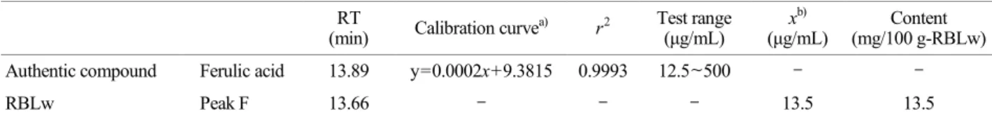
neuroprotective effect in rats (Baek et al., 2014), we analyzed the ferulic acid content in RBLw with HPLC. As shown in Fig. 2A, authentic ferulic acid was observed at a retention time of 13.89 min. As shown in Fig. 2B, the retention time (13.66 min) of peak F was almost identical with that of ferulic acid. Accordingly, we propose that peak F is a com- pound corresponding to ferulic acid. The concentration of peak F, as calculated from the calibration curve, was 13.5 mg /100 g RBLw (Table 2), which corresponded to 37.1% of the total phenolic content (Table 1).
Effect of RBLw on ADP-induced human platelet aggre- gation
The concentration of ADP that induced maximal platelet aggregation was approximately 20 μM (Fig. 3A). Therefore, 20 μM of ADP was used as the platelet agonist in this study.
When PRP (108/mL) were activated with ADP (20 μM), the aggregation rate increased to 56.3 ± 1.0%. However, various concentrations of RBLw (10 to 500 μg/mL) significantly inhibited ADP-stimulated platelet aggregation in a dose- dependent manner (Fig. 3B).
Effect of RBLw on TXA2 production
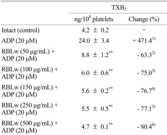
The amount of TXA2 (determined as TXB2) in intact platelets was 4.2 ± 0.2 ng/108 platelets, which was markedly increased to 24.0 ± 3.4 ng/108 platelets by ADP (20 μM).
RBLw (50~500 μg/mL), however, potently reduced the production of TXA2 (Fig. 4, Table 3) in a dose-dependent manner. These results show that RBLw may inhibit the activity of COX-1 or TXAS in a dose-dependent manner to suppress the production of TXA2 in ADP-induced platelet aggregation.
Table 2. Calibration curve and content of ferulic acid in RBLw RT
(min) Calibration curvea) r2 Test range (μg/mL)
xb) (μg/mL)
Content (mg/100 g-RBLw) Authentic compound Ferulic acid 13.89 y=0.0002x+9.3815 0.9993 12.5~500 - -
RBLw Peak F 13.66 - - - 13.5 13.5
a, b)
y, peak areas of analytes; x, concentrations of analytes in 100 mg/mL RBLw.
Fig. 3. Effect of RBLw on ADP-induced human platelet aggre- gation. (A) The concentration threshold of ADP on platelet aggre- gation. (B) Effect of RBLw pretreatment on ADP-induced platelet aggregation. Measurement of platelet aggregation was performed as described in the "Materials and Methods" section. Inhibition rate by RBLw was expressed as the percentage of the ADP-induced aggregation rate. The data are expressed as the mean ± S.E.M. (n
= 4). **P < 0.001 versus the ADP-stimulated platelets.
Determination of enzyme source of COX-1 and TXAS
To determine whether RBLw is involved in the inhibition of COX-1 and TXAS, enzyme sources having COX-1 or TXAS were needed. We separated F1-, and F2-cellular
fractions from platelet lysates (homogenates), and measured the activity of cytochrome c reductase, an endoplasmic reticulum (microsomes) marker enzyme. As shown in Fig.
5A, we found that the F1-cellular fraction has the highest specific activity of cytochrome c reductase. Next, we deter- mined which fraction from platelet lysates expresses both COX-1 and TXAS proteins. As the result, remarkably high expressions of COX-1 (72 kDa) and TXAS (60.5 kDa) were observed in F1-cellular fraction (Fig. 5B). Because these are in accordance with that F1-cellular fraction has the highest activity of cytochrome c reductase, the F1-cellular fraction is likely the microsomal fraction. This microsomal fraction was used to determine the activity of COX-1 and TXAS.
Table 3. Effects of RBLw on TXB2 production in ADP-induced platelet aggregation
TXB2
ng/108 platelets Change (%)
Intact (control) 4.2 ± 0.2 -
ADP (20 μM) 24.0 ± 3.4 + 471.41)
RBLw (50 μg/mL) +
ADP (20 μM) 8.8 ± 1.2** - 63.32) RBLw (100 μg/mL) +
ADP (20 μM) 6.0 ± 0.6** - 75.03) RBLw (150 μg/mL) +
ADP (20 μM) 5.6 ± 0.2** - 76.74) RBLw (250 μg/mL) +
ADP (20 μM) 5.5 ± 0.5** - 77.15) RBLw (500 μg/mL) +
ADP (20 μM) 4.7 ± 0.1** - 80.46) Data were from Fig. 4. 1) Change (%) = (ADP-intact) / intact × 100; 2) to 6) Change (%) = [ADP-(RBLw+ADP) / ADP] × 100.
The data are expressed as the mean ± S.E.M. (n = 4). **P < 0.001 versus the ADP-stimulated platelets.
Fig. 5. Determination of enzyme source of cyclooxygenase-1 (COX-1) and thromboxane A2 synthase (TXAS). (A) Cytochrome c reductase activities in homogenates, F1-, and F2-cellular fraction.
NADPH-cytochrome c reductase, the marker enzyme for dense tubular system in platelets (100 μg of total protein) was assayed as described in "Materials and Methods". (B) Western blot analysis of COX-1 and TXAS in homogenates, F1-, F2-cellular fraction.
Western blotting was performed as described in "Materials and Methods". H: homogenates; F1: F1-cellular fraction (microsomal fraction); F2: F2-cellular fraction. **P<0.001 compared with that of homogenates.
Fig. 4. Effects of RBLw on ADP-induced TXA2 production. Meas- urement of TXB2, a stable metabolite of TXA2, was performed as described in "Materials and Methods". The data are expressed as the mean ± S.E.M. (n = 4). aP<0.05 versus non-stimulated platelets,
**P<0.001 versus the ADP-stimulated platelets.
Effect of RBLw on COX-1 activity
To determine if the inhibitory effect of TXA2 production by RBLw was due to the direct suppression of COX-1, a cell-free enzyme assay method was used. As shown in Fig.
6A, COX-1 activity in the absence of RBLw (control) was 18.3 ± 2.1 nmol/mg-protein/min. However, RBLw dose- dependently inhibited COX-1 activity. Aspirin (500 μM), a
positive control of COX-1, inhibited COX-1 activity to 13.7
± 2.5 nmol/mg-protein/min (Fig. 6A), which was weak compared with that by RBLw (150 μg/mL).
Effect of RBLw on TXAS activity
To determine whether the inhibitory effect on TXA2 pro- duction by RBLw was due to the direct suppression of TXAS, the aforementioned cell-free enzyme assay method was also used. As shown in Fig. 6B, TXAS activity by RBLw- untreated platelets was 655.4 ± 55.7 ng/mg-protein/min.
However, RBLw (250 μg/mL) almost did not inhibit TXAS activity. In addition, ozagrel (11 nM), a positive control of TXAS, potently inhibited TXAS activity (Fig. 6B).
DISCUSSION
TXA2 is a strong agonist on resting platelets as a positive feedback promoter on activated platelets (Ruggeri, 2002;
Jennings, 2009), and also acts as a vasoconstrictor and a bronchoconstrictor (FitzGerald, 1991) in an autacoidal action.
Thus, a compound that can inhibit TXA2 production- associated enzymes (COX-1 or TXAS), TXA2 production or TXA2 action could be applied as an anti-thrombotic agent (Clutton et al., 2001). Presently, we tried to explain the inhibitory mechanism of RBLw in TXA2 production by determining the activities of COX-1 and TXAS. In this study, we established that RBLw inhibits COX-1 activity to attenuate TXA2 production in ADP-induced platelet aggre- gation. Presently, it is unknown whether ferulic acid of RBLw is directly or indirectly involved in inhibition of COX-1 activity. In the current study, because COX-1 activity was determined with microsomal fraction prepared from washed platelets, and was inhibited by RBLw, it is suggested that RBLw is involved in direct inhibition of COX-1 activity.
Accordingly, considering our previous report that authentic ferulic acid inhibited ADP-induced platelet aggregation (Kim et al., 2014), it is inferred that ferulic acid of RBLw may directly involve in inhibition of COX-1 activity to suppress the production of TXA2, a potent stimulator in platelet aggregation.
RBLw inhibited potently COX-1 activity rather than TXAS activity (Fig. 6A, B), which is in accord with reports Fig. 6. Effects of RBLw on cyclooxygenase-1 (COX-1) and throm-
boxane A2 synthase (TXAS) activities. (A) Effect of RBLw on COX-1 activity in F1-cellular fraction (microsomal fraction). COX-1 activity was performed as described in "Materials and Methods".
(B) Effect of RBLw on TXAS activity in F1-cellular fraction (microsomal fraction). TXAS activity was performed as described in "Materials and Methods". The data are expressed as the mean ± S.E.M. (n = 4). *P<0.05, **P<0.001 compared with that of non- treated F1-cellular fraction (microsomal fraction).
that the inhibition of agonists-induced platelet aggregation is dependent on inhibition of COX-1 rather than TXAS (Lewis and Watts, 1982; Jang et al., 2002). Because these results suggest that inhibition of TXA2 by RBLw is respon- sible for the suppression of microsomal specific COX-1, it is shown that RBLw may be used as a COX-1 inhibitor. In addition, TXA2 binds to its receptor in platelets, and sub- sequently mobilizes Ca2+ to activate platelets (Ohkubo et al., 1996; Cattaneo et al., 1991; Su et al., 1999). This means that TXA2 and Ca2+ are involved in thrombogenesis by intensifying platelet aggregation. Therefore, the inhibitory effect of ADP-elevated TXA2 production by RBLw could be related to the attenuation of Ca2+ to suppress the thrombo- genesis. It is known that aspirin is involved in inhibition of COX-1 activity via acetylation of COX-1 protein (Roth et al., 1975; DeWitt et al., 1990). At present, it is unknown how RBLw inhibits COX-1 activity, and whether RBLw acetylates COX-1 protein in the same as aspirin. With regard to this, further studies are necessary.
In conclusion, RBLw that inhibits TXA2 production via attenuation of COX-1 activity may have therapeutic poten- tial for the treatment or prevention of platelet aggregation- mediated diseases, such as thrombosis, myocardial infarction, atherosclerosis, and ischemic cerebrovascular disease.
Acknowledgements
This study was supported by a grant from the High Value- added Food Technology Development Program [112074-3], iPET (Korea Institute of Planning and Evaluation for Tech- nology in Food, Agriculture, Forestry and Fisheries).
Conflict of interest
The authors declare no conflict of interest.
REFERENCES
Baek SE, Kim JY, Song WT, Lee SH, Hong JH, Lee CK, Kang SG. Neuroprotective effect of rice bran extract supplemented with ferulic acid in the rat model of ischemic brain injury.
Animal Cells and Systems. 2014. 18: 93-100.
Carey F, Menashi S, Crawford N. Localization of cyclo-oxygenase and thromboxane synthase in human platelet intracellular
membranes. Biochem J. 1982. 204: 847-851.
Cattaneo M, Tenconi PM, Lecchi A, Mannucci PM. In vitro effects of picotamide on human platelet aggregation, the release reaction and thromboxane B2 production. Thromb Res. 1991.
62: 717-724.
Clutton P, Folts JD, Freedman JE. Pharmacological control of platelet function. Pharmacol Res. 2001. 44: 255-264.
DeWitt DL, el-Harith EA, Kraemer SA, Andrews MJ, Yao EF, Amstrong RL, Smith WL. The aspirin and heme-binding sites of ovine and murine prostaglandin endoperoxide synthases. J Biol Chem. 1990. 265: 5192-5198.
FitzGerald GA. Mechanisms of platelet activation: thromboxane A2 as an amplifying signal for other agonists. Am J Cardiol.
1991. 68: 11B-15B.
Goufo P, Trindade H. Rice antioxidants: phenolic acids, flavonoids, anthocyanins, proanthocyanidins, tocopherols, tocotrienols, γ-oryzanol, and phytic acid. Food Sci Nutr. 2014. 2: 75-104.
Gresele P, Deckymyn H, Nenci GG, Vermylen J. Thromboxane synthase inhibitors, thromboxane receptor antagonists and dual blockers in thrombotic disorders. Trends Pharmacol Sci. 1991.
12: 158-163.
Hamberg M, Svensson J, Samuelsson B. Thromboxanes: a new group of biologically active compounds derived from prosta- glandin endoperoxides. Proc Natl Acad Sci USA. 1975. 72:
2994-2998.
Hong JH, Kim JY, Baek SE, Ingkasupart P, Park HJ, Kang SG.
Effects of rice bran extracts rermented with Lactobacillus plantarum on neuroprotection and cognitive improvement in a rat model of ischemic brain injury. Biomed Sci Lett. 2015.
21: 92-102.
Hou Z, Qin R, Ren G. Effect of anthocyanin-rich extract from black rice (Oryza sativa L. Japonica) on chronically alcohol-induced liver damage in rats. J Agric Food Chem. 2010. 58: 3191-3196.
Jang EK, Azzam JE, Dickinson NT, Davidson MM, Haslam RJ.
Roles for both cyclic GMP and cyclic AMP in the inhibition of collagen-induced platelet aggregation by nitroprusside. Br J Haematol. 2002. 117: 664-675.
Jennings LK. Role of platelets in atherothrombosis. Am J Cardiol.
2009. 103(3 Suppl): 4A-10A.
Jun HI, Song GS, Yang EI, Youn Y, Kim YS. Antioxidant activities and phenolic compounds of pigmented rice bran extracts. J Food Sci. 2012. 77: C759-C764.
Kim HH, Hong JH, Ingkasupart P, Lee DH, Park HJ. Inhibitory effects of water extract from rice bran due to cAMP-dependent
phosphorylation of VASP (Ser157) on ADP-induced platelet aggregation. Biomed Sci Lett. 2014. 20: 129-138.
Kim HH, Lee DH, Hong JH, Ingkasupart P, Nam GS, Ok WJ, Kim MJ, Yu YB, Kang HC, Park HJ. Inhibitory effects of rice bran water extract fermented Lactobacillus plantarum due to cAMP-dependent phosphorylation of VASP (Ser157) on human platelet aggregation. Biomed Sci Lett. 2015. 21: 103-114.
Lagarde M, Menashi S, Crawford N. Localisation of phospholipase A2 and diglyceride lipase activities in human platelet intra- cellular membranes. FEBS Lett. 1981. 124: 23-26.
Lewis GP, Watts IS. Prostaglandin endoperoxides, thromboxane A2 and adenosine diphosphate in collagen-induced aggregation of rabbit platelets. Br J Pharmacol. 1982. 75: 623-631.
Li Z, Delaney MK, O'Brien KA, Du X. Signaling during platelet adhesion and activation. Arteroscler Thromb Vasc Biol. 2010.
30: 2341-2349.
Malmsten C, Hamberg M, Svensson J, Samuelsson B. Physio- logical role of an endoperoxide in human platelets: hemostatic defect due to platelet cyclooxygenase deficiency. Proc Natl.
Acad Sci USA. 1975. 72: 1446-1450.
Moriyama T, Takamura H, Narita H, Tanaka K, Matsuura T, Kito M. Elevation of cytosolic free Ca2+ is directly evoked by thromboxane A2 in human platelets during activation with collagen. J Biochem. 1988. 103: 901-902.
Ohkubo S, Nakahata N, Ohizumi Y. Thromboxane A2-mediated
shape change: independent of Gq-phospholipase C-Ca2+ path- way in rabbit platelets. Br J Pharmacol. 1996. 117: 1095-1104.
Patrono C. Aspirin: New cardiovascular uses for an old drug. Am J Med. 2001. 110: 62S-65S.
Qureshi AA, Sami SA, Khan FA. Effects of stabilized rice bran, its soluble and fiber fractions on blood glucose levels and serum lipid parameters in humans with diabetes mellitus Types I and II. J Nutr Biochem. 2002. 13: 175-187.
Roth GJ, Stanford N, Majerus PW. Acetylation of prostaglandin synthase by aspirin. Proc Natl Acad Sci USA. 1975. 72: 3073 -3076.
Ruggeri ZM. Platelets in atherothrombosis. Nat Med. 2002. 8: 1227 -1234.
Samuelsson B, Goldyne M, Granstrom E, Mamberg M, Hammarstrom S, Malmsten C. Prostaglandin and throm- boxanes. Annu Rev Biochem. 1978. 47: 997-1029.
Schwartz SM, Heinmark RL, Majesky MW. Developmental mech- anisms underlying pathology of arteries. Physiol Rev. 1990.
70: 1177-1209.
Singleton VL, Rossi JA. Colorimetry of total phenolics with phos- phomolybdicphosphotungstic acid reagents. Am J Enol Vitic.
1965. 16: 144-158.
Su CY, Shiao MS, Wang CT. Differential effects of ganodermic acid S on the thromboxane A2-signaling pathways in human platelets. Biochem Pharmacol. 1999. 58: 587-595.