ISSN 0378-6471 (Print)⋅ISSN 2092-9374 (Online)
http://dx.doi.org/10.3341/jkos.2015.56.12.1899
Original Article
큰 황반원공에서 시행한 내경계막 자가이식술
Autologous Transplantation of Internal Limiting Membrane for the Treatment of Large Macular Hole
김경호1,2⋅정재우1⋅박성후3⋅변익수1,2,4⋅이지은3,4
Kyong Ho Kim, MD1,2, Jae Woo Jung, MD1, Sung Who Park, MD3, Ik Soo Byon, MD1,2,4, Ji Eun Lee, MD, PhD3,4 양산부산대학교병원 안과학교실1, 양산부산대학교병원 의생명융합연구소2, 부산대학교병원 안과학교실3, 부산대학교 의학전문대학원
안과학교실4
Department of Ophthalmology, Pusan National University Yangsan Hospital1, Yangsan, Korea
Research Institute for Convergence of Biomedical Science and Technology, Pusan National University Yangsan Hospital 2, Yangsan, Korea Department of Ophthalmology, Pusan National University Hospital 3, Busan, Korea
Department of Ophthalmology, Pusan National University School of Medicine4, Yangsan, Korea
Purpose: To evaluate the surgical outcome of autologous transplantation of internal limiting membrane (ILM) for the treatment of large macular hole.
Methods: Twenty-five gauge pars plana vitrectomy was performed for the treatment of patients with full thickness macular hole larger than 400 μm. ILM was stained using 0.025% brilliant blue G. ILM around the hole was circumferentially peeled as large as 2.5 disc diameter (DD) in size and then transplanted inside the hole. ILM was peeled out additionally approximately 1.5 DD in size. Fluid-air exchange and gas injection were performed. After surgery, the hole was scanned using spectral domain optical coherence tomography.
Results: A total of 5 eyes were included in the present study. The mean age was 65.0 ± 11.8 years (52-77) and mean best cor- rected visual acuity (log MAR) was 0.80 ± 0.27. The mean refractive error was -2.0 ± 2.2 diopter, mean horizontal size of hole was 701.4 ± 129.4 μm, mean vertical size was 630.2 ± 202.8 μm, mean hole base size was 1,043.4 ± 225.0 μm and hole height was 464.4 ± 218.9 μm. At the first day after surgery, transplanted ILM was detected inside the hole in all 5 eyes and complete closure of the hole occurred in 4 eyes. One hole was closed between postoperative days 4 and 7. Foveal contour improved gradually but photoreceptor integrity did not during the follow-up period. Two eyes showed visual improvement but 3 did not after surgery.
Conclusions: Macular hole was closed successfully and quickly using the autologous ILM transplantation technique. Based on our results, the autologous ILM should be considered the treatment of choice for large macular holes.
J Korean Ophthalmol Soc 2015;56(12):1899-1905
Key Words: Autologous internal limiting membrane implantation, Macular hole
■Received: 2015. 5. 22. ■ Revised: 2015. 8. 5.
■Accepted: 2015. 9. 25.
■Address reprint requests to Ik Soo Byon, MD
Department of Ophthalmology, Pusan National University Yangsan Hospital, #20 Geumo-ro, Mulgeum-eup, Yangsan 50612, Korea
Tel: 82-55-360-2592, Fax: 82-55-360-2161 E-mail: [email protected]
* This study was supported by a 2015 research grant from Pusan National University Yangsan Hospital.
ⓒ2015 The Korean Ophthalmological Society
This is an Open Access article distributed under the terms of the Creative Commons Attribution Non-Commercial License (http://creativecommons.org/licenses/by-nc/3.0/) which permits unrestricted non-commercial use, distribution, and reproduction in any medium, provided the original work is properly cited.
황반원공(macular hole)은 중심와부분의 전층망막결손과 중심와 주변의 낭포성 변화를 일으켜 중심시력 저하를 일 으키는 질환이다.1 중심와 부근 유리체와 망막의 견인력이 주된 발병원인으로 생각되며, 원공 발생 후에는 섬유아세 포증식과 근섬유모세포의 수축으로 시간이 지날수록 원공 이 커지게 된다.2,3
황반원공의 치료는 내경계막제거술이 소개된 이후, 수술 성 공률은 약 80-90%에 이를 정도로 우수하다.4 하지만 400 μm
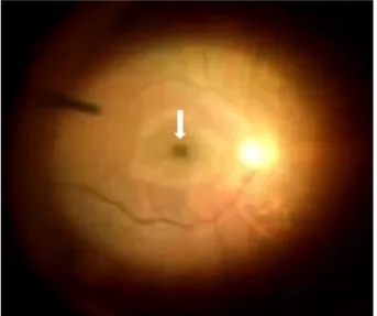
Figure 1. Fundus image during the autologous transplantation
of the internal limiting membrane (ILM). ILM stained by 0.025% Brilliant Blue G dye was placed in the macula hole (white arrow).Table 1. Characteristics of patients
Casenumber Sex Age
(years) Laterality Refractive error (diopter)
Axial length (mm)
Hole size (horizontal/vertical, μm)
Hole height (μm)
Hole base (μm)
Other ocular disorder
1 F 52 OD -1.0 22.66 830/830 320 1,328 DR
2 F 54 OD -5.0 26.61 725/637 845 1,172 Retinal tear,
pseudophakia
3 M 77 OD -3.75 23.75 711/441 382 759 Cataract
4 M 66 OS 0 23.04 485/411 448 890 Cataract
5 M 76 OD -0.25 23.35 756/832 327 1,068 Dry AMD, cataract
F= female; M = male; OD = oculus dexter; OS = oculus sinister; DR = diabetic retinopathy; AMD = age-related macular degeneration.
이상의 큰 황반원공에서는 수술성공률이 상대적으로 낮아 서 자가혈소판농축액 주입술, 자가혈청 주입술, 국소레이저 등이 함께 시행되기도 한다.5,6
최근에는 벗겨낸 내경계막을 뒤집어서 원공을 덮는 변형 된 내경계막제거술을 이용하여 원공폐쇄와 시력호전에 도 움이 되었다는 보고들이 있다.7-10 이 술기는 벗겨낸 내경계 막이 원공의 경계면과 부착이 유지되어야 하는데, 수술 술 기가 일반적인 내경계막 제거술보다 어렵고, 내경계막 절 편(flap)을 만드는 도중에 절편이 원공의 경계에서 떨어지 게 되면 원공을 덮을 수 없게 되어 계획된 수술술기를 시행 할 수 없게 된다.
이에 저자들은 크기가 큰 황반원공 환자에서 고식적인 방법으로 원공 주변의 내경계막을 망막면으로부터 벗겨낸 뒤, 제거한 내경계막 절편을 원공 안으로 직접 이식하는 술 기를 시행하고 결과를 알아보고자 하였다.
대상과 방법
2014년 8월부터 2014년 12월까지 양산부산대학교병원 안과를 방문한 환자 중에서 안저검사와 빛간섭단층촬영에 서 중심와 부분에 감각신경망막의 전층 결손이 확인된 황 반원공 환자를 대상으로 하였다. 그중에서 원공 크기가 400 μm 이상인 환자에서 내경계막 자가이식술을 시행하였다.
수술 전 시력, 굴절값, 안구길이를 조사하였으며 빛간섭단 층촬영영상으로 원공의 가로 및 세로 크기, 높이, 원공 바 닥면의 최대지름을 조사하였다. 시력은 스넬렌 시력표를 이용하여 측정하였다. 평균시력은 스넬렌시력을 logarithm of the minimum angle of resolution (logMAR)으로 변환한 뒤 계산하였다. 굴절값은 구면대응치(spherical equivalent) 로 환산하였다. 수술 후에는 빛간섭단층촬영으로 중심와의 해부학적 모양과 시세포층의 회복을 조사하였다. 중심와의 모양은 술 후 망막의 감각신경층의 결손 없이 황반원공이 폐쇄되는 경우를 제I형 폐쇄(type I closure), 결손이 있는 경우를 제II형 폐쇄(type II closure)로 구분하였다.11,12 빛간 섭단층촬영 영상은 스펙트럼영역 빛간섭단층촬영 장비
(Cirrus OCT, Carl zeiss Meditec, Dublin, CA, USA)와 파 장가변광원 빛간섭단층촬영 장비(DRI OCT-1, Topcon, Tokyo, Japan)를 사용하였으며, 두 장비의 영상 중에서 화 질이 우수한 것을 분석하였다.
수술방법은 25게이지 평면부 유리체절제술을 시행하며, 완전한 뒷유리체박리를 유도하고 유리체를 제거하였다. 0.025% Brilliant Blue G (BBG)를 이용하여 내경계막을 염 색한 후 원공 주변의 내경계막을 2.5 유두직경 크기로 벗긴 뒤 황반원공 안으로 제거한 내경계막 절편을 집어넣었다. 제거된 내경계막에서 바깥쪽으로 1.5 유두직경 크기의 내 경계막을 추가적으로 제거하였다(Fig. 1). 원공 속에 이식된 내경계막 절편이 이탈되지 않도록 천천히 액체-공기 교환 술을 시행하였다. 공기-가스교환을 하고 수술을 종료하였 다. 백내장을 동시에 수술하는 경우에는 유리체절제술에 앞서 투명각막절개창을 만들고 수정체유화술로 수정체를 제거한 뒤 인공수정체를 수정체낭에 삽입하였다.
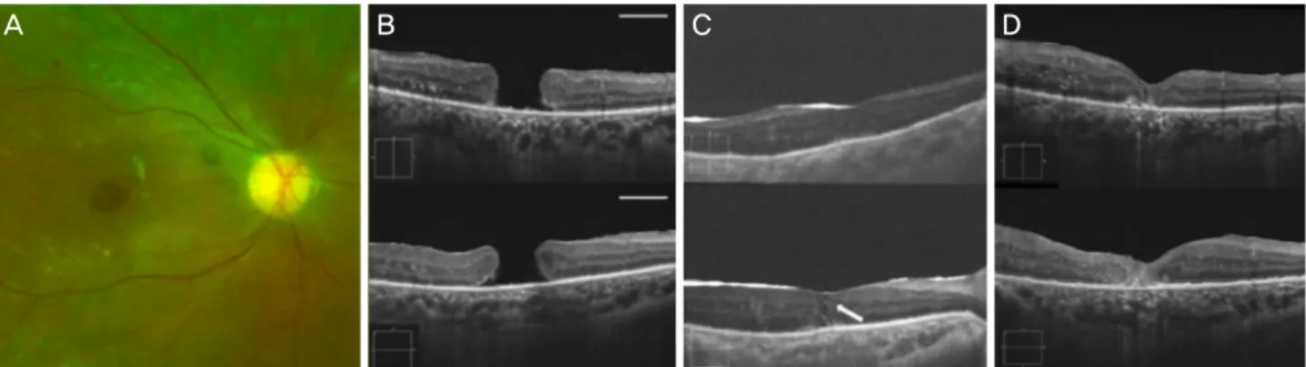
Figure 2. A case of full thickness macular hole (MH) in 52-year-old female. Preoperative color fundus photograph (A) and OCT (B,
horizontal white line: 1 mm scale bar) of the right eye demonstrated diabetic retinopathy and large macular hole. One day after sur- gery, transplanted internal limiting membrane was seen inside hole (white arrow), MH was closed (C). At 3 months, type 1 closure of hole was detected in OCT scan image. Snellen vision improved from 0.1 to 0.4 (D). OCT = optical coherence tomography.Table 2. Results of autologous internal limiting membrane transplantation
Casenumber
Time from symptom onset to surgery (days)
POD (months)
Baseline visual acuity (Snellen)
Postopertive visual acuity (Snellen)
Time to close hole after surgery (days)
Type of
closure IS/OS integrity
1 26 4 0.1 0.4 1 Type I Disrupted
2 120 2 0.1 0.1 1 Type I Disrupted
3 270 6 0.3 0.3 1 Type I Disrupted
4 23 3 0.3 0.3 1 Type I Disrupted
5 180 6 0.1 0.15 4-7 Type I Disrupted
POD = postoperative day; IS/OS = inner segment/outer segment junction of the photoreceptor layer.
결 과
대상 환자 5안 중 3안은 남자, 2안은 여자였다. 평균 나이는 65.0 ± 11.8 (52-77)세였다. 수술 전 평균교정시력(logMAR)은 0.80 ± 0.27이었고, 평균 구면대응 굴절값은 -2.0 ± 2.2디옵터 였다. 원공 크기는 평균 수평직경 701.4 ± 129.4 μm, 수직 직 경 630.2 ± 202.8 μm였다. 원공 높이는 464.4 ± 218.9 μm, 원공 바닥의 최대직경은 1,043.4 ± 225.0 μm였다. 수술 후 평균 경과관찰 기간은 4.2 (2-6)개월이었다. 증례1은 당뇨 망막증이, 증례5는 비삼출성 나이관련황반변성이 동반된 상태였다(Table 1).
수술 후 시행한 빛간섭단층촬영영상에서 4안은 첫날부터 원공이 폐쇄되었으며, 1안은 4-7일 사이에 폐쇄되었다. 중 심와의 모양은 5안 모두 제I형 폐쇄였으며 경과관찰 기간 동 안 더욱 개선되었다. 중심와 부분의 시세포층은 5안 모두에 서 경과관찰 기간 동안 회복되지 않았다. 수술 후 5안 중 2안 에서 시력의 개선이 있었으나 3안에서는 변화가 없었다 (Table 2).
증례1은 황반부종을 동반한 증식당뇨망막증으로 레이저 광응고술과 유리체강내 아바스틴 주입술을 시행 받고 경과관 찰 중이던 55세 여자 환자였다. 내원 2주 전 발생한 우안 시력 저하로 최대교정시력은 0.1로 측정되었다. 빛간섭단층촬영에
서 원공의 크기는 가로와 세로가 830 μm, 높이 320 μm, 바닥 면의 지름 1,328 μm였다. 백내장 수술과 함께 25게이지 평 면부 유리체절제술, 내경계막 자가이식술, 가스주입술을 시 행하였다. 수술 후 1일째 시행한 빛간섭단층촬영에서 이식 된 내경계막 절편과 황반원공의 폐쇄가 관찰되었고, 수술 후 3개월째에는 중심와의 모양이 더욱 개선된 상태였다. 교 정시력은 0.4로 호전되었으나 시세포층의 내절/외절 경계 는 회복되지 못한 상태였다(Fig. 2).
증례2는 1년 전 우안 백내장 수술을 받은 인공수정체안 의 54세 여자 환자였다. 4개월 전부터 우안의 변시증과 중 심시력저하가 있어 내원하였다. 우안 최대교정시력은 0.1 이었다. 안저검사에서 7시와 11시 방향에 망막열공을 동반 한 황반원공이 관찰되었다. 빛간섭단층촬영에서 원공의 크 기는 가로 725 μm, 세로 637 μm, 높이 845 μm, 바닥면 지 름 1,172 μm였다. 25게이지 평면부 유리체절제술, 내경계 막 자가이식술, 가스주입술을 시행하였다. 수술 중 12시, 1 시, 2시에도 망막열공이 관찰되어 7시, 11시 부위와 함께 레이저광응고술을 추가로 시행하였다. 수술 후 첫날 빛간 섭단층촬영에서 원공의 폐쇄가 관찰되었다. 수술 후 2개월 째 황반원공의 폐쇄는 제I형으로 유지되었으나 시세포층 내절/외절 경계는 회복되지 않았으며, 시력도 0.1에서 개선 되지 않았다(Fig. 3).
A B C D
Figure 3. A case of closure of a macular hole (MH) in 54-year-old female. Preoperative color fundus photograph (A) and optical co-
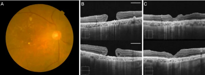
herence tomography (OCT) (B, horizontal white line: 1 mm scale bar) shows the full thickness MH with perifoveal cyst. One day after surgery, the MH had closed and transplanted inner limiting membrane is seen inside the hole (white arrow) (C). At 1 month, OCT scan shows type 1 closure MH (D).Figure 4. A case in 76-year-old male. Preoperative color fundus photograph (A) and optical coherence tomography (OCT) (B, hori-
zontal white line: 1 mm scale bar) showing the multiple soft drusen and the large macular hole. At 6 months, OCT scan image showed the improvement of foveal configuration (type 1 closure of hole), but photoreceptor layer remained disrupted (C). His snell- en vision slightly improved from 0.1 to 0.15.증례3은 9개월 된 우안 시력저하로 내원한 77세 남자였다.
최대교정시력은 우안 0.3이었다. 안저검사에서 우안에 크기 가 큰 황반원공이 관찰되었다. 빛간섭단층촬영에서 크기 가 로 711 μm, 세로 441 μm, 기저부 759 μm, 높이 382 μm의 황반원공이 진단되었다. 백내장 수술과 함께 25게이지 평 면부 유리체절제술, 내경계막 자가이식술, 가스주입술을 시 행하였다. 수술 후 4일째 시행한 빛간섭단층촬영에서 원공 속 내경계막 절편과 원공의 폐쇄가 관찰되었다. 수술 후 6 개월까지 원공폐쇄는 유지되었으며 중심와의 모양도 더욱 개선되었다. 하지만 시세포층의 내절/외절 경계는 회복되지 못하였다. 시력은 0.3으로 술 전과 차이가 없었다.
증례4는 약 3주 전부터 발생한 좌안의 중심시력 저하로 내원한 66세 남자 환자였다. 좌안의 최대교정시력은 0.3이 었다. 안저검사와 빛간섭단층촬영에서 가로 485 μm, 세로
441 μm, 높이 448 μm, 바닥면 기저부 지름이 890 μm인 황 반원공이 진단되었다. 백내장 수술과 함께 25게이지 평면 부 유리체절제술, 내경계막 자가이식술, 가스주입술을 시행 하였다. 수술 후 1일째 시행한 빛간섭단층촬영에서 원공 속 내경계막 절편과 원공의 폐쇄가 관찰되었다. 수술 후 3개월 최대교정시력은 0.3으로 술 전 시력과 차이는 없었다. 중심 와 모양은 제I형의 형태로 폐쇄되었으나, 시세포층은 회복 되지 못하였다.
증례5는 76세 남자로서 6개월 전부터 발생한 우안 시력 저하를 호소하였다. 우안 최대교정시력은 0.1이었다. 안저 검사에서 전층황반원공과 함께 황반부에 다수의 연성드루 젠이 동반된 비삼출성 나이관련 황반변성이 관찰되었다. 원공의 크기는 가로 756 μm, 세로 832 μm, 높이 327 μm, 원공 바닥 지름 1,068 μm였다. 백내장 수술과 함께 25게이
A B C D
A B C
지 평면부 유리체절제술, 내경계막 자가이식술, 가스주입술 을 시행하였다. 수술 후 1일째부터 빛간섭단층촬영에서 원 공 속에 이식된 내경계막 절편과 함께 원공의 폐쇄가 관찰 되었다. 수술 후 3개월째에는 우안 시력 0.15로 호전되었으 며 원공폐쇄는 유지되었다. 술 후 6개월까지 중심와는 제I 형으로 폐쇄되었으나 시세포층 내절/외절 경계는 회복되지 못하였다(Fig. 4).
고 찰
특발성 황반원공의 수술적 치료는 내경계막 제거술이 소 개된 이후로 약 90%에 달하는 해부학적 성공률과 시력 개선 을 얻을 수 있게 되었다.13,14 하지만 원공의 크기가 크고 오 래된 경우에는 원공 페쇄가 상대적으로 어려워 400 μm 이상 의 Gass 분류 3, 4단계인 원공은 내경계막 제거술을 시행한 유리체절제술로도 약 69%의 수술 성공률을 보인다고 보고 된 적이 있다.15 본 연구에서는 중심와 주변의 내경계막을 벗 겨낸 뒤, 제거된 내경계막 절편을 원공 속으로 직접 이식하 여 크기가 큰 황반원공 환자 5안에서 모두 성공적인 원공의 폐쇄를 달성하였다. 내경계막 자기이식술은 Morizane et al16 에 의해 내경계막 제거술을 시행한 뒤 원공 폐쇄에 실패한 황반원공 환자의 이차수술에서 남아있는 내경계막을 떼어 원공 속에 이식하는 것으로 시도된 바 있다. 이러한 술기를 사용하여 Morizane et al16은 10안 중 9안에서 원공폐쇄에 성공하였다고 보고하였다. 이 연구에서는 이식한 내경계막 이 이탈되지 않도록 관류액을 잠근상태에서 저분자 점탄물 질(low molecular weight viscoelastic material)을 이식편 위 에 도포하여 안정화시켰는데, 점탄물질을 사용하지 않고서 내경계막 절편을 원공 속에 이식한 본 연구의 술기와는 차 이가 있다.
최근 크기가 큰 황반원공과 고도근시에서 발생한 황반원 공에서 내경계막을 원공 경계면까지만 벗기고 뒤집어 원공 을 덮는 방법(Inverted internal limiting membrane [ILM]
flap technique)으로도 좋은 수술결과를 보고한 바 있다.7-10 이 술기는 내경계막을 벗기면서 내경계막 절편이 망막면에 서 떨어지게 되면 원공을 덮을 수 없게 되므로 원공의 경계 부분에서는 내경계막이 떨어지지 않게 주의하여야 하며, 덮은 절편이 원공면에서 미끄러져 이탈되기도 하는 단점이 있다. 이에 비해, 내경계막 자가이식술은 일반적인 내경계 막 제거술과 동일한 술식으로 내경계막을 일정 크기로 벗 겨낸 뒤 원공 속으로 넣으면 되므로, 수술방법이 비교적 간 편하다. 하지만 내경계막 절편을 원공 속으로 이식하는 과 정에서 원공바닥면의 망막색소상피층에 기계적 손상을 줄 수 있어 주의가 필요하다.
원공이 폐쇄되는 기전은 아직 명확히 알려지진 않았으 나, 수술을 통해 전후 방향 및 접선 방향의 견인력을 제거 하여 원공경계면이 서로 접근하게 하고, 교세포 및 기타 다 른 세포들의 증식을 자극하여 신경망막의 결손된 부분을 메우게 되는 것이다. 눈속 충전물은 원공으로의 액체유입 을 막고 교세포의 이주를 촉진하는 역할을 하게 된다. 내경 계막의 제거는 교세포의 증식을 촉진하게 한다.17-19 이런 과정에서 뮬러세포(Muller cell) 증식은 원공의 폐쇄에 필수 적인데, 내경계막으로 원공을 덮거나 원공 안에 이식하는 경우에는 내경계막 표면에 있는 뮬러세포(Muller cell)를 함 께 원공으로 이주시키게 되며, 뮬러세포(Muller cell)가 증 식할 때 내경계막이 골격체로서의 역할을 하여 원공폐쇄를 돕는다고 생각된다.7,16
수술 후 원공이 폐쇄되는 기간은 원공의 크기에 따라 비 례하는데, 작은 원공은 24시간 내에 폐쇄되기도 하지만 크 기가 큰 원공은 14일 이상 걸린다고 알려져 있다.20-22 본 연 구에서는 대상 환자가 모두 400 μm 이상의 크기가 큰 원공 임에도 불구하고, 5안 중 4안에서 술 후 첫날부터 원공의 폐쇄를 얻을 수 있었으며, 1안은 4-7일 사이에 폐쇄되었다.
이를 통해 내경계막을 이식한 경우에는 크기가 큰 원공임 에도 불구하고 원공폐쇄까지의 기간이 상당히 짧은 것을 확인할 수 있었다.
크기가 큰 원공의 경우에는 수술 후 폐쇄에 성공하더라도 중심와의 형태가 정상과 차이가 있는 경우가 많다. 중심와의 형태에 따라 술 후 망막의 감각신경층의 결손 없이 황반원공 이 폐쇄되는 경우는 제I형 폐쇄(type I closure), 결손이 있는 경우는 제II형 폐쇄(type II closure)로 구분하였다.11,12 크기가 400 μm 이상인 큰 원공에서는 제I형 폐쇄(type I closure)가 56% 정도라고 하였다.23 본 연구에서는 5안 모두 중심와의 형태가 제I형 폐쇄로 되어 우수한 해부학적 결과를 보였다.
황반원공 수술 후 시력은 수술 전 원공의 크기, 시세포층의 상태, 술 전 시력, 증상 발생부터 수술일까지의 기간이 영향 을 주는 것으로 알려저 있다.24-26 본 연구에서는 5안 모두 성공적으로 원공이 폐쇄되었음에도 불구하고 시력개선에 있어서는 다양한 결과를 보였다.
본 연구는 적은 환자를 대상으로 시행된 후향적 연구로 서, 내경계막 자가이식술기에 따른 시력의 결과는 결론을 내릴 수 없었다. 하지만 크기가 큰 황반원공에서 내경계막 자가이식술은 빠른 원공의 폐쇄와 우수한 해부학적 결과를 얻을 수 있었다. 본 연구를 통해 내경계막 자가이식술은 지 금까지 알려진 수술방법에 더하여 새로운 술기로서 시도해 볼 수 있을 것으로 생각되며, 향후 많은 환자를 대상으로 내경계막 자가이식술에 대한 시력예후와 합병증 등에 대한 연구가 필요하다.
REFERENCES
1) La Cour M, Friis J. Macular holes: classification, epidemiology, natural history and treatment. Acta Ophthalmol Scand 2002;80:
579-87.
2) Johnson RN, Gass JD. Idiopathic macular holes. Observations, stages of formation, and implications for surgical intervention.
Ophthalmology 1988;95:917-24.
3) Yooh HS, Brooks HL Jr, Capone A Jr, et al. Ultrastructural features of tissue removed during idiopathic macular hole surgery. Am J Ophthalmol 1996;122:67-75.
4) Tatham A, Banerjee S. Face-down posturing after macular hole surgery: a meta-analysis. Br J Ophthalmol 2010;94:626-31.
5) Konstantinidis A, Hero M, Nanos P, Panos GD. Efficacy of autolo- gous platelets in macular hole surgery. Clin Ophthalmol 2013;
7:745-50.
6) Kung YH, Wu TT. The effect of autologous serum on vitrectomy with internal limiting membrane peeling for idiopathic macular hole. J Ocul Pharmacol Ther 2013;29:508-11.
7) Michalewska Z, Michalewski J, Adelman RA, Nawrocki J.
Inverted internal limiting membrane flap technique for large mac- ular holes. Ophthalmology 2010;117:2018-25.
8) Kuriyama S, Hayashi H, Jingami Y, et al. Efficacy of inverted in- ternal limiting membrane flap technique for the treatment of mac- ular hole in high myopia. Am J Ophthalmol 2013;156:125-31.e1.
9) Michalewska Z, Michalewski J, Dulczewska-Cichecka K, Nawrocki J. Inverted internal limiting membrane flap technique for surgical repair of myopic macular holes. Retina 2014;34:664-9.
10) Shin MK, Park KH, Park SW, et al. Perfluoro-n-octane-assisted single-layered inverted internal limiting membrane flap technique for macular hole surgery. Retina 2014;34:1905-10.
11) Kang SW, Ahn K, Ham DI. Types of macular hole closure and their clinical implications. Br J Ophthalmol 2003;87:1015-9.
12) Shukla SY, Afshar AR, Kiernan DF, Hariprasad SM. Outcomes of chronic macular hole surgical repair. Indian J Ophthalmol 2014;
62:795-8.
13) Tognetto D, Grandin R, Sanguinetti G, et al. Internal limiting mem- brane removal during macular hole surgery: results of a multicenter retrospective study. Ophthalmology 2006;113:1401-10.
14) Haritoglou C, Reiniger IW, Schaumberger M, et al. Five-year fol- low-up of macular hole surgery with peeling of the internal limiting
membrane: update of a prospective study. Retina 2006;26:618-22.
15) Freeman WR, Azen SP, Kim JW, et al. Vitrectomy for the treatment of full-thickness stage 3 or 4 macular holes. Results of a multi- centered randomized clinical trial. The Vitrectomy for Treatment of Macular Hole Study Group. Arch Ophthalmol 1997;115:11-21.
16) Morizane Y, Shiraga F, Kimura S, et al. Autologous transplantation of the internal limiting membrane for refractory macular holes. Am J Ophthalmol 2014;157:861-9.e1.
17) Wender J, Iida T, Del Priore LV. Morphologic analysis of stage 3 and stage 4 macular holes: implications for treatment. Am J Ophthalmol 2005;139:1-10.
18) Costa RA, Cardillo JA, Morales PH, et al. Optical coherence to- mography evaluation of idiopathic macular hole treatment by gas-assisted posterior vitreous detachment. Am J Ophthalmol 2001;132:264-6.
19) Smiddy WE, Feuer W, Cordahi G. Internal limiting membrane peeling in macular hole surgery. Ophthalmology 2001;108:1471-6;
discussion 1477-8.
20) Yamashita T, Yamashita T, Kawano H, et al. Early imaging of mac- ular hole closure: a diagnostic technique and its quality for gas-fil- led eyes with spectral domain optical coherence tomography.
Ophthalmologica 2013;229:43-9.
21) Kasuga Y, Arai J, Akimoto M, Yoshimura N. Optical coherence to- mograghy to confirm early closure of macular holes. Am J Ophthalmol 2000;130:675-6.
22) Tan MH, Chen FK. “Doctor, why is my macular hole still open?”
Graefes Arch Clin Exp Ophthalmol 2014;252:165-7.
23) Ip MS, Baker BJ, Duker JS, et al. Anatomical outcomes of surgery for idiopathic macular hole as determined by optical coherence tomography. Arch Ophthalmol 2002;120:29-35.
24) Michalewska Z, Michalewski J, Cisiecki S, et al. Correlation be- tween foveal structure and visual outcome following macular hole surgery: a spectral optical coherence tomography study. Graefes Arch Clin Exp Ophthalmol 2008;246:823-30.
25) Leonard RE 2nd, Smiddy WE, Flynn HW Jr, Feuer W. Long-term visual outcomes in patients with successful macular hole surgery.
Ophthalmology 1997;104:1648-52.
26) Kumagai K, Ogino N, Demizu S, et al. Variables that influence vis- ual acuity after macular hole surgery. Jpn J Ophthalmol 2001;
45:112.
= 국문초록 =
큰 황반원공에서 시행한 내경계막 자가이식술
목적: 크기가 큰 황반원공의 수술적 치료로서 내경계막 자가이식술을 시행한 환자 5명의 수술방법과 결과를 알아보고자 한다.
대상과 방법: 크기가 400 μm 이상인 큰 황반원공 환자를 대상으로 유리체절제술을 시행하고 0.025% Briliant blue G로 내경계막을 염색하였다. 원공 주변의 내경계막을 약 2.5 유두직경의 크기로 벗겨낸 뒤, 원공 속에 내경계막 절편을 이식하고, 추가적으로 약 1.5 유두직경 정도의 내경계막을 더 제거하였다. 액체공기교환을 하고 가스를 충전하였다. 수술 후 빛간섭단층영상으로 원공 폐쇄를 조사 하였다.
결과: 5안의 평균 나이는 65.0 ± 11.8 (52-77)세였다. 평균 수술 전 교정시력(logMAR)은 0.80 ± 0.27이었고, 평균 구면대응 굴절값 은 -2.0 ± 2.2디옵터였다. 원공 크기는 평균 수평직경 701.4 ± 129.4 μm, 수직 직경 630.2 ± 202.8 μm였다. 기저부는 1,043.4
± 225.0 μm, 높이는 464.4 ± 218.9 μm였다. 4안은 수술 후 1일째, 1안은 수술 후 4-7일 사이에 원공이 완전히 폐쇄되었다. 경과관찰 기간 동안 중심와의 해부학적 모양은 더욱 개선되었다. 2안에서는 시력호전이 되었으나, 3안은 술 전과 차이가 없었다.
결론: 내경계막 자가이식술을 통해 수술 후 빠른 기간에 성공적인 원공의 폐쇄를 얻을 수 있었다. 크기가 큰 황반원공에서 시행해 볼 만한 유용한 수술 술기라고 생각한다.
<대한안과학회지 2015;56(12):1899-1905>