pISSN: 0378-6471 eISSN: 2092-9374 DOI : 10.3341/jkos.2011.52.4.407
= 증례보고 =
2.8 mm 이하의 절개창 크기의 동축 백내장수술에서 난시 유발과 각막 고위수차 비교
문상정⋅이동준⋅이경헌 성모안과병원
목적: 동축 미세절개 백내장수술(절개창: 1.8 mm)과 소절개 백내장수술(절개창: 2.2 mm, 2.8 mm) 후 난시 유발과 각막 전면과 후면의 고위수차를 비교 분석하였다.
대상과 방법: 100눈에서 32눈은 1.8 mm, 38눈은 2.2 mm, 30눈은 2.8 mm 크기로 이측에 투명각막절개를 통한 백내장 수술을 하였다.
전향적으로 술 전에 각막곡률계로 각막난시, 각막난시를 측정하였고, 술 후 1개월에 각막난시, 각막 수차, 3개월에 각막난시를 측정하 여수술 유발 난시를 계측하였다.
결과: 수술 유발 난시량은 1.8 mm 절개에서 0.41 ± 0.30, 2.2 mm 절개에서 0.47 ± 0.21, 2.8 mm 절개에서 0.71 ± 0.50디옵터로 1.8 mm 절개에서 가장 적은 난시가 유발되었다(p=0.002). 절개에 따른 각막 전면의 코마수차, 구면수차와 후면의 트레포일수차, 구면수차의 변화량은 세 군간에 차이가 없었다.
결론: 백내장수술 1개월 후 각막 전면과 후면의 고위수차의 변화는 각막절개의 크기와 연관이 없으나 각막절개의 크기에 따른 수술 유발 난시는 2.8 mm 절개보다 1.8 mm와 2.2 mm 절개에서 유의하게 작았다.
<대한안과학회지 2011;52(4):407-413>
■ 접 수 일: 2010년 2월 4일 ■ 심사통과일: 2010년 11월 9일
■ 게재허가일: 2011년 2월 15일
■ 책 임 저 자: 이 경 헌
부산시 해운대구 우2동 1078-7 성모안과병원
Tel: 051-743-0775, Fax: 051-743-0776 E-mail: [email protected]
* 본 논문의 요지는 2009년 대한안과학회 제102회 학술대회에서 구연으로 발표되었음.
수정체유화기기의 뛰어난 역동성, 초음파 에너지의 제어 기술, 새로운 인공수정체 발달이 지속적으로 이루어지고 있 으며, 이를 통해 수술 후 시기능은 더욱 빠르게 회복될 수 있다. 특히 접힘인공수정체의 발달은 백내장수술에서 인공 수정체의 삽입을 용이하게 하며 절개창의 크기를 작게 만 드는 데 많은 기여를 하였다. 2.8 mm나 3.0 mm 절개창을 통한 백내장수술에서부터 2.0 mm 이하의 동축 미세절개 수술까지 가능하며, 이축(bimanual or biaxial) 백내장 술기 로는 더 작은 절개로 백내장 수술을 할 수 있다.1-5
그러나 이축 백내장 수술은 인공수정체 삽입 시 각막절 개 확장이 필요하고, 작은 크기의 관류관 때문에 전방의 안 정성이 떨어지고 무엇보다 술기에 익숙해지기 위한 학습 곡선이 가파르다는 단점이 있다.6-9이에 반해 동축 미세절 개 백내장수술은 이축 백내장수술의 장점과 수술 과정과 술 후 염증 감소, 창상 조직의 빠른 회복으로 절개 부위의
누출의 빈도가 적어 안내염 발생을 줄일 수 있다. 또한 수 술 유발 난시가 적게 일어나는 장점이 있다.10,11
시력 검사, 대비 감도, 수차 분석을 통해 시기능 정도와 시력의 질을 객관적, 과학적으로 평가 할 수 있다. 이중 수 차 분석은 굴절교정 수술에 많이 이용되고 있으며, 최근에 는 백내장수술 이후의 광학적 질 평가를 위해 이용된다.
Wavefront를 분석하여 백내장수술에 의한 저위수차와 고 위수차 변화와 인공수정체에 따른 안구의 수차 변화를 평 가할 수 있다.12,13
3.0 mm 이상의 절개 창을 통한 백내장수술이 수술 유발 에 미치는 영향에 대한 연구는 활발이 이루어져 있으나 2.8 mm 이하의 절개를 이용한 동축 백내장수술 후 각막 난시 유발, 수차에 대한 연구는 상대적으로 부족하고 1.8 mm 미 세절개 후 동축으로 이루어진 백내장수술이 난시 유발, 각 막 수차에 미치는 연구는 거의 없다.12,14최근 국내 연구로 는 2.2 mm 절개창과 2.8 mm 절개창 백내장 수술에서의 수 술 유발 난시와 각막 고위 수차 비교에서 양 군 간에 큰 차이가 없다는 보고와 1.8 mm, 2.8 mm 절개창 비교에서 구면 수차, 고위 수차, 전체 수차는 차이가 없지만 수술 유 발 난시는 양 군 간에 통계적으로 차이가 있음을 보고하였
다.15,16본 연구는 1.8 mm 동축 백내장수술을 포함하여 2.2
mm, 2.8 mm 소절개 백내장 수술 후 수술 유발 난시, 각막 의 전면과 후면의 트레포일수차, 코마수차, 구면수차를 비
Table 1. Demographics of patients and surgical results after cataract surgery
Dermographics of patientsBaseline variables 1.8 mm 2.2 mm 2.8 mm p-value
No. eyes 32 38 30
Age (mean±SD, yr) 64.5 ± 10.5 61.8 ± 15.3 69.4 ± 10.0 0.045*
UCVA† (mean±SD, logMAR) 0.71 ± 0.36 0.70 ± 0.33 0.53 ± 0.21 0.053
BCVA‡ (mean±SD, logMAR) 0.40 ± 0.33 0.45 ± 0.37 0.41 ± 0.21 0.836
SE§ (mean±SD, diopter) -1.71 ± 5.98 0.31 ± 1.85 0.02 ± 2.09 0.119
Endothelial cell density (mean±SD, cell/mm2) 2850 ± 419 2618 ± 433 2724 ± 345 0.063 Surgical results after cataract surgery
UCVA (mean±SD, logMAR) 0.34 ± 0.32 0.26 ± 0.30 0.15 ± 0.20 0.029*
BCVA (mean±SD, logMAR) 0.05 ± 0.09 0.05 ± 0.90 0.06 ± 0.10 0.881
SE (mean±SD, diopter) -1.05 ± 1.05 -0.45 ± 0.78 -0.32 ± 0.51 0.000*
Endothelial cell density (mean±SD, cell/mm2) 2516 ± 466 2303 ± 381 2339 ± 329 0.221
*p-value<0.05; †uncorrected visual acuity; ‡best corrected visual acuity; §spherical equivalent.
교 분석하였다.
대상과 방법
본 연구는 2009년 3월부터 2009년 8월까지 100안을 무 작위로 분류하여 3 종류의 절개창으로 한 술자(L.K.H)에 의해 동축 수정체유화술 시행 후 절개 크기에 따라 각기 다 른 인공수정체를 삽입한 안을 대상으로 하였다. 1.8 mm 절 개창은 32안으로 MI60 (Bausch & Lomb, Rochester, NY, USA), 2.2 mm 절개창은 38안으로 Acrysof IQ (Alcon, Fort Worth, TX, USA), 2.8 mm 절개창은 30안으로 Akreos AO (Bausch & Lomb)를 각각 삽입하였다. 2.25 디옵터의 이하의 각막 난시가 있는 눈을 대상으로 하였고 이전에 굴절수술을 포함하는 각막 수술을 받은 눈, 각막 혼 탁 등의 이유로 각막곡률계 측정이 불가능한 눈, 수술 중 각막 난시에 영향을 주는 시술을 받거나 각 그룹의 인공수 정체가 아닌 다른 종류의 인공수정체가 삽인된 눈은 연구 에서 제외하였다. 수술은 모든 눈에서 0.5% proparacaine hydrochloride (Alcaine, Alcon)로 국소 마취 후 점탄 물질 로 전낭을 채우고 이측부에 투명각막절개를 하였다. 낭집게 를 이용하여 5.0에서 5.5 mm의 수정체낭원형절개를 만들 고 수력분리술과 수력분층술을 시행한 후 Akahoshi pre- chopper를 이용하여 수정체 핵을 분할한 다음에 1.8 mm 절개 창에서는 Stellaris (Bausch & Lomb), 나머지 눈들에 서 Infiniti (Alcon, Fort Worth)을 이용하여 수정체유화술, 피질 흡인을 시행하였다. 이후 점탄물질로 전방을 다시 채 우고 인공수정체를 삽입하고 관류흡인으로 점탄물질을 제 거하고 적절한 안압을 확인한 후 각막봉합을 시행하지 않 고 기질수화로 창상을 폐쇄하였다.
각막난시는 술 전, 술 후 1개월, 3개월에 자동각막곡률계 RK-5 (Canon, Kyoko, Japan)로 검사하고 벡터 방식으로
수술 유발 난시(SIA calculator, http://doctor-hill.com)를 계산하였다.17 각막수차는 술 전, 술 후 1개월에 Pentacam (Occlus, Wetzlar, Germany)로 직경 6.0 mm의 각막을 검 사한 후 Zernike polynomial mode에서 수차를 구하였으며 이 중 트레포일수차, 코마수차, 구면수차, 트레포일 RMS (root mean square), 코마 RMS를 각막의 전면과 후면으로 나누어 조사하였다. 그 외 나안시력, 최대교정시력, 각막내 피세포검사를 시행하였다. 통계적인 분석은 SPSS 17.0 for Windows (SPSS Inc. Chicago, IL, USA)를 이용하였고, 각 그룹에서 수술 전후의 난시 변화는 repeated measure ANOVA (analysis of variance)로, 고위 수차 변화는 paired t-test 로 분석 하였다. 세 그룹 간의 비교에서는 ANOVA (analysis of variance) 방식으로 통계 처리하였으며, 통계적 유의성 은 p<0.05로 하였다.
결 과
대상안 전체는 89명 100안이었고 이 중 남자는 30명, 여 자는 59명이었다. 평균 나이는 65.0세이었다. 1.8 mm 절개 창은 32안, 2.2 mm 절개창은 38안, 2.8 mm 절개는 30안 이었다. 2.2 mm 절개군은 나이가 61.8세로 나이가 적었고, 1.8 mm 절개는 인공수정체의 목표치가 -1.0디옵터 이상 인 경우가 6안이 포함되어 술 후 나안시력이 차이가 났으나 최대교정시력은 세 군 간에 차이가 없었다(Table 1). 각각 의 군에서 술 전, 술 후 1개월, 3개월에 각막 난시 양은 3 그룹 간의 비교에서 통계적 차이가 없었다(Table 2).
벡터로 분석한 수술 유발 난시는 술 후 1개월째 1.8 mm 그룹에서 0.41디옵터, 2.2 mm 그룹에서 0.47디옵터, 2.8 mm 그룹에서 0.71디옵터로 세 군 간의 비교에서 통계적으 로 유의한 차이가 있었고(Fig. 1), 각막 절개크기가 작을수 록 유발되는 난시가 작아 1.8 mm 절개에서 가장 작았고
Table 2. Summary of data of keratometric astigmatisms and corneal meridian
Astigmatism 1.8 mm 2.2 mm 2.8 mm p-value
Preop Astigmatism* (mean±SD, diopter) 0.72 ± 0.38 0.70 ± 0.38 0.76 ± 0.53 0.846
Steep axis 87 ± 52 106 ± 51 90 ± 43 0.236
1 month Astigmatism* (mean±SD, diopter) 0.78 ± 0.53 0.59 ± 0.44 0.90 ± 0.55 0.520
Steep axis 99 ± 49 108 ± 37 91 ± 40 0.249
3 months Astigmatism* (mean±SD, diopter) 0.76 ± 0.50 0.71 ± 0.48 0.97 ± 0.61 0.122
Steep axis 90 ± 45 109 ± 37 111 ± 26 0.045†
*Corneal astigmatism; †p-value<0.05.
Table 3. Comparison of high-order aberrations and root mean square of anterior corneal surface between preoperation and 1 month
of cataract surgery within group respectivelyGroups Zernike terms
1.8 mm (mean±SD) 2.2 mm (mean±SD) 2.8 mm (mean±SD)
preop postop p-value preop postop p-value preop postop p-value
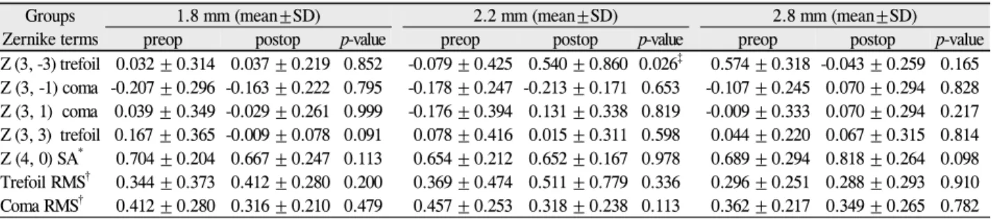
Z (3, -3) trefoil 0.032 ± 0.314 0.037 ± 0.219 0.852 -0.079 ± 0.425 0.540 ± 0.860 0.026‡ 0.574 ± 0.318 -0.043 ± 0.259 0.165 Z (3, -1) coma -0.207 ± 0.296 -0.163 ± 0.222 0.795 -0.178 ± 0.247 -0.213 ± 0.171 0.653 -0.107 ± 0.245 0.070 ± 0.294 0.828 Z (3, 1) coma 0.039 ± 0.349 -0.029 ± 0.261 0.999 -0.176 ± 0.394 0.131 ± 0.338 0.819 -0.009 ± 0.333 0.070 ± 0.294 0.217 Z (3, 3) trefoil 0.167 ± 0.365 -0.009 ± 0.078 0.091 0.078 ± 0.416 0.015 ± 0.311 0.598 0.044 ± 0.220 0.067 ± 0.315 0.814 Z (4, 0) SA* 0.704 ± 0.204 0.667 ± 0.247 0.113 0.654 ± 0.212 0.652 ± 0.167 0.978 0.689 ± 0.294 0.818 ± 0.264 0.098 Trefoil RMS† 0.344 ± 0.373 0.412 ± 0.280 0.200 0.369 ± 0.474 0.511 ± 0.779 0.336 0.296 ± 0.251 0.288 ± 0.293 0.910 Coma RMS† 0.412 ± 0.280 0.316 ± 0.210 0.479 0.457 ± 0.253 0.318 ± 0.238 0.113 0.362 ± 0.217 0.349 ± 0.265 0.782
*Spherical aberration; †root mean square; ‡p-value<0.05.
Figure 1. Inter-group comparison of surgically induced astig-
matism (SIA) at 1 and 3 months after cataract surgery. SIA is statistically different among 1.8-mm, 2.2-mm and 2.8-mm in- cision group at 1 and 3 months respectively.Figure 2. Mean changes (μm) in high order aberrations and root
mean square of anterior corneal surface betweenbaseline and 1 month after operation. Only Z (3, -3) is different in statistical comparison among 3 groups. *p<0.05; †root mean square.2.8 mm 절개에서 가장 컸다. 또한 술 후 3개월째 수술 유 발 난시도 3 그룹 간 통계적인 차이가 있었다(Fig. 1). 그러 나 모든 그룹 내에서 1개월과 3개월 간에 유발 난시의 크기 는 통계적인 차이가 없었다.
각막 전면의 수차는 개별 그룹의 수술 전후 비교에서 차 이가 없었다(Table 3). 수차 변화량의 그룹 간 비교에서 술 전과 술 후 1개월째 비교에서 코마수차, 구면수차는 차이가 없었다. Z (3, -3) 트레포일수차는 2.2 mm 절개에서 증가 하였으나 세 그룹 간 트레포일 RMS는 유의한 차이가 없었 다(Fig. 2).
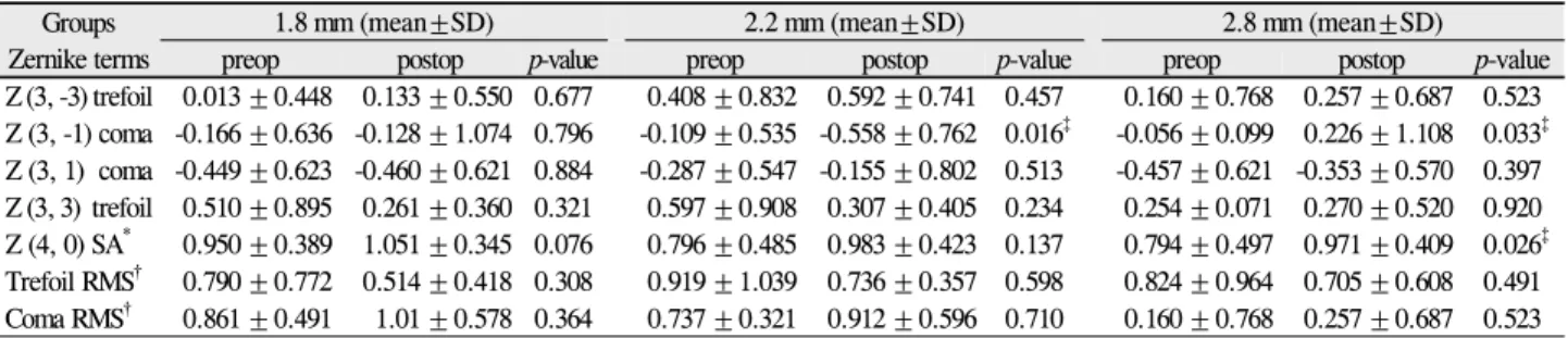
술 전후 그룹 내 비교에서 각막 후면의 고위수차 대부분 은 통계적으로 유의한 변화가 일어나지 않았다(Table 4).
그러나 수술 전후로 2.2 mm 절개의 Z (3, -1) 코마수차는
감소하였고, 2.8 mm 절개의 Z (3, -1) 코마수차와 Z (4, 0) 구면수차는 증가하였다. 각막 후면 수차의 변화량은 2.2 mm 절개의 Z (3, -1) 코마수차에서 유의하게 감소하였지 만, 코마 RMS는 차이가 없었다(Fig. 3).
고 찰
백내장수술은 흡인과 관류를 위한 각막 절개를 달리하는 이축 백내장수술과 초음파 첨단부의 관류와 흡인장치를 분 리시키지 않은 동축 백내장수술로 나눌 수 있고, 2.0 mm 전후 크기를 기준으로 하여 미세절개 백내장수술과 소절개 백내장수술로 분류한다.18 이축 미세절개 백내장수술은 절 개창의 크기가 작아 최대교정시력에 도달하는 시간이 빠르
Table 4. Comparison of high-order aberrations and root mean square of posterior corneal surface between baseline and 1 month after
phacoemulsificationGroups Zernike terms
1.8 mm (mean±SD) 2.2 mm (mean±SD) 2.8 mm (mean±SD)
preop postop p-value preop postop p-value preop postop p-value
Z (3, -3) trefoil 0.013 ±0.448 0.133 ±0.550 0.677 0.408 ±0.832 0.592 ±0.741 0.457 0.160 ± 0.768 0.257 ±0.687 0.523 Z (3, -1) coma -0.166 ±0.636 -0.128 ±1.074 0.796 -0.109 ±0.535 -0.558 ±0.762 0.016‡ -0.056 ± 0.099 0.226 ±1.108 0.033‡ Z (3, 1) coma -0.449 ±0.623 -0.460 ±0.621 0.884 -0.287 ±0.547 -0.155 ±0.802 0.513 -0.457 ± 0.621 -0.353 ±0.570 0.397 Z (3, 3) trefoil 0.510 ±0.895 0.261 ±0.360 0.321 0.597 ±0.908 0.307 ±0.405 0.234 0.254 ± 0.071 0.270 ±0.520 0.920 Z (4, 0) SA* 0.950 ±0.389 1.051 ±0.345 0.076 0.796 ±0.485 0.983 ±0.423 0.137 0.794 ± 0.497 0.971 ±0.409 0.026‡ Trefoil RMS† 0.790 ±0.772 0.514 ±0.418 0.308 0.919 ±1.039 0.736 ±0.357 0.598 0.824 ± 0.964 0.705 ±0.608 0.491 Coma RMS† 0.861 ±0.491 1.01 ±0.578 0.364 0.737 ±0.321 0.912 ±0.596 0.710 0.160 ± 0.768 0.257 ±0.687 0.523 Paired t-test is used between preoperative data and postoperative data at 1 month within group respectively.
*Spherical aberration; †root mean square; ‡p-value<0.05.
Figure 3. Mean changes (μm) in high order aberrations and root
mean square of posterior corneal surface betweenbaseline and 1 month after operation. Only Z (3, -1) is different in statistical comparison among 3 groups. *p<0.05; †root mean square.며, 효율 초음파 사용 시간이 짧고 유발되는 난시와 고위수 차가 작다는 장점이 있다.6,8,9그러나 절개창에 더 많은 스 트레스로 창상의 구조가 변화하여 창상 누출의 위험성이 증가되고, 일반적인 크기의 인공수정체를 삽입할 때 절개창 크기를 확장시켜야 하며 각막내피세포의 소실이 많은 단점 이 있지만 무엇보다 전통적 백내장수술에 익숙한 수술의에 게는 다소 익숙하지 않는 기술로 학습 곡선이 가파르
다.19-22이번 반해 동축 미세절개 백내장 수술은 작은 절개
를 통해 얻는 이축 백내장 수술의 장점을 가지면서 인공수 정체 삽입을 위한 절개창의 확장 과정을 생략할 수 있다.5,22 백내장수술에서 난시의 변화는 술 후 시력의 중요한 요 소이다. 백내장 수술에서 난시에 영향을 주는 인자로는 절 개의 크기, 위치, 방향과 모양, 봉합사 장력의 크기 등이 있 으며, 축의 이동은 절개의 크기, 위치, 창상 회복과 연관되 며, 난시 크기 변화는 절개의 크기, 절개의 위치와 모양에 따라 다르다.23-25그리고 절개창 크기에 따른 Phaco probe, 흡입과 관류(irrigation and aspiration) probe, 인공수정체 삽입을 위한 카트리지에 의해 절개창에 가해지는 스트레스 에 의한 차이가 난시에 영향을 미칠 수 있다. Phaco probe
는 세 그룹 모두 직경 0.9 mm를 사용하였고, 흡입과 관류 probe 역시 세 그룹 동일하게 직경 0.3 mm를 사용하였다.
인공수정체 삽입은 1.8 mm 군에서는 MI60 (Bausch &
Lomb) 전용 주사기를 사용하였고, 2.2 mm 군에서 카트리 지 D 시스템에 인공수정체를 올려놓고 Monarch III 주사기 에 장착한 후 절개창을 터널로 이용하여 인공수정체를 삽 입하였다. 2.8 mm 군에서 인공수정체는 일회용 PI-28 (Bausch & Lomb) 주사기를 절개창에 넣고 삽입하였다. 본 연구에서는 직난시가 있는 눈들에서 같은 모양, 방향과 위 치에 절개 창의 크기만을 차이를 두고 백내장수술을 하였 으므로 단순히 난시 크기의 변화보다 수술 유발 난시의 비 교가 술 후 난시의 절대 크기 변화를 더욱 잘 반영한다. 벡 터 방식으로 분석한 본 연구의 수술 유발 난시는 2.8 mm 이하의 절개에서 비례적으로 증가하여 절개 크기가 비교적 작은 절개의 백내장 수술에서도 각막 크기에 따라 난시는 영향을 받음을 보여준다. 그러나 본 연구에서 유발 난시와 나안시력을 반영하지 못하였는데, 2.8 mm 절개의 나안시 력이 1.8 mm 절개와 유사하게 매우 우수하였고, 90% 이상 에서 1디옵터 이내의 적은 난시를 가진 눈에서 유발된 난시 이므로 직접적으로 나안 시력에 미치는 영향은 미미한 것 으로 사료된다. 하지만 각막 절개가 작을수록 유발되는 난 시가 적어 1.8 mm 절개가 각막의 저위수차인 난시에서 더 우수한 성적을 보였다.
수차는 렌즈 등의 광학계에서 상을 맺을 때 원거리에 오 는 평행 광선은 광학계를 지난 다음 한 점에 맺혀야 하지만, 한 점에 모이지 않아 빛 번짐이 있거나 일그러져 보이는 현 상이다. 전체 안구수차는 각막수차와 수정체 등의 안구 내 수차에 따라 결정되고, 각막수차에 맞춰 안구 내 수차가 보 상을 하여 전체 안구수차가 낮은 수준으로 유지된다.26,27전 체 수차에서 고위수차의 비중은 10-20%이지만, 일반적인 방법으로 고위수차가 효율적으로 교정되지 못하면 눈부심, 달무리, 별 모양의 광채, 다중 형상, 흐려 보임, 대비 감도 감소 등의 시기능의 질적 저하를 일으키고 많은 고위 수차
들 중에서 트레포일수차, 코마수차, 구면수차 등의 3차와 4 차 수차가 고위 수차의 대부분을 차지한다.28-30백내장수술 의 목표가 환자의 시력에 대한 높은 만족이라는 측면에서 수차의 최소화는 백내장수술에서 중요하게 고려해야 할 요 소이다.
백내장 수술에서 각막 절개 크기가 각막 전면의 수차에 영향을 미치는지에 대해 이견이 있다.7,12,18,31-33
백내장 수 술을 받은 눈과 정상 눈의 고위수차 비교에서 수술 눈의 수 차가 증가하며 특히, 코마수차, 트레포일수차가 변화가 보 고되고 있지만 항상 일치되는 결과는 아니다.32,33 1.5 mm 양손 백내장수술과 3.0 mm 소절개 백내장수술에서 Z (3, -3), Z (3, 3) 트레포일수차, Z (4, -4), Z (4, 4) 테트라 포일수차가 변화하지만 코마수차와 구면수차는 유의한 차 이가 없음을 Tong et al12이 발표하였다. 그러나 Yao et al34 은 1.6 mm 미세절개와 3.2 mm 절개 백내장수술 후 시기능 비교에서 구면수차, 코마수차, 트레포일수차는 변화가 없 고, MTF (modulation transfer function)에서 차이로 시기 능의 질에서 차이가 난다는 발표를 하였다. 국내 연구로는 2.2 mm 와 2.8 mm 절개창 비교에서, 1.8 mm와 2.8 mm의 절개창 크기에 따른 각막 전면 고위수차 비교에서, 절개창 크기에 따른 수차는 변화가 없는 일치된 결과가 발표되었
다.15,16동축 백내장수술인 본 연구에서는 1.8 mm, 2.8 mm
절개는 변화가 없었고, 2.2 mm 절개의 Z (3, -3) 트레포 일수차에서만 통계적 차이가 있었다. 특히 그룹간의 수차 변화량 비교에서 대부분의 수차가 0.1 μm 이하로 변화하 여, 각막 전면은 2.8 mm 이하 절개 백내장수술 이후 술 전 상태와 유사한 구조를 가지는 것으로 예상된다.
백내장수술 후 각막 후면의 수차에 대한 연구는 널리 알 려져 있지 않다. Wang et al13은 3.2 mm 절개 후 수정체유 화술에서 Pentacam (Oclus, Wetzlar, Germany)을 이용한 각막 중심 6 mm의 각막 전면과 후면의 수차에 대한 변화를 연구하였고, 술 후 3개월째 각막 전면에서는 0.013 μm으로 약간 증가하고, 후면에서는 0.17 μm으로 통계적으로 유의 하게 증가함을 발표하였는데, 후면의 전체 고위수차뿐만 아 니라 구면수차, 코마수차, 트레포일 수차를 포함하는 대부 분의 수차에서도 통계적으로 차이가 있었다. 본 연구에서 역시 각막전면보다 각막후면에서 고위수차의 변화가 더 많 았다(Fig. 2, 3). 각막 전면에 비해 후면의 변화가 많은 이 유는 절개창이 전면보다 후면에서 각막 중심에 더 근접하 기 때문으로 생각된다. 그러나 2.2 mm 절개의 Z (3, -1) 코마수차, 2.8 mm 절개의 Z (3, -1) 코마수차, Z (4, 0)의 구면수차에서만 유의하게 변화하였다. Wang et al13와 본 연구의 결과 차이는 prechopper를 이용한 핵 분할 후 최소 한의 초음파 에너지 사용으로 수술을 진행하여, 각막 후면
의 질적 변화가 본 연구에서 더 적게 일어났을 것으로 추정 된다.
본 연구는 대상눈을 무작위 분류로 백내장수술을 하였으 나 2.2 mm 절개 군에서 다른 군에 비해 다소 젊어, 환자 나 이면에서 분류가 이상적이지 못하다. 나이에 따른 각막의 탄성도가 달라 난시에 영향을 줄 수 가능성도 있다. 각막 후면의 변화량에서 2.2 mm 절개의 Z (3, -1) 코마수차만 술전과 비교할 때 유의하게 통계적 변화가 있어 나이에 의 한 수차의 변화인지, 그룹의 특성에 차이가 있어 수정체유 화술 과정에서 각막 후면의 수차에 영향을 미칠 수 있는 다 른 인자가 있는지에 대한 의문은 이 연구에서는 알 수가 없 었지만, 같은 Infiniti (Alcon)로 수정체를 유화한 2.2 mm, 2.8 mm 절개에서 2.2 mm 에서 2.8 mm 절개 보다 각막에 주어지는 스트레스가 더욱 커서 2.2 mm 군에서 후면 수차 변화가 일어난 것으로 추정한다.
결론적으로 1.8 mm, 2.2 mm, 2.8 mm 동축 백내장수술 에서 수술 유발 난시는 절개 크기에 따라 증가하며, 수술 1 개월과 3개월간에는 유발 난시는 변화가 없었다. 그리고 고 위수차의 변화는 각막 후면에서 전면보다 변화가 많았지만 대부분의 수차들의 유의한 차이가 없어 2.8 mm 이하 절개 의 백내장수술은 트포레일수차, 코마수차, 구면수차에는 영 향을 미치지 않는 것으로 생각된다.
본 연구는 무작위 분류, 전향적 방법으로 이루어졌지만 대상 경과 관찰 기간이 짧은 한계가 있어 각막수차에 변화 에 대한 지속적인 연구가 필요할 것으로 보인다.
참고문헌
1) Shepherd JR. Induced astigmatism in small incision cataract surgery. J Cataract Refract Surg 1989;15:85-8.
2) Steinert RF, Brint SF, White SM, Fine IH. Astigmatism after small incision cataract surgery. A prospective, randomized, multicenter comparison of 4- and 6.5-mm incisions. Ophthalmology 1991;98:
417-23; discussion 423-4.
3) Soscia W, Howard JG, Olson RJ. Microphacoemulsification with WhiteStar. A wound-temperature study. J Cataract Refract Surg 2002;28:1044-6.
4) Donnenfeld ED, Olson RJ, Solomon R, et al. Efficacy and wound- temperature gradient of whitestar phacoemulsification through a 1.2 mm incision. J Cataract Refract Surg 2003;29:1097-100.
5) Dosso AA, Cottet L, Burgener ND, Di Nardo S. Outcomes of co- axial microincision cataract surgery versus conventional coaxial cataract surgery. J Cataract Refract Surg 2008;34:284-8.
6) Kurz S, Krummenauer F, Gabriel P, et al. Biaxial microincision versus coaxial small-incision clear cornea cataract surgery.
Ophthalmology 2006;113:1818-26.
7) Elkady B, Alió JL, Ortiz D, Montalbán R. Corneal aberrations after microincision cataract surgery. J Cataract Refract Surg 2008;
34:40-5.
8) Denoyer A, Denoyer L, Marotte D, et al. Intraindividual com- parative study of corneal and ocular wavefront aberrations after biaxial microincision versus coaxial small-incision cataract surgery. Br J Ophthalmol 2008;92:1679-84.
9) Lee AG, Greenlee E, Oetting TA, et al. Assessing cataract surgical competency. Ophthalmology 2007;114:1415-6.
10) Osher RH. Microcoaxial phacoemulsification Part 2: clinical study. J Cataract Refract Surg 2007;33:408-12.
11) Osher RH, Injev VP. Microcoaxial phacoemulsification Part 1: lab- oratory studies. J Cataract Refract Surg 2007;33:401-7.
12) Tong N, He JC, Lu F, et al. Changes in corneal wavefront aberra- tions in microincision and small-incision cataract surgery. J Cataract Refract Surg 2008;34:2085-90.
13) Wang J, Tang X, Zhang S, Li LH. Changes in high order aberra- tions of anterior and posterior surfaces of cornea before and after phacoemulsification. Zhonghua Yan Ke Za Zhi 2008;44:1066-71.
14) Lee KM, Kwon HG, Joo CK. Microcoaxial cataract surgery out- comes: comparison of 1.8 mm system and 2.2 mm system. J Cataract Refract Surg 2009;35:874-80.
15) Lee SY, Chung JL, Hong JP, et al. Comparative study of two asphe- ric, aberration-free intraocular lenses in cataract surgery. J Korean Ophthalmol Soc 2009;50:1520-6.
16) Hwang SJ, Choi SK, Oh SH, et al. Surgically induced astigmatism and corneal higher order aberrations in microcoaxial and conven- tional cataract surgery. J Korean Ophthalmol Soc 2008;49:1597- 602.
17) Holladay JT, Moran JR, Kezirian GM. Analysis of aggregate surgi- cally induced refractive change, prediction error, and intraocular astigmatism. J Cataract Refract Surg 2001;27:61-79.
18) Alió J, Rodríguez-Prats JL, Galal A, Ramzy M. Outcomes of mi- croincision cataract surgery versus coaxial phacoemulsification.
Ophthalmology 2005;112:1997-2003.
19) Linebarger EJ, Hardten DR, Shah GK, Lindstrom RL. Phacoe- mulsification and modern cataract surgery. Surv Ophthalmol 1999;
44:123-47.
20) Dick HB, Schwenn O, Krummenauer F, et al. Inflammation after sclerocorneal versus clear corneal tunnel phacoemulsification.
Ophthalmology 2000;107:241-7.
21) Lundström M. Endophthalmitis and incision construction. Curr
Opin Ophthalmol 2006;17:68-71.
22) Weikert MP. Update on bimanual microincisional cataract surgery.
Curr Opin Ophthalmol 2006;17:62-7.
23) Dam-Johansen M, Olsen T. Induced astigmatism after 4 and 6 mm scleral tunnel incision. A randomized study. Acta Ophthalmol Scand 1997;75:669-74.
24) Mendívil A. Frequency of induced astigmatism following phacoe- mulsification with suturing versus without suturing. Ophthalmic Surg Lasers 1997;28:377-81.
25) Lyhne N, Krogsager J, Corydon L, Kjeldgaard M. One year fol- low-up of astigmatism after 4.0 mm temporal clear corneal and su- perior scleral incisions. J Cataract Refract Surg 2000;26:83-7.
26) Artal P, Guirao A, Berrio E, Williams DR. Compensation of cor- neal aberrations by the internal optics in the human eye. J Vis 2001;
1:1-8.
27) Artal P, Guirao A. Contributions of the cornea and the lens to the aberrations of the human eye. Opt Lett 1998;23:1713-5.
28) Mester U, Dillinger P, Anterist N. Impact of a modified optic de- sign on visual function: clinical comparative study. J Cataract Refract Surg 2003;29:652-60.
29) Guirao A, Redondo M, Geraghty E, et al. Corneal optical aberra- tions and retinal image quality in patients in whom monofocal in- traocular lenses were implanted. Arch Ophthalmol 2002;120:
1143-51.
30) Castejón-Mochón JF, López-Gil N, Benito A, Artal P. Ocular wave-front aberration statistics in a normal young population.
Vision Res 2002;42:1611-7.
31) Yao K, Tang X, Ye P. Corneal astigmatism, high order aberrations, and optical quality after cataract surgery: microincision versus small incision. J Refract Surg 2006;22:S1079-82.
32) Guirao A, Tejedor J, Artal P. Corneal aberrations before and after small-incision cataract surgery. Invest Ophthalmol Vis Sci 2004;
45:4312-9.
33) Marcos S, Rosales P, Llorente L, Jiménez-Alfaro I. Change in cor- neal aberrations after cataract surgery with 2 types of aspherical in- traocular lenses. J Cataract Refract Surg 2007;33:217-26.
34) Yao K, Tang XJ, Huang XD, Ye PP. Clinical evaluation on the bi- manual microincision cataract surgery. Zhonghua Yan Ke Za Zhi 2008;44:525-8.
=ABSTRACT=
Induced Astigmatism and High-Order Aberrations after 1.8-mm, 2.2-mm and 3.0-mm Coaxial Phacoemulsification Incisions
Sang Jeong Moon, MD, Dong Jun Lee, MD, Kyung Hun Lee, MD
Sungmo Eye Hospital, Busan, Korea
Purpose: To study theeffect of micro incision (1.8 mm) and small incision (2.2 mm and 2.8 mm) coaxial phacoemulsifica- tion on surgically induced astigmatism (SIA) and high-order aberrations (HOA) of anterior and posterior corneal surface.
Methods: The present randomized clinical study included 32 eyes having a 1.8-mm, 38 eyes having a 2.2-mm, and 30 eyes having a 2.8-mm corneal incision. SIAs were measured at 1 and 3 months postoperatively. HOAs included coma, tre- foil, and spherical aberration. The coma–root mean square (RMS) and trefoil-RMS were evaluated at 1 month after the cat- aract operation.
Results: Surgically induced astigmatisms were 0.41 ± 0.30 diopter (D) in the 1.8-mm incision group, 0.47 ± 0.21 D in 2.2-mm group and 0.71 ± 0.50 D in the 2.8-mm group. The SIA of the 1.8-mm group was smaller than the other groups (p
= 0.002). There was no statistically significant difference in coma, spherical aberration of the corneal anterior surface and trefoil, or spherical aberration of the posterior surface among the 3 groups at 1 month after surgery.
Conclusions: Incision size contributes to postoperative corneal astigmatism. Phacoemulsification cataract surgery with less than 2.8-mm incision does not significantly influence the corneal aberrationsof anterior and posterior corneal surfaces.
J Korean Ophthalmol Soc 2011;52(4):407-413
Key Words: Coaxial microincisional cataract surgery, High-order aberration, Surgically induced astigmatism
Address reprint requests to Kyung Hun Lee, MD Sungmo Eye Hospital
#1078-7 Woo 2-dong Haeundae-gu, Busan, Korea
Tel: 82-51-743-0775, Fax: 82-51-743-0776, E-mail: [email protected]