591 대한안과학회지 2017년 제 58 권 제 5 호
J Korean Ophthalmol Soc 2017;58(5):591-594 ISSN 0378-6471 (Print)⋅ISSN 2092-9374 (Online)
https://doi.org/10.3341/jkos.2017.58.5.591
Case Report
전이 눈꺼풀 병변으로 진단된 원발유방암 1예
Eyelid Mass as Initial Presentation of Breast Cancer: A Case Report
이유림⋅양희정⋅백지선⋅양석우
Youlim Lee, MD, Hee jung Yang, MD, Ji-sun Paik, MD, PhD, Suk-Woo Yang, MD, PhD
가톨릭대학교 의과대학 서울성모병원 안과 및 시과학교실
Department of Ophthalmology and Visual Science, Seoul St. Mary’s Hospital, College of Medicine, The Catholic University of Korea, Seoul, Korea
Purpose: We report a case in which eyelid mass was the initial presentation of breast cancer. The diagnosis of breast cancer was made after lid biopsy.
Case summary: A 41-year-old female patient presented with a painful mass on the left lower lid after 1 month. There was a pink- ish mass in the lateral portion of the tarsal conjunctiva, and computed tomography revealed a mass with an irregular margin on the lower lid connected to the left lacrimal gland. Biopsy was performed at the tarsal conjunctiva of the left eye, and histopatho- logic examination was suggestive of malignant epithelial cell neoplasm, especially metastatic carcinoma in the breast. The pa- tient was diagnosed as having invasive ductal cancer, for which she is currently receiving chemotherapy.
Conclusions: Metastases to the eyelid are very rare, accounting for less than 1% of all malignant eyelid lesions. We report a pa- tient who presented with an eyelid mass as the initial presentation of breast cancer, which was diagnosed with metastatic breast cancer.
J Korean Ophthalmol Soc 2017;58(5):591-594
Keywords: Breast cancer, Eyelid mass, Metastatic cancer
■Received: 2016. 12. 22. ■ Revised: 2017. 3. 9.
■Accepted: 2017. 4. 19.
■Address reprint requests to Suk-Woo Yang, MD, PhD Department of Ophthalmology, The Catholic University of Korea Seoul St. Mary’s Hospital, #222 Banpo-daero, Seocho-gu, Seoul 06591, Korea
Tel: 82-2-3779-1848, Fax: 82-2-761-6869 E-mail: [email protected]
ⓒ2017 The Korean Ophthalmological Society
This is an Open Access article distributed under the terms of the Creative Commons Attribution Non-Commercial License (http://creativecommons.org/licenses/by-nc/3.0/) which permits unrestricted non-commercial use, distribution, and reproduction in any medium, provided the original work is properly cited.
유방암에서 안와 전이가 생기는 경우는 매우 드물며 한 연구에서는 유방암 환자의 0.2%에서 안와로의 전이가 생 기는 것으로 보고하였다.1 안와 전이의 가장 흔한 원인은 유방암이며 폐암, 전립선암, 소화기계암, 피부암 등도 안와 로 전이되는 경우가 있다.2
그중에서도 특히 전이암이 눈꺼풀에만 국한된 경우는 매 우 드물며, 눈꺼풀의 악성 병변의 1% 이하를 차지하는 것 으로 되어있고3 통증이나 시력저하를 동반하는 경우도 있
으나 종괴 자체에 의한 증상만을 나타내는 경우도 많아 다 래끼나 다른 안질환으로 오진이 내려지는 경우가 드물지 않다.1 저자들은 이전에 유방암을 진단받은 과거력이 없이 눈꺼풀의 종괴를 주소로 내원한 환자에서 조직검사로 원발 병소인 유방암을 진단한 사례가 있어 이를 보고하고자 한 다. 이는 눈꺼풀 종괴를 주소로 내원한 환자에서 조직 검사 를 시행하여 원발 유방암을 진단한 국내의 첫 증례이다.
증례보고
특별한 과거력이 없는 41세 여자 환자가 1달 전부터 발 생한 왼쪽 아래눈꺼풀의 종괴를 주소로 내원하였다. 이학 적 검사에서 경계가 불분명한 종괴가 왼쪽 아래눈꺼풀 부 위에서 촉지되었고 주변 피부, 근육층 모두 뻣뻣하고 단단 한 양상이었으며 압통 또한 동반되었다(Fig. 1). 초진 시 교 정시력은 우안 0.8, 좌안 0.63이며 안압은 우안 12 mmHg,
592
- 대한안과학회지 2017년 제 58 권 제 5 호 -
A B
Figure 1. Anterior segment photographs of the mass at initial presentation. (A) An irregular palpable mass (black arrow) is seen on
the lateral side of the left lower eyelid. (B) Atypical upper and lower lid swelling is seen on left eye.A B
Figure 2. Non-enhance Orbit computed tomography, showing Ill-defined, soft tissue density lesion in left lower eyelid, connected
to left lacrimal gland. (A) Coronal view. (B) Axial view.A B
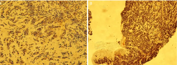
Figure 3. Overview dissection of biopsy of eyelid mass. Epithelioid cell proliferation and positive staining of (A) GCDFP-15 and (B)
GATA-3 was seen, suggestive of breast origin metastatic carcinoma (A: GDCFP-15, ×40) (B: GATA-3, ×10).좌안 35 mmHg로 좌안에 도르졸라미드-티몰롤 복합제제를 점안 중인 상태였다. 전안부 검사에서 좌안의 가쪽 아래 눈 꺼풀판결막에 경계가 불분명한 분홍색의 덩어리가 관찰되
었으며 안와 컴퓨터단층촬영(computed tomography, CT)에 서 눈물샘과 이어지는 좌안 아래눈꺼풀에 경계가 불분명한 종괴 소견을 보였다(Fig. 2). 정확한 진단 및 치료를 위해
593 - 이유림 외 : 눈꺼풀 병변으로 진단된 원발유방암 -
국소 마취하에 좌안 눈꺼풀판결막에서 결막 덩어리의 절제 생검을 시행하였다. 조직검사에서 악성 상피세포 화생소견 및 유방암의 면역조직화학표지자로 이용되는 GCDFP-15 및 GATA-3 양성 소견을 보여 유방 기원의 전이암종일 가 능성이 높다는 병리 조직 결과가 나왔다(Fig. 3). 이후 유방 외과에 협진 진료를 시행하여 유방에 단단한 덩어리를 발 견하였고 유방종괴에 대한 생검과 전신검사를 시행하였으 며 다발성 간과 골전이를 동반한 양측 유방의 침윤성 관상 피암을 진단받고 현재 항암치료를 시행하는 중이다.
고 찰
눈꺼풀에는 다양한 종양이 생길 수 있고 그중 악성은 1/3 정도를 차지한다.4,5 눈꺼풀에서 가장 흔한 악성 종양은 바 닥세포암으로 전체 눈꺼풀 악성 종양의 56.2% 가량을 차지 하며 편평상피암은 23.3% 가량을 차지한다. 악성흑색종은 1% 이하이며 전이암의 경우 눈꺼풀 악성 종양의 1% 이하 로 매우 적은 수를 차지한다.6,7
다른 장기에 비해 안와는 전이암이 생기는 경우가 매우 드문 것으로 알려져 있으나 눈에 생기는 전이암 중 가장 흔 한 원인은 유방암으로 알려져 있다. 또한 안와에 전이암종 이 생긴 환자는 그 전에 다른 장기로의 전이가 먼저 발병하 는 경우가 더 흔하며8 이것은 악성 종양의 말기에 주로 생 기고 보통 나쁜 예후의 지표로 생각된다.9
눈꺼풀에 생긴 전이암은 비특이적 증상을 나타내는 경우 가 많고 무통성의 피부 종괴에서 염증의 유무와 관계 없이 전반적인 눈꺼풀 부종만을 나타내는 경우도 있다.3 위, 아 래눈꺼풀이 침범되는 비율은 같은 것으로 되어있고 눈꺼풀 의 겉면과 결막 부분 모두를 침범할 수 있다.10
Ferry and Font11의 연구에서 유방암 환자의 9%에서 전 이로 인한 안구의 증상을 보이는 것으로 보고되었으며,호 발부위는 안와, 맥락막이 가장 흔한 것으로 보고되었고 눈 꺼풀, 섬모체 등에서도 역시 호발할 수 있다고 한다.2 증상 은 침범 부위에 따라 안구함몰, 안구 운동의 제한, 시력저 하 등으로 나타날 수 있고 눈꺼풀로 유방암의 전이가 생긴 경우에는 발적을 동반한 경결, 결절의 증상이 흔한 것으로 보고되었다.4
눈꺼풀에 전이된 유방암의 경우 전이암 중에서도 말기인 경우가 많아 전신으로의 전이를 고려해야 하고 치료는 전 신적 항암치료가 주가 되며, 일반적으로 고식적인 치료를 위주로 진행하게 된다. 치료의 목적은 주로 삶의 질을 높이 고 생존 기간을 길게 하는 것에 있으며 중앙 생존 기간은 18개월에서 24개월로 알려져 있다.12,13
국내에서 원발 유방암의 안와로의 전이에 의한 압박 시
신경병증, 안구돌출 및 맥락막 전이에 대한 증례 보고들이 있었으나 모두 유방암을 진단받고 치료받은 과거력이 있는 환자들이었다.14,15 본 증례는 눈꺼풀 종괴를 주소로 내원한 환자에서 조직 검사를 시행하여 원발성 유방암을 진단한 국내의 첫 증례이다.
저자들은 유방암 과거력이 없는 눈꺼풀 종괴를 주소로 내원한 환자에서 조직 검사를 통하여 원발성 유방암을 진 단한 국내에 보고된 바 없는 증례를 경험하였기에 보고하 고자 한다. 이는 특별한 병력이 없는 환자일지라도 눈꺼풀 종괴를 주소로 내원하였을 때 전이암의 가능성을 반드시 고려해야 하며 여성 환자의 경우 유방암에 대한 병력의 청 취와 문진이 필요함을 시사한다.
REFERENCES
1) Fonseca NL Jr, Lucci LM, Cha SB, et al. Metastatic eyelid disease associated with primary breast carcinoma: case report. Arq Bras Oftalmol 2009;72:390-3.
2) Goldberg RA, Rootman J. Clinical characteristics of metastatic or- bital tumors. Ophthalmology 1990;97:620-4.
3) Bianciotto C, Demirci H, Shields CL, et al. Metastatic tumors to the eyelid: report of 20 cases and review of the literature. Arch Ophthalmol 2009;127:999-1005.
4) Shields JA, Shields CL, Scartozzi R. Survey of 1264 patients with orbital tumors and simulating lesions: The 2002 Montgomery Lecture, part 1. Ophthalmology 2004;111:997-1008.
5) Tomizawa Y, Ocque R, Ohori NP. Orbital metastasis as the initial presentation of invasive lobular carcinoma of breast. Intern Med 2012;51:1635-8.
6) Cook BE Jr, Bartley GB. Epidemiologic characteristics and clinical course of patients with malignant eyelid tumors in an incidence co- hort in Olmsted County, Minnesota. Ophthalmology 1999;106:746-50.
7) Park HN, Jung SK, Cho WK, et al. Clinicopathological character- istics of malignant eyelid tumor in Korea. J Korean Ophthalmol Soc 2014;55:348-53.
8) Tamura M, Tada T, Tsuji H, et al. Clinical study on the metastasis to the eyes from breast cancer. Breast Cancer 2004;11:65-8.
9) Rubio FA, Pizarro A, Robana G, et al. Eyelid metastasis as the pre- senting sign of recurrent carcinoma of the breast. Br J Dermatol 1997;137:1026-7.
10) Mansour AM, Hidayat AA. Metastatic eyelid disease. Ophthalmology 1987;94:667-70.
11) Ferry AP, Font RL. Carcinoma metastatic to the eye and orbit. I. A clinicopathologic study of 227 cases. Arch Ophthalmol 1974;92:
276-86.
12) Kiely BE, Soon YY, Tattersall MH, Stockler MR. How long have I got? Estimating typical, best-case, and worst-case scenarios for pa- tients starting first-line chemotherapy for metastatic breast cancer:
a systematic review of recent randomized trials. J Clin Oncol 2011;29:456-63.
13) Zhang GJ, Adachi I, Yin DF, et al. Eyelid metastasis from breast cancer showing marked response to chemotherapy. Jpn J Clin Oncol 1995;25:10-5.
594
= 국문초록 =
전이 눈꺼풀 병변으로 진단된 원발유방암 1예
목적: 눈꺼풀의 종괴를 주소로 내원한 환자에서 조직검사를 시행하여 원발유방암을 진단한 사례를 경험하였기에 이를 보고하고자 한다.
증례요약: 41세 여자 환자가 한 달 전부터 발생한 왼쪽 아래눈꺼풀의 통증을 동반한 종괴를 주소로 내원하였다. 좌안 가쪽 아래 눈꺼풀 판결막에서도 분홍색의 덩어리가 발견되었으며 안와 컴퓨터 단층촬영에서 눈물샘과 이어지는 왼쪽 아래눈꺼풀에 경계가 불분명한 종괴 소견을 보였다. 좌안 눈꺼풀판결막에서 결막 덩어리의 생검을 시행하였으며 조직검사에서 악성 상피성 종양, 특히 유방 기원의 전이암일 가능성이 높다는 결과를 받았다. 유방외과에서 협진 진료를 시행하였으며 양측 유방의 침윤성 관상피암을 진단 받고 현재 항암치료를 시행하는 중이다.
결론: 눈꺼풀의 전이암은 매우 드물고 전체 악성 눈꺼풀 병변의 1% 이내를 차지한다. 눈꺼풀의 종괴를 주소로 내원한 환자에서 원발암 을 진단한 사례를 경험하여 이에 대하여 보고하고자 한다.
<대한안과학회지 2017;58(5):591-594>
- 대한안과학회지 2017년 제 58 권 제 5 호 -
14) Jung JW, Jin HC, Kim KS, Kim YC. A case of compressive optic neuropathy due to breast cancer metastasis. J Korean Ophthalmol Soc 2010;51:1161-5.
15) Kim HJ, Kim YH. Two cases of metastatic breast cancer to the choroid. J Korean Ophthalmol Soc 1990;31:984-7.