E
형 간염 바이러스에 의한 제 1형 인터페론 신호전달분자 활성 억제
명진종*
전북대학교인수공통전염병연구소
Received: September 19, 2018 / Revised: September 28, 2018 / Accepted: October 8, 2018
서 론
E형간염바이러스(Hepatitis E virus, HEV)는 7200가량
의 염기로 구성된 단일 양성 가닥 RNA 바이러스이며,
Hepeviridae 과에속한다[1, 2]. HEV 유전체는총 7개의비 구조단백질과 2개의구조단백질을 encoding하고있다. 비 구조단백질은개방형해독틀 1 (open reading frame 1)에서 만들어지며 이중에는 methyltransferase (MeT), Y-domain, papain-like cysteine protease (PCP 혹은 PLP), hypervariable region (HVR), X-domain, RNA helicase, RNA-dependent RNA polymerase (RdRp) 등이포함된다. 구조단백질은두 종류가있으며, ORF2 및 ORF3에서만들어진다. HEV는현 재까지 7개의유전자형(genotypes, GT)이보고되어있고, 이 중에 4 개의유전자형이사람에게감염할수있는것으로알 려져 있다[3]. GT1과 GT2는사람에게만감염하며, GT3와 GT4는사람과돼지, 사슴및맷돼지등동물에게도감염하 므로인수공통전염병원체로분류된다[4].
바이러스가감염하면 숙주세포는방어를위하여다양한 신호전달물질을분비하게되는데, 이중에서가장먼저발 현되는것이제 1형인터페론(type I interferon)이다[5−8].
세포질로침입한 바이러스의 RNA 유전체를인지하는것 은 retinoic acid-inducible gene I (RIG-I), melanoma differentiation-associated protein 5 (MDA5) 및 Toll-like receptors (TLRs)가알려져있다. RIG-I는비교적짧고끝이 뭉툭한(blunt) RNA를인지하는데, 이 RNA의끝에세개의
인산이 결합되어 있어야 인지가 가능하다[9, 10]. 반면에
MDA5는이런화학적조성의제한이없고비교적긴 RNA
를인지하는것으로알려져있다. RIG-I와 MDA5가침입한
바이러스의 RNA를인지하면활성화되고, 활성화된 RIG- I와 MDA5는미토콘드리아표면에서 mitochondrial antiviral- signaling protein (MAVS)를활성화한다[11, 12]. 활성화된 MAVS는 다시 하위의 I kappa B kinase epsilon (IKKε)/
TANK-binding kinase 1 (TBK1)을활성화한다. 활성화된이 들단백질은세포질내에비활성상태로존재하는 interferon regulatory factor 3 (IRF3)를인산화를유도한다[13−15].
인산화된 IRF3는이합체(dimer)를형성하고핵안으로이
동하여제 1형인터페론의발현을활성화한다. 발현된인 터페론은발현한세포및이웃세포에서다양한인터페론 유도단백질(interferon-stimulated genes, ISGs)의발현을 Hepatitis E Virus Inhibits Activation of Signaling Molecules Involved in Induction of Type I Interferon
Jinjong Myoung*
Korea Zoonosis Research Institute, Chonbuk National University, Iksan 54531, Republic of Korea
Hepatitis E virus (HEV) infection accounts for 20 million annual infections worldwide. HEV can be fatal in approximately 20−30% of pregnant women. HEV infections are normally self-limiting and mostly asymp- tomatic. However, in patients with insufficient immunity, such as acquired immunodeficiency syndrome patients, chronic and often fatal infections may ensue. Therefore, it is likely that host immune responses, especially interferon responses, play a critical role in HEV infection control. Here, we report that an HEV- encoded non-structural protein down-regulates type I interferon response. In addition, some other immune genes involved in the induction of type I interferon may be regulated as well. Detailed molecular mecha- nisms are currently being studied.
Keywords: Hepatitis E virus, type I interferon, protease
*Corresponding author
Tel: +82-63-900-4055, Fax: +82-63-900-4012 E-mail: Jinjong.myoung@jbnu.ac.kr
© 2018, The Korean Society for Microbiology and Biotechnology
유도함으로써바이러스의증식을억제한다. 따라서병원성 바이러스들은이과정중의하나혹은둘이상을저해함으로
써생존을꾀한다[4]. 예를들어 C형간염바이러스가발현하
는단백질중에서비구조단백질 NS3/4A와 5A는간접적혹 은직접적방식으로제 1형인터페론의발현을억제하는것 으로알려져있다[16−23].
반면에 HEV가인터페론을저해하는기작은알려진것이
거의없다[3, 4]. 면역능이충분한숙주에감염한경우 HEV
는급성감염을일으키며대부분의경우증상이없다. 그러 나고령환자나임산부혹은다른이유로면역능이약화된환
자에감염하는경우에는만성감염을유도할수있고[24, 25],
특히임산부에감염한경우사망률이 20−30%에이르는것 으로알려져있다. 따라서 HEV의증식을제어함에있어면 역계의역할이큰것으로추정해볼수있다. 본연구진은이 전연구에서 HEV가감염한세포에서제 1형인터페론의프 로모터활성이크게감소함을보였다. 이것은 HEV의구조 혹은비구조단백질중하나혹은그이상의단백질이제 1 형인터페론의발현을저해함을의미한다[3, 26, 27]. 흥미롭 게도 methyltransferase가 RIG-I에의한 인터페론발현을 억제하는것을증명하였다. 이연구에서 PCP도 RIG-I 활성 화에의한인터페론발현유도도억제되는것으로보인다.
이것은이전의다른연구자의연구에서 PCP에의해 RIG-I
의 탈유비퀴틴화(de-ubiquitination)가 저해 되고 따라서 RIG-I의활성화가저해되는결과와일치한다[28−30]. 또한 본연구진은후속연구에서 PCP에의하여 MDA5 의존성제 1형인터페론의발현이억제됨을보였다(논문투고중). 따
라서 PCP가제 1형인터페론발현활성화에중요한 MDA5
와 RIG-I를억제하는것으로보인다. 그러나 MDA5와 RIG-
I 활성화다음단계인 MAVS의활성화는분명히확인되지
않았다(논문투고중). 또한바이러스감염을모방(mimic)하 는 제 1형 인터페론 유도체인 polyinosinic-polycytidylic acid (polyI:C) 처리에의한세포내신호전달을 PCP가억제 할수있는지도불분명하다.
따라서본연구진은 HEV 단백질에의하여 poly(I:C) 의존 성제 1형인터페론활성화억제여부를분석하였다. 흥미롭 게도 PCP가 poly(I:C)에의하여활성화되는제 1형인터페 론발현을유의미하게억제함을관찰하였다. 또한 RIG-I와
poly(I:C)를함께처리한경우에도 PCP는농도의존성저해
작용을보였다. 뿐만아니라 MAVS와 poly(I:C)가함께처리
된경우에도 PCP 농도의존성저해가관찰되는것으로보
아, PCP가세포내다양한신호전달물질을효과적으로저
해하는것으로추정된다. 이런연구는향후 HEV에효과적 인항바이러스제제개발에기초적인정보를제공할것으로 기대된다.
재료 및 방법
세포 배양 및 시약
Human embryonic kidney 293 T (HEK293T) 세포는모 세포인 HEK293 세포에 Simian virus 40 (SV40)의 large T 항원을안정적으로발현하는세포이며, SV40 프로모터를포 함하는플라스미드의 세포 내증폭을 유도하여형질주입 (transfection)의 효율을 높여 준다. HEK293T 세포는 American Type Culture Collection (ATCC)에서구입하였 다. HEK293T 세포는 Dulbecco’s Modified Eagle Medium (DMEM)에 10% FBS (Gibco, USA)와 1% 페니실린/스트렙
토마이신 (Gibco, USA)를첨가하여만든배양배지에서유지
하였다[4, 31−33]. 세포는 5% CO2가공급되는환경에서 37℃ 에서배양하였다[16, 34−36]. Poly(I:C)는고분자 RNA(high molecular weight poly(I:C))를 invivogen에서 구입하여사 용하였으며, transfection reagent-poly(I:C) 복합체로구성되 어있어, 별도의처리없이세포배양액에직접처리하였다. Anti-Flag 항체는 Sigma-Aldrich (USA)에서구입하였고, 그 외의 항체(anti-mouse IgG-HRP, anti-GAPDH IgG, HA- ubiquitin)들은 Cell Signaling (USA)에서구입하였다.
플라스미드 제작 및 형질주입(transfection)
MDA5, MAVS 및 RIG-I를발현하는플라스미드는한림대
학교황순봉교수로부터제공받았다. HEV의각유전자를발 현하는플라스미드와 IFN-β-luciferase 및β-galactosidase 발현플라스미드들은이전연구에서사용한것이고제작방 법은다른논문에기술되었다[3]. 형질전환을위하여사용된 시약은 polyethylenimme (PEI)이며, Sigma-Aldrich에서구 입하여사용하였다. 형질전환을위한 DNA:PEI 복합체생성 은상온에서 30 min 진행하였으며, 이때 DNA:PEI 비율은 1:2였다[4, 37, 38]. HEK293T 세포는형질주입 24 h 전에 6-well plate에 8 × 10⁵/well의농도로분주하였다. 형질주입
전에확인한세포의밀도는약 70% 였으며모든실험에서
동일한조건에서수행하였다. 생성된 DNA:PEI 복합체는 2 ml의배양배지가들어있는 6-well plate에한방울씩천 천히주입해주었다. 형질주입을수행한후 24 h 지난후형
질주입의효율은 60% 이상이었다.
단백질 발현 분석
단백질의발현은 sodium dodecyl sulfate-polyacrilamide gel electrophoresis (SDS-PAGE) 분석법과 Western blotting 법을이용하여분석하였다[39, 40]. 세포용해(lysis)는 cell lysis buffer를이용하였다. 원심분리로상층액을취한후, 단 백질의총량을 PierceTm BSA Protein Assay Kit (Thermo
Scientific, USA)을이용하여정량하였다. 동일양의단백질 을 10% SDS-PAGE에서분리하여 nitrocellulose membrane에 전달하여 Western blotting을시행하였다. Anti-Flag 항체는 1:5000, anti-mouse IgG 항체는 1:2000, anti-GAPDH 항체 는 1:1000으로각각희석하여사용하였다.
루시퍼라아제 활성분석
HEK293T 세포는 6-well plate에 8 × 10⁵/well의 농도로 분주하였다. 분주후 24 h이지난후그림에서표시된바와 같이다양한 DNA를함께형질주입하였다. 형질주입의효율
을측정하여 50% 이상일때루시퍼라아제활성분석을실시
하였다. 형질주입후 24 h 후에배양액을제거하고, 세포분 쇄 완충용액(reporter lysis 5X buffer, Promega, USA)를 넣고세포를수거하였다. 세포막등은 4℃, 13,000 ×g에서
5 min 원심분리하여제거하고상층액을이용하여루시퍼라
아제의활성을분석하였다. 분석을위해서는 luciferase assay system (Promega, USA)를사용하였다. 발광(luminescence) 값은 Glomax (Promega, USA)를이용하여분석하였고, 각
각의발광값은β-galactosidase 활성도를측정하여보정하
였다. 통계분석
실험군과 대조군의 평균은 정규분포 가정에 의하여
Student’s t-test를이용하여통계적유의성을분석하였다.
실험은최소한두번반복하였고, 대표적인데이터를제시 하였다.
결과 및 고찰
HEV 단백질의 발현 및 RIG-I 활성 저해 단백질 발굴
Flag tag이붙어있는 HEV 단백질을형질주입으로발현
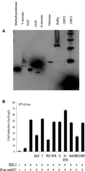
하고 24 h 이후에발현양을 SDS-PAGE-Western blotting으 로분석하였다(Fig. 1A). HVR 및 ORF3가가장높은양으로 발현되었고, MeT, PCP, helicase 및 RdRp가비교적높은정 도로발현되었다. 흥미로운것은 ORF2는발현양이낮았으 나제 1형인터페론의발현을대조군대비 50% 정도억제하
였다(Fig. 1B). RIG-I만발현하였을때는처리하지않은대
조군에비하여 IFN-β발현유도정도는 5−6배에그쳤지만
(Fig. 1B, 두번째열), poly(I:C) 500 ng/ml을처리하였을때 는 50배이상 증가하였다. 따라서 RIG-I 유전자의발현과
poly(I:C) 처리는서로상승작용을일으키는것으로사료된
다. 그러나 Methyltransferase, PLP (혹은 PCP) 및 ORF2 를함께발현하였을때는 IFN-β의발현유도가현저하게감 소하였으며, 이중 PCP의효과가가장높은것으로보인다 (~60% 저해; Fig. 1B). 따라서, PCP는이전연구에서본연
구팀이밝힌것처럼 RIG-I만처리한경우를저해할뿐만아
니라[3], RIG-I와 poly(I:C)가상승작용을일으킨경우에도효
과적으로저해할수있는것으로사료된다. 반면에 HVR, X,
X + HVR, ORF3 등은 RIG-I에의한제 1형인터페론반응 에영향을미치지않았다.
Fig. 1. HEV proteins inhibits RIG-I-mediated induction of type I interferon. (A) Plasmids, encoding each HEV gene, were transfected into HEK293T cells. At 24 h post-transfection, protein expression was evaluated by SDS-PAGE analysis followed by Western blotting. (B) HEK293T cells were co-transfected with IFN- β-luc, β-galactosidase, and RIG-I-expressing plasmids with sub- sequent treatment with 500 ng/ml poly(I:C) for 24 h. Luciferase activity of each sample was evaluated and plotted.
PCP에 의한 RIG-I의 농도의존성 저해
RIG-I와 poly(I:C)에의한 IFN-β의발현유도를 PCP가농 도의존적으로억제하는지분석하기위하여 0, 0.5, 1, 2 μg 의 PCP 발현플라스미드를처리하였을때(Fig. 2), PCP의발 현양에따라제 1형인터페론발현이점진적으로억제되는 것을관찰하였다. 이것은 PCP가 RIG-I에의한세포내신호 전달을효과적/특이적으로억제한다는것을시사한다. 이전
연구에서 PCP가 RIG-I의탈유비퀴틴화를저해하여 RIG-I
의활성화를억제하는것으로알려져있다[28−30]. 향후연
구에서 PCP에의한탈유비퀴틴화의범위와대상을밝힌다 면 PCP의작용기전을보다효과적으로이해할수있을것 이라고사료된다.
PCP에 의한 MAVS의 농도의존성 저해
서론에서밝힌바와같이, 활성화된 RIG-I는미토콘드리
아의외막에존재하는 MAVS를활성화여응집(aggregation) 을유도함으로써하위신호전달을활성화한다[11, 12]. 따라
서 PCP에의하여 RIG-I가억제된다면하위신호전달물질
인 MAVS에의한신호전달도억제될가능성이높다. 이것
을확인하기위하여 MAVS를발현하는 DNA를세포에전달
하고, 다양한농도의 PCP를함께형질주입하였다(0, 0.5, 1, 2, 3 μg; Fig. 3). PCP를낮은농도로발현하였을때유의적 인감소를확인할수없었으나, 농도가높아짐에따라유의 미한억제를확인할수있었다(Fig. 3). 흥미롭게도 MAVS 단 백질의양은 PCP가높은정도로발현될때에도감소하지않 았다(Fig. 3; low panels). 이것은 PCP에의한 MAVS의억 제가단백질생성이후과정에서일어난다는것을시사한다
고사료된다. PCP에의하여 MAVS의기능이억제되는과정
이 RIG-I의예에서보듯이탈유비퀴틴저해에의한것인지
를밝히는것은향후 PCP의구체적인분자기작을밝히는 데도움을줄것으로사료된다.
요 약
E형간염바이러스는전세계적으로매년 2천만건의감염 을일으키는것으로알려져있다. 흥미로운것은임산부에서 특히높은치사율을보이는데, 무려 20−30%의사망률을보 인다는점이다. 면역능이있는감염환자에서는 E형간염바 이러스감염은별다른증상없이자연치유되지만, 면역능이 낮은에이즈환자나고령환자에서는만성감염을일으킬수도 있으며사망에이르게할수도있다. 따라서, E형간염바이 러스의증식억제에면역반응의중요성이높으며, 특히인터 페론반응이중요한역할을할것으로사료된다. 본연구팀 은 E형간염바이러스의프로티아제가 RIG-I 의존적제 1형 인터페론반응을유의미하게억제하는것을관찰하였다. 또 한바이러스프로티아제가 RIG-I의하위신혼전달분자의
하나인 MAVS의활성도농도의존적으로억제하는것으로
분석하였다. E형간염바이러스에의한제 1형인터페론반 응억제의구체적인작용기작을밝힌다면효과적인항바이 Fig. 2. HEV PCP inhibits RIG-I-mediated activation of type I
interferon in a dose-responsive manner. HEK293T cells were co-transfected with IFN-β-luc, β-galactosidase, and RIG-I- expressing plasmids as well as with an increasing amount of PCP- expressing construct (0, 0.5, 1, 2 μg). At 6 h post-transfection, poly(I:C) was treated at 500 ng/ml for 18 h. Cells were harvested
for luciferase reporter assay. Fig. 3. HEV PCP inhibits MAVS-mediated activation of type I interferon in a dose-responsive manner. HEK293T cells were co-transfected with IFN-β-luc, β-galactosidase, and RIG-I- expressing plasmids as well as with an increasing amount of MAVS-expressing construct (0, 0.5, 1, 2, and 3 μg). At 24 h post- transfection, cells were subjected to luciferase reporter assay (top panels). In the same samples, expression of MAVS, PCP and GAPDH was determined by SDS-PAGE analysis followed by West- ern blotting (bottom panels).
러스제제의개발의초석을놓을수있을것으로기대된다.
Acknowledgments
This research was supported by Basic Science Research Program through the National Research Foundation (NRF) funded by the Min- istry of Education (2017R1A6A1A03015876).
Conflict of Interest
The authors have no financial conflicts of interest to declare.
References
1. Forni D, Cagliani R, Clerici M, Sironi M. 2018. Origin and disper- sal of Hepatitis E virus. Emerg. Microbes. Infect. 7: 11.
2. Tam AW, Smith MM, Guerra ME, Huang CC, Bradley DW, Fry KE, et al. 1991. Hepatitis E virus (HEV): molecular cloning and sequencing of the full-length viral genome. Virology 185: 120- 131.
3. Kang S, Choi C, Choi I, Han KN, Roh SW, Choi J, et al. 2018. Hep- atitis E Virus Methyltransferase Inhibits Type I Interferon Induc- tion by Targeting RIG-I. J. Microbiol. Biotechnol. 28: 1554-1562.
4. Kang S, Myoung J. 2017. Host Innate Immunity against Hepati- tis E Virus and Viral Evasion Mechanisms. J. Microbiol. Biotech- nol. 27: 1727-1735.
5. Kim N, Now H, Nguyen NTH, Yoo JY. 2016. Multilayered regula- tions of RIG-I in the anti-viral signaling pathway. J. Microbiol.
54: 583-587.
6. Schmidt ME, Varga SM. 2017. Modulation of the host immune response by respiratory syncytial virus proteins. J. Microbiol.
55: 161-171.
7. Theofilopoulos AN, Baccala R, Beutler B, Kono DH. 2005. Type I interferons (α/β) in immunity and autoimmunity. Annu. Rev.
Immunol. 23: 307-336.
8. Zitvogel L, Galluzzi L, Kepp O, Smyth MJ, Kroemer G. 2015.
Type I interferons in anticancer immunity. Nat. Rev. Immunol.
15: 405-414.
9. Loo YM, Fornek J, Crochet N, Bajwa G, Perwitasari O, Martinez- Sobrido L, et al. 2008. Distinct RIG-I and MDA5 signaling by RNA viruses in innate immunity. J. Virol. 82: 335-345.
10. Takeuchi OandAkira S. 2010. Pattern recognition receptors and inflammation. Cell 140: 805-820.
11. Kawai T, Takahashi K, Sato S, Coban C, Kumar H, Kato H, et al.
2005. IPS-1, an adaptor triggering RIG-I- and Mda5-mediated type I interferon induction. Nat. Immunol. 6: 981-988.
12. Seth RB, Sun L, Ea CK, Chen ZJ. 2005. Identification and charac- terization of MAVS, a mitochondrial antiviral signaling protein that activates NF-κB and IRF 3. Cell. 122: 669-682.
13. Grandvaux N, Servant MJ, tenOever B, Sen GC, Balachandran S, Barber GN, et al. 2002. Transcriptional profiling of interferon regulatory factor 3 target genes: direct involvement in the reg-
ulation of interferon-stimulated genes. J. Virol. 76: 5532-5539.
14. Honda K, Takaoka A, Taniguchi T. 2006. Type I interferon [cor- rected] gene induction by the interferon regulatory factor family of transcription factors. Immunity 25: 349-360.
15. Liu S, Cai X, Wu J, Cong Q, Chen X, Li T, et al. 2015. Phosphoryla- tion of innate immune adaptor proteins MAVS, STING, and TRIF induces IRF3 activation. Science 347: aaa2630.
16. Wi J, Jeong MS, Hong HJ. 2017. Construction and characteriza- tion of an Anti-Hepatitis B Virus preS1 humanized antibody that binds to the essential receptor binding site. J. Microbiol.
Biotechnol. 27: 1336-1344.
17. Bode JG, Ludwig S, Ehrhardt C, Albrecht U, Erhardt A, Schaper F, et al. 2003. IFN-α antagonistic activity of HCV core protein involves induction of suppressor of cytokine signaling-3.
FASEB J. 17: 488-490.
18. Kang SM, Won SJ, Lee GH, Lim YS, Hwang SB. 2010. Modulation of interferon signaling by hepatitis C virus non-structural 5A protein: implication of genotypic difference in interferon treat- ment. FEBS Lett. 584: 4069-4076.
19. Lan KH, Lan KL, Lee WP, Sheu ML, Chen MY, Lee YL, et al. 2007.
HCV NS5A inhibits interferon-alpha signaling through sup- pression of STAT1 phosphorylation in hepatocyte-derived cell lines. J. Hepatol. 46: 759-767.
20. Li K, Foy E, Ferreon JC, Nakamura M, Ferreon AC, Ikeda M, et al.
2005. Immune evasion by hepatitis C virus NS3/4A protease- mediated cleavage of the Toll-like receptor 3 adaptor protein TRIF. Proc. Natl. Acad. Sci. USA 102: 2992-2997.
21. Li XD, Sun L, Seth RB, Pineda G, Chen ZJ. 2005. Hepatitis C virus protease NS3/4A cleaves mitochondrial antiviral signaling pro- tein off the mitochondria to evade innate immunity. Proc. Natl.
Acad.Sci. USA 102: 17717-17722.
22. Lin W, Kim SS, Yeung E, Kamegaya Y, Blackard JT, Kim KA, et al.
2006. Hepatitis C virus core protein blocks interferon signaling by interaction with the STAT1 SH2 domain. J. Virol. 80: 9226- 9235.
23. Taylor DR, Shi ST, Romano PR, Barber GN, Lai MM. 1999. Inhibi- tion of the interferon-inducible protein kinase PKR by HCV E2 protein. Science 285: 107-110.
24. Jilani N, Das BC, Husain SA, Baweja UK, Chattopadhya D, Gupta RK, et al. 2007. Hepatitis E virus infection and fulminant hepatic failure during pregnancy. J. Gastroenterol. Hepatol. 22: 676-682.
25. Navaneethan U, Al Mohajer M, Shata MT. 2008. Hepatitis E and pregnancy: understanding the pathogenesis. Liver Int. 28:
1190-1199.
26. Krain LJ, Nelson KE, Labrique AB. 2014. Host immune status and response to hepatitis E virus infection. Clin. Microbiol. Rev.
27: 139-165.
27. Zhou X, Xu L, Wang W, Watashi K, Wang Y, Sprengers D, et al.
2016. Disparity of basal and therapeutically activated inter- feron signalling in constraining hepatitis E virus infection.
J. Viral Hepat. 23: 294-304.
28. Karpe YA, Lole KS. 2011. Deubiquitination activity associated with hepatitis E virus putative papain-like cysteine protease.
J. Gen. Virol. 92: 2088-2092.
29. Nan Y, Yu Y, Ma Z, Khattar SK, Fredericksen B, Zhang YJ. 2014.
Hepatitis E virus inhibits type I interferon induction by ORF1 products. J. Virol. 88: 11924-11932.
30. Oshiumi H, Miyashita M, Matsumoto M, Seya T. 2013. A distinct role of Riplet-mediated K63-Linked polyubiquitination of the RIG-I repressor domain in human antiviral innate immune responses. PLoS Pathog. 9: e1003533.
31. Choi S, Park H, Minelko M, Kim EK, Cho MR, Nam JH. 2017.
Recombinant adeno-associated virus expressing truncated IK cytokine diminishes the symptoms of inflammatory arthritis.
J. Microbiol. Biotechnol. 27: 1892-1895.
32. Hamid FB, Kim J, Shin CG. 2017. Characterization of prototype foamy virus infectivity in transportin 3 knockdown human 293t cell line. J. Microbiol. Biotechnol. 27: 380-387.
33. Kim MJ, Lee SY, Kim HJ, Lee JS, Joo IS, Kwak HS, et al. 2016.
Development of a one-step duplex RT-PCR method for the simultaneous detection of VP3/VP1 and VP1/P2B regions of the Hepatitis A Virus. J. Microbiol. Biotechnol. 26: 1398-1403.
34. Lee JM, Cho JB, Ahn HC, Jung W, Jeong YJ. 2017. A novel chemical compound for inhibition of SARS coronavirus helicase.
J. Microbiol. Biotechnol. 27: 2070-2073.
35. Lee JM, Kim J, Ryu I, Woo HM, Lee TG, Jung W, et al. 2017. An
aptamer-based electrochemical sensor that can distinguish influenza virus subtype H1 from H5. J Microbiol Biotechnol. 27:
2037-2043.
36. Lim S, Cha S, Jang JH, Yang D, Choe J, Seo T. 2016. Alterations in acetylation of histone H4 Lysine 8 and trimethylation of lysine 20 associated with lytic gene promoters during Kaposi's sar- coma-associated herpesvirus reactivation. J. Microbiol. Biotech- nol. 27: 189-196.
37. Elkholy YS, Hegab AS, Ismail DK, Hassan RM. 2016. Evaluation of a novel commercial quaternary ammonium compound for eradication of mycobacteria, HCV and HBV in egypt. J. Microbiol.
54: 39-43.
38. Jeong H, Seong BL. 2017. Exploiting virus-like particles as inno- vative vaccines against emerging viral infections. J. Microbiol.
55: 220-230.
39. Kim JH, Lee CH, Lee SW. 2016. Hepatitis C virus infection stimu- lates transforming growth factor-β1 expression through up- regulating miR-192. J. Microbiol. 54: 520-526.
40. Shin JS, Ku KB, Jang Y, Yoon YS, Shin D, Kwon OS, et al. 2017.
Comparison of anti-influenza virus activity and pharmacokinetics of oseltamivir free base and oseltamivir phosphate. J. Microbiol.
55: 979-983.