진주종성 중이염 환자의 진주종에서 iNOS 존재
충북대학교 의과대학 이비인후과학교실
유인선·추무진·신시옥·최영석·염창섭
Existence of iNOS in the Human Cholesteatoma
In-Seon Yoo, MD, Moo-Jin Choo, MD, See-Ok Shin, MD, Young-Seok Choi, MD and Chang-Seop Yum, MD
Department of Otolaryngology, College of Medicine, Chungbuk National University, Cheongju, Korea
- -
- - ABSTRACT - - - -
Background and Objectives:Bone resorption and hyperkeratosis are the most characteristic findings of chronic otitis media with cholesteatoma. Overexpression of IL-1 in cholesteatoma was reported having a significant role in the pathogenesis of cholesteatoma. Also, overproduction of nitric oxide (NO) stimulated the production of the cyclooxygenase and PGE2. Therefore, NO and inducible nitric oxide synthase (iNOS) may be suggested having a role of the pathogenesis of cholesteatoma. This study was designed to identify the existence of iNOS in human cholesteatoma. Materials and Methods:Ten fresh human cholesteatoma tis- sues and one postauricular skin were used. Dot and Western blot analysis were used for detection of iNOS.
Results:Dot blot showed different degrees of response. The bands of iNOS about 137 kDa area existed in the Western blot analysis. Conclusion:These findings suggest that iNOS may present in human cholesteat- oma and NO may have play a role in pathogenesis of chronic otitis media with cholesteatoma. ((((J Clinical Otolaryngol 1999;10:190-194))))
KEY WORDS:Cholesteatoma·Nitric oxide·Nitric oxide synthase.
서 론
진주종의 생성기전에 대하여 여러 가지 기전이 알려 졌지만 그 기전에 관계없이 골흡수와 상피의 과증식은 진주종의 가장 특징적인 소견인데, 활성화된 파골세포 와 그 수적증가가 진주종성중이염에서의 골흡수기전에 서 중요한 역할을 한다.1) 많은 수의 파골세포활성인자
가 알려져 있는데, interleukin-1(IL-1)과 prosta- glandin E2(PGE2)는 그중 가장 강력한 활성인자들이 다.2) 여러 저자들이 IL-1의 발현이 진주종에서 증가되 는 결과를 보고하면서 IL-1이 진주종의 발생기전에 중 요한 역할을 할 것이라고 주장하였다.3)4) IL-1β는 ni- tric oxide (NO)의 생성과 함께 cyclooxygenase(COX) 의 생성을 자극하여 PGE2의 생성을 증가시킨다. 또한 NOS inhibitor가 직접적으로 COX를 억제하지는 않지 만, NOS inhibitor에 의한 NO 생성의 억제는 prost- aglandins의 생성을 억제시키는 것으로 알려졌다.5) 이 는 내인성으로 생성된 NO가 COX경로를 조절하며, NOS 와 COX가 모두 유발된 환경에서는 직접적으로 NO에 의한 pro-inflammatory prostaglan-dins가 생성되어 논문접수일:1999년 8월 1일
심사완료일:1999년 10월 6일
교신저자:유인선, 361-763 충북 청주시 흥덕구 개신동 산48 충북대학교 의과대학 이비인후과학교실
전화:(0431) 269-6157・팩스:(0431) 265-6157 E-mail:mjchoo@med.chungbuk.ac.kr
결과적으로 강화된 염증반응을 일으키는 것으로 생각되 어진다.5)6)
이상의 여러 소견들을 종합하여 보면 NO가 진주종의 생성기전에 관여할 가능성이 높으므로 dot blot과 We- stern blot analysis를 이용하여 사람의 진주종에서 iNOS 의 존재를 확인하여 NO와 진주종성중이염과의 관련성 을 밝히고자 한다.
대상과 방법
1997년 10월부터 충북대학교병원 이비인후과에서 진주종성 중이염으로 수술을 시행받은 10례의 환자에 서 수술 중에 제거한 진주종 조직에서 진주종 상피조 직를 채취하였다. 1례에서는 약간의 후이개피부를 얻 어 이전에 채취한 진주종 상피조직과 함께 -70℃에서 급랭 보관하였다. Dot blot을 위해서는 4례의 진주종 상피조직과 1례의 후이개피부를 준비하였다. 각각의 조직을 사기사발에서 액화질소(liquid nitrogen)를 첨 가하여 동결상태에서 부순 후에 분해완충액(lysis bu- ffer)에서 30분 보관한 다음 원심분리하여 상층액을 채 취하여 Coomassie Blue방법을 이용하여 단백질정량 을 하였다. Western blot analysis를 위해서는 6례의 진주종조직을 준비하였다. 먼저 homogenizer로 조직 을 처리한 다음에 ultrasonic homogenizer(Sonic- ator, B.Braun 2000, USA)로 균일화시킨 후에 분해 완충액에서 30분 동안 반응시켰다. 그 후에 12,000 rpm 에서 30분 동안 원심분리하였다. 상층액을 채취하여 단 백질정량을 하였다.
Dot blot
검체부하는 단백질정량 결과에 따라 각각 50 ㎍의 단 백질을 포함하도록 하였으며 air dry 후에 5% milk로 차단하였다. 일차항체로 1;1,000으로 희석한 단클론성 mouse anti-iNOS(Transduction Lab. Lexington, KY, USA)를 상온에서 1시간 반응시켰다. 그 후 이차항체 인 peroxydase labeled anti-mouse IgG(Amersham Life Science, Buckinghamshir, England)를 상온에서 1시간 처리하고 ECL kit(Amersham Life Science, Buckinghamshir, England)를 이용하여 반응결과를 관
찰하였다. 음성 대조군은 postauricular skin, albumin 을 사용하였고 양성 대조군은 INF-γ와 LPS로 자극된 대식세포로부터의 lysate(Transduction Lab. Lexin- gton, KY)를 사용하였다.
SDS-PAGE & Western blot analysis
원심분리 후에 각각의 조직에서 단백질정량 결과에 따 라 100 μg의 단백질을 포함하는 양의 상층액을 취하 여 4×SDS gel-loading buffer(200 mM Tris-HCl, 400mM dithiothreitol, 8% SDS, 0.4% bromophenol blue, 40% glycerol)를 첨가하여 100℃에서 5분간 끓 였다. 5% stacking gel과 8% resolving gel을 사용하 여 SDA-PAGE를 30 mA에서 3시간 동안 시행하였다.
전기영동된 gel을 transfer buffer에서 nitrocellulose paper(Biorad, Hercules, CA, USA)로 100V에서 2시 간 동안 이동시켰다. 그 후 0.1% Tween 20과 3% mi- lk가 포함된 TBS로 1시간 동안 차단하였고, 1;1,000 으로 희석시킨 단클론성 mouse anti-iNOS(Transd- uction Lab. Lexington, KY, USA)로 상온에서 1시간 동안 처리를 하였다. 그 후 5분씩 3번 세척한 후에 1;
1,000으로 희석시킨 이차항체(peroxydase labeled anti-mouse IgG)로 1시간 처리하였다. 그 다음 10분 씩 3회 세척을 하였으며 ECL kit(Amersham Life Sc- ience, Buckinghamshir, England)로 2분 동안 반응시 킨 다음 X-ray film에 10분 동안 노출시킨 후에 관찰 하였다.
결 과
Dot blot analysis
단백질정량 결과 각각의 조직에서 0.5~1.9 μg/μl의 단백질 함량을 보였다(Table 1). 각 조직을 단백질정량 결과에 따라 50 μg의 단백질을 포함하도록 loading한 후에, 1:1,000으로 처리한 일차항체와 1:2,000으로 Table 1. Protein amount of each specimen in the dot bl- ot analysis
Sample number 1 2 3 4 5 6
Amount(μg/μl) 0.5 1.5 1.9 0.7 0.5 0.5 1-4;cholesteatoma tissues, 5-6;same human speci- men of postauricular skin
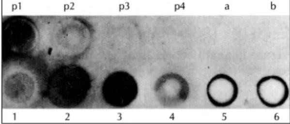
희석한 이차항체를 처리한 dot blot에서 2, 3번 진주종 조직은 양성 대조군으로 사용한 mouse macrophage lysate 100 μg의 반응에 비해 더 강한 양성반응을 보 였고, 4번 진주종 조직은 중간 양성반응을, 1번 진주종 조직은 약한 양성반응을 보였다. 후이개피부조직은 변연 에서만 양성반응을 보였고, albumin과 saline에서는 반 응을 보이지 않았다(Fig. 1).
Western blot analysis
단백질정량 결과 각각의 조직에서 2~8 μg/μl의 단 백질 함량을 보였다(Table 2). 각 조직을 단백질정량 결과에 따라 각각 100 μg의 단백질을 포함하도록 lo- ading한 후에, 1;1,000으로 희석한 일차항체와 이차항 체를 처리한 Western blot 결과 6례의 진주종 모두에 서 130 kDa 부근에서 band를 관찰할 수 있었다(Fig. 2).
5례에서는 정확히 인지되는 band를 관찰할 수 있었고
1번 한 례에서는 약한 band를 보였다.
고 찰
Nitric oxide(NO)는 인체의 여러 기관에서 다양한 세 포들이 기능을 수행함에 있어서 중요한 세포매개체(cell mediator)이다.7) NO는 nitric oxide synthase(NOS) 에 의해 L-arginine으로부터 생성되며 부산물로 L-ci- trulline을 만든다.8) 여러 가지 NOS의 isoform이 알려 졌는데 neural NOS(nNOS)는 신경조직에서 증명되었 으며 신경전달에 관여하는 것으로 보고되었다.9) 다른 isoform인 endothelial NOS(eNOS)는 혈관 내피에서 발견되었으며 혈관 평활근의 조절에 관여하는 것으로 보 여진다.10) 또 다른 isoform은 inducible NOS(iNOS) 로 대식세포와 호흡기상피 등의 다양한 조직에서 발견 되었으며 면역자극과 세포독성 등을 포함한 여러 가지 세포기능에 관여하는 것으로 알려졌다.11)12) 임상적으로 는 건선(psoriasis) 환자의 피부에서 활성화된 각질세포 (keratinocyte)가 iNOS의 발현을 증가시킨다는 보고 가 있으며,13) 두경부 편평상피암종에서 NOS 활성이 증 가되어 있다고 하였다.14)
NO의 골흡수 기전에 관한 여러 실험보고가 있다. NO 가 파골세포의 활성의 조절에 관여한다는 사실이 보고되 었는데, in vitro실험에서 NO는 직접적으로 파골세포에 작용하여 골흡수를 감소시키는데 이는 NO에 의한 파골 세포의 형태를 변화와 관련이 있으며 cGMP나 세포내 칼슘의 변화에는 의존하지 않는다고 하였다.15) 조골세 포(osteoblast)는 단백질분해효소, cytokines 등의 다 양한 요소를 분비함으로서 파골세포의 골흡수를 조절 한다. 동물실험에서 osteoblast-like cell line은 IFN- γ과 LPS의 자극 하에서 NO를 생성하는데, IL-1α는 NO의 생성을 유발시키며, TGF-β2는 이를 억제시킨 다고 하였다.16) 그리고 NO 생성의 경쟁적 억제물질인 NG-monomethyl-arginine(L-NMMA)의 주입에 의 해서 이러한 NO의 골흡수 억제효과가 방지된다고 하 였다.16)
그리고 NO와 세포고사(apoptosis)와의 연관성이 제 기되어 왔는데 과량으로 생성된 NO는 DNA에 손상을 주거나, 조직에서 p53을 축적시켜 세포고사를 유발시키 Table 2. Protein amount of each specimen in Western
blot analysis
Sample number 1 2 3 4 5 6
Amount (μg/μl) 2 5 8 4 5 5 1-6;human cholesteatoma tissues
Fig. 1. Result of the dot blot analysis (p;positive contr- ols, p1;100 μg of mouse macrophage lysate (MML), p2;50 μg of MML, p3;10 μg of MML, p4;5μg of MML, a;saline, b;bovin serum albumin, 1-4;human chol- esteatoma tissues, 5, 6;postauricular skin).
Fig. 2. Result of Western blot analysis (1-6;human ch- olesteatoma tissues).
며,17) 소량으로 생성되는 NO는 caspase의 활성을 억 제하거나 heat shock protein 32(HSP 32), HSP 70 의 유도 등을 통하여 세포고사를 억제할 수 있다.18) 진 주종에서 상피세포의 과증식을 관찰할 수 있는데 이는 NO가 상피세포의 고사를 억제하는데 관여하여 발생한 결과일 수 있다.
현재까지 진주종 상피조직에서 iNOS의 존재를 보고 한 경우는 없었다. 저자들은 이번 연구를 시작하면서 진 주종 상피조직이 어느 정도의 단백질을 함유하고 있는 지 가름할 수가 없었다. 또한 iNOS는 진주종 상피조직 의 단백질 중에서 상대적으로 소량이 존재할 것으로 추 측되었다. Dot blot은 비교적 적은 양의 단백질의 존재 를 확인할 수 있는 실험방법이므로 먼저 이 방법을 시행 하였다. Dot blot 결과 iNOS가 존재함을 확인할 수 있 었다. 반응의 결과는 약한 양성반응에서 강한 양성반응 까지 다양하게 나타났으며 후이개피부는 주로 변연에 서만 양성반응을 보였다. 변연에서만 반응를 보인 이유 는 모세관현상과 nitrocellulose paper가 눌리면서 변연 에서 많은 양의 단백질과 항체가 충분히 반응하여 이러 한 현상이 생겼을 것으로 생각된다. 조직에 liquid nit- rogen을 부어 사기사발에서 매우 잘게 부수고 갈아보려 고 하였지만 미세하게 분해할 수는 없었고 다음 단계로 옮기는 과정에서 상당량의 조직을 잃게 되었다. 따라서 이 방법은 소량의 단백질을 검사하는 방법으로는 적당 하지 않다고 판단되어 Western blot analysis에서는 조 직을 동결상태에서 메스를 사용하여 잘게 절개하고 조 직을 분해완충제에 담아 30분 보관한 후에 homoge- nizer을 사용하고 sonicator를 이용하여 더 미세하게 분 해하였다. 원심분리과정부터는 이전 방법과 동일하였다.
후자의 방법을 사용한 결과 이전의 방법보다 단백질정량 결과 더 많은 단백질을 얻을 수 있었다. 또한 dot blot 를 시행하면서 anti-iNOS의 농도를 결정할 수 있었다.
Western blot analysis에서는 dot blot를 시행하면서 얻은 결과를 토대로 하여 iNOS의 존재를 확인할 수 있 었다.
본 연구에서는 우선 dot blot 방법을 통하여 진주종 에서 iNOS를 발현을 확인할 수 있었고 그 정도가 조직 마다 다름을 알 수 있었다. Western blot 결과 137 kDa 지역에서 형성되는 band를 통하여 진주종의 상피조직
에서 iNOS의 존재를 확인할 수 있었다. 또한 진주종에 서 iNOS가 control보다 다량 발현됨을 확인할 수 있었 으며, 진주종의 발생기전에 있어 IL-1의 발현이 증가되 어 NO와 COX를 활성화시키면 이들에 의해 과도하게 발현된 PGE 2가 파골세포를 자극하여 심각한 골흡수를 유발시킨다고 추측된다.
결 론
사람에서 발생한 진주종의 발생기전에 있어 NO가 연 관될 것이라고 가설 하에 진주종 상피조직을 대상으로 iNOS에 대한 단클론성 항체를 사용한 dot blot에서 4 례 모두에서 양성반응을 보였으며, Western blot에서 6례 모두에서 130kDa 부근에서 band를 보여 진주종 의 상피조직에서 iNOS의 존재를 확인할 수 있었고, 진 주종의 생성기전에서 IL-1과 연관되어 NO가 관여할 것으로 사료된다.
중심 단어
:진주종・iNOS.REFERENCES
1) Chole RA. Cellular and subcellular events of bone resor- ption in human and experimental cholesteatoma: the role of osteoclasts. Laryngoscope 1984;94:76-95.
2) Garrett I, Mundy G. Relationship between interleukin-1 and prostaglandins in resorbing neonatal calvaria. J Bo- ne Mine Res 1989;4:789-94.
3) Ahn JM, Huang CC, Abramson M. Interleukin-1 causing bone destruction in middle ear cholesteatoma. Otolaryn- gol Head Neck Surg 1990;103:527-36.
4) Schilling V, Negri B, Bujia J. Possible role of Interleukin 1 alpha and Interleukin 1 beta in the pathogenesis of ch- olesteatoma of the middle ear. Am J Otol 1992;13:350-5.
5) Salvemini D, Seibert K, Masferrer JL, Settle SL, Misko TP, Currie MG, Needleman P. Nitric oxide and the cycl- ooxygenase pathway. Advances in prostaglandin, thromb- oxane, and leukotriene research. 1995;23:491-3.
6) Corbett JA, Kwon G, Turk J, McDaniel ML. IL-1β ind- uces the coexpression of both nitric oxide synthase and cyclooxygenase by islets of langerhans: activation of cyc- looxygenase by nitric oxide. Biochem 1993;32:13767-70.
7) Moncada S, Higgs EA. Endogenous nitric oxide: physio- logy, pathology, and clinical relevance. Eur J Clin Invest 1992;21:361-74.
8) Moncada S, Palmer RMJ, Higgs EA. Biosynthesis of nitric oxide from L-arginine: a pathway for the regulation of cell function and communication. Biochem Pharmacol 1989;
11:1709-15.
9) Hope BT, Michael GJ, Knigge KM, Vincent SR. Neur- onal NADPH diaphorase is a nitric oxide synthase. Proc Natl Acad Sci 1991;88:2811-4.
10) Palmer RMJ, Ferrige AG, Moncada S. Nitric oxide release accounts for the biologic activity of endothelium-derived relaxing factor. Nature 1987;327:524-6.
11) Adcock IM, Brown CR, Kwon O, Barnes PJ. Oxidative stress induces NFkB DNA binding and inducible NOS mRNA in human epithelial cells. Biochem Biophys Rese- arch Commun 1994;199:1518-24.
12) Lyons CR, Orloff GJ, Cunningham JM. Molecular cloning and functional expression of an inducible nitric oxide sy- nthase from a murine macrophage cell line. J Biol Chem 1992;267:6370-4.
13) Sirsjo A, Karlsson M, Gidlof A, Rollman O, Torma H.
Increased expression of inducible nitric oxide synthase in psoriatic skin and cytokine-stimulated keratinocytes. Br J Dermatol 1996;134:643-8.
14) Rosbe KW, Prazma J, Peter P, Mins W, Ball SS, Weissler MC. Immunohistochemical characterization of nitric ox-
ide synthase activity in squamous cell carcinoma of the head and neck. Otolaryngol Head Neck Surg 1995;113:
541-9.
15) MacIntyre I, Zaidi M, Towhidul Alam ASM, Datta HK, Moonga BS, Lidbury PS, Hecker M, and Vane JR. Osteo- clastic inhibition: an action of nitric oxide not mediated by cGMP. Proc Natl Acad Sci 1991;88:2936-40.
16) Lowik CW, Nibbering PH, Ruit M, Papapoulos SE. Indu- cible production of nitric oxide in osteoblast-like cells and in fetal mouse bone explants is associated with suppress- ion of osteoclastic bone resorption. J Clin Invest 1994;93:
1465-72.
17) Nguyen T, Brunson D, Crespi CL. DNA damage and mu- tation in human cells exposed to nitric oxide in vitro. Proc Natl Acad Sci 1992;89:3030-4.
18) Kim YM, de Vere ME, Watkins SC. Nitric oxide protects cultured rat hepatocytes from tumor necrosis factor-alpha- induced apoptosis by inducting heat shock protein 70 ex- pression. J Biol Chem 1997;272:1402-11.