Abstract
The purpose of this study was to evaluate the guided bone regeneration, using 1-ethyl-3-(3-dimethyl aminopropyl) carbodiimide (EDC), cross-linked type-I collagen membrane in bony defects around the installation site of the implant. In two case studies, presented buccal and palatal dehiscence defects were solved using EDC collagen membrane. Defects were measured clinically at the time of the 1st surgery and were compared with defects at the 2nd surgery. No clinical complication was observed. EDC collagen membrane improved the ability of space maintenance and showed biocompatibility and low cytotoxicity. In conclusion, using an implant with EDC collagen membrane has satisfactory results with regard to esthetic and functional aspects.
Key Words: case report, collagen membrane, dental implant, guided bone regeneration
1-ethyl-3-(3-dimethyl aminopropyl)
Carbodiimide로 가교화된 제1형 콜라겐 차폐막의
골유도재생 효과: 증례보고
이재홍, 유 훈, 김유경, 신현기, 임현창, 김영택, 이중석, 정의원, 최성호
연세대학교 치과대학 치주과학교실, 치주조직재생연구소
Guided Bone Regeneration Using Type-I Collagen Membrane Cross-Linked by
1-ethyl-3-(3-dimethyl aminopropyl) Carbodiimide in Two Implant Dehiscence
Cases
Jae-Hong Lee, Hoon You, You-Kyung Kim, Hyun-Ki Shin, Hyun-Chang Lim, Young-Taek Kim, Jung-Seok Lee, Ui-Won Jung, Seong-Ho Choi
Department of Periodontology, Research Institute for Periodontal Regeneration, Yonsei University College of Dentistry, Seoul, Korea ISSN 1229-5418 Implantology 2015; 19(1): 16~25
Copyright © 2015. The Korean Academy of Oral & Maxillofacial Implantology
This is an Open Access article distributed under the terms of the Creative Commons Attribution Non-Commercial License (http://creativecommons.org/licenses/by-nc/3.0/) which permits Reprint requests: Seong-Ho Choi
Department of Periodontology, Yonsei University College of Dentistry, 50, Yonsei-ro, Seodaemun-gu, Seoul 120-752, Korea
Tel: 82-2-2228-3189, Fax: 82-2-392-0398 E-mail: [email protected]
Received for publication: March 10, 2015 Accepted for publication: March 14, 2015
교신저자: 최성호 (120-752) 서울시 서대문구 연세로 50 연세대학교 치과대학 치주과학교실 Tel: 82-2-2228-3189, Fax: 82-2-392-0398 E-mail: [email protected] 원고접수일: 2015년 3월 10일 게재확정일: 2015년 3월 14일
하여 분해 속도를 늦춘 콜라겐 차폐막들이 임상에서 성
공적으로 사용되고 있다7,8. 이 중, EDC로 가교화된 콜라
겐 차폐막은 수용성 carbodiimide 계통으로, glutaralde-hyde나 polyepoxides와는 달리 linkage의 일부분으로 남 지 않고 세포 독성이 낮은 수용성 요소 유도체로 변화되 어 제거가 쉬우며 높은 생체친화성을 가지고 있다7,9. 또 한, 반응 효율성을 증가시키고 수화를 지연시키기 위하 여 N-hydroxysuccinimde를 추가하여 EDC로 가교화된 콜라겐 차폐막은 glutaraldehyde로 가교화한 차폐막과 유사하게 늦은 분해 속도를 갖는다9. 본 연구에 사용한 EDC로 가교화된 제1형 콜라겐 차폐막은 porcine 피부에 서 추출한 순도 99.83%의 아텔로콜라겐으로 제작되었으 며, 세포 차단 효과 및 효소 저항성이 높고 골 형성을 위 한 충분한 흡수 지연 시간을 가지고 있는 것으로 알려져 있다. 따라서, 협측 열개 결손을 가진 상악 전치부와 상 악동 골이식술이 동반된 구개측 열개 결손을 가진 구치 부에 EDC로 가교화된 콜라겐 차폐막을 적용한 단일치 임플란트의 증례를 통하여 본 차폐막의 임상적 유용성을 평가해보고자 한다.
II
증례보고
1. Case 1
23세 남자 환자가 외상으로 인해 상악 우측 중절치가 부러졌다는 주소로 본원에 내원하여 응급으로 치수절단 술 시행하였다(Fig. 1). 다음 내원 시 구개측 파절선이 치 은 연하로 깊게 존재하여 지대치로써 예후가 불량할 것 으로 판단되어 즉시 임플란트 식립을 계획하였으나, 발 치와 협측 결손이 발견되어 구개 상피를 이용한 발치와 보존술을 시행하였다(Fig. 2). 연조직의 치유가 이루어진 1달 후 골유도재생술을 동반한 조기 임플란트 식립을 결I
서론
조
직유도재생술은 긴 상피 접합과 같은 원하지 않는 조직의 이주를 막고, 상실되거나 소실된 치주조직 및 치조골의 재생을 주요한 목적으 로 한다1. 조직유도재생술에 사용되는 비흡수성 차폐막 과 흡수성 차폐막이 결손된 치주조직의 치유 기간 동안 골 열개, 골 천공, 수평 및 수직적인 형태의 골 결손에 임 상적으로 성공적으로 사용되어 왔다. 초기에는 expanded-polytetrafluoroethylene (e-PTFE) 와 같은 비흡수성 차폐막이 주로 사용되었는데, 세포의 이동을 막고 공간을 유지, 형성하는 데 이상적인 재료로 사용되어 왔다1. 그러나 차폐막을 제거하는 데 추가적인 술식이 필요하고, 치유 기간 동안 차폐막의 노출이 빈번 하였다. Trombelli 등2은 골 결손에서의 e-PTFE 차폐막 의 노출 빈도를 66%로 보고하였다. 이러한 의도하지 않 은 차폐막의 노출은 세균의 감염을 통한 염증 반응을 일 으키고 궁극적으로는 조직 재생의 실패를 가져올 수 있 다3. 이에 반해, 흡수성 콜라겐 차폐막은 이차 수술이 필요 하지 않으며, 많은 연구에서 비흡수성 차폐막에 뒤지지 않는 임상 결과를 보여주고 있다4,5. 그러나 흡수성 콜라 겐 차폐막은 분해되는 과정에서 주변 조직에 불필요한 염증 반응을 야기할 수 있으며, 낮은 강도로 인한 붕괴와 효소 및 세균에 의한 빠른 흡수가 단점으로 지적되고 있 다6. 따라서 중등도 이상의 넓은 결손에서는 차폐막의 붕 괴를 막기 위하여 결손부를 지지해 줄 수 있는 골이식재 를 사용하며, 다양한 방법(ultraviolet radiation, hexa-methylene diisocyanate, combined glutaraldehyde and irradiation, diphenylphosphoryl-azide, 1-ethyl-3-[3-dimethyl aminopropyl] carbodiimide [EDC])으로 가교화Case Report
Fig. 1.
Preoperative intraoral photographs. A maxillary right central incisor fractured by trauma. (A) Anterior view. (B) Occlusal view. (C) Periapical radiograph.Jae-Hong Lee et al. : Guided Bone Regeneration Using Type-I Collagen Membrane Cross-Linked by 1-ethyl-3-(3-dimethyl aminopropyl) Carbodiimide in Two Implant Dehiscence Cases. Implantology 2015
Fig. 2.
Photographs of a socket seal technique. (A) Buccal bone is not intact and immediate implantation is not possible. (B) Taking a soft tissue punch from the palatal donor site. (C) Socket closure with soft tissue graft.Jae-Hong Lee et al. : Guided Bone Regeneration Using Type-I Collagen Membrane Cross-Linked by 1-ethyl-3-(3-dimethyl aminopropyl) Carbodiimide in Two Implant Dehiscence Cases. Implantology 2015
Fig. 3.
Clinical situation after 4 weeks. (A) Biologically well integrated and clinically healthy gingiva in the area where the implant will later be placed. (B, C) Intraoperative view at the time of implant insertion.Jae-Hong Lee et al. : Guided Bone Regeneration Using Type-I Collagen Membrane Cross-Linked by 1-ethyl-3-(3-dimethyl aminopropyl) Carbodiimide in Two Implant Dehiscence Cases. Implantology 2015
A B C
A B C
Case Report
정하였다. 수직 및 수평 절개를 가한 후 전층 판막으로 박리하였으며, 6 mm의 협측 열개 골 결손을 관찰할 수 있었다(Fig. 3). Lekholm-Zarb 분류에 따른 골질과 골량 은 D3/B로 판단하였고 Regular CrossFit (diameter 4.1×12 mm; Straumann, Waldenburg, Switzerland) 매 식체를 20 N으로 식립하였다(Fig. 4). 자가골을 먼저 매 식체 위에 이식한 후 Osteon (Dentium, Seoul, Korea) 0.5 cc 및 Rapi-Gide (Dalim Tissen, Seoul, Korea)를 사 용하는 샌드위치 테크닉을 사용하여 골유도재생술을 시 행하였다10(Fig. 5). 봉합 후 1~2 mm 가량의 연조직 열개 및 차폐막의 노출이 관찰되었다(Fig. 6). 1주일 뒤 임시 치아를 인접 자연치를 이용하여 접착하였으며, 수술 2주 뒤 발사를 시행하였다. 3개월 뒤 이차 수술을 시행하였 으며, 부족한 협측 연조직을 보강하기 위하여 구개에서 채득한 상피하결합조직을 이식하였다(Fig. 7). 이차 수술 당시 perio-test value는 -2였으며, 술 후 4개월 뒤 보철 을 완료하였다. 수술 후 관찰기간 동안 특별한 합병증은 발생하지 않았으며, Nordland-Tarnow 분류에 따라 class I의 치간 유두의 소실이 관찰되지만 협측 연조직은 잘 유지되었으며, 보철물 주위 치은 형태화 교합은 양호 하였다(Fig. 8).
Fig. 4.
Intraoral photographs of implant placement. (A, B) Correct positioning of the implant. (C) After implant placement.Jae-Hong Lee et al. : Guided Bone Regeneration Using Type-I Collagen Membrane Cross-Linked by 1-ethyl-3-(3-dimethyl aminopropyl) Carbodiimide in Two Implant Dehiscence Cases. Implantology 2015
Fig. 5.
(A) After implant insertion in the correct position, autologous bone is placed into the buccal defect. (B) Filling the buccal gap between the alveolar wall and the implant with Osteon (Dentium) granules. (C) Bone graft materials are covered with a Rapi-Gide (Dalim Tissen) membrane.Jae-Hong Lee et al. : Guided Bone Regeneration Using Type-I Collagen Membrane Cross-Linked by 1-ethyl-3-(3-dimethyl aminopropyl) Carbodiimide in Two Implant Dehiscence Cases. Implantology 2015
A B C
Case Report
2. Case 2
46세 여자 환자가 상실된 상악 좌측 제1대구치 수복을 주소로 내원하였다. 발치는 2개월 전에 시행되었으며, 특이할 만한 전신적 병력은 없었다(Fig. 9). 스텐트를 이 용한 cone-beam computed tomography 촬영 영상을 바 탕으로 구개부의 2 mm 열개 및 4 mm의 높이가 부족할 것으로 판단하였으며, 오스테오톰을 이용한 상악동 거상 술을 계획하였다(Fig. 10). 수술 부위 전층 판막을 거상 후 제조자의 가이드라인에 따라 골 삭제를 진행하였으 며, 치조정 접근법을 통해 6 mm의 상악동 거상을 포함 한 골이식술을 시행 후 TS III (diameter 5.0×10 mm; Osstem, Seoul, Korea) 매식체를 식립하였다(Fig. 11). Lekholm-Zarb 분류에 따른 골질과 골량은 D2/C로 판단 하였고 최종 식립 토크는 30 N이었다. 상악동 골이식재 와 구개부 열개에 Bio-Oss (Geistlich Biomaterials, Bern, Switzerland)를 0.5 g 사용하였으며, 구개부 열개 에는 골이식 후 흡수성 콜라겐 차폐막 Rapi-Gide를 사용 하여 골유도재생술을 시행한 후 일차 봉합하였다(Fig. 12). 3개월 뒤 이차 수술을 시행하였으며, 임상적으로 확 인하였을 때 2 mm의 구개측 열개 결손은 회복되었고
Fig. 6.
(A, B) Flaps are sutured to allow soft and hard tissue healing and maturation. A small portion of the membrane was still exposed. (C) Postoperative periapical radiograph.Jae-Hong Lee et al. : Guided Bone Regeneration Using Type-I Collagen Membrane Cross-Linked by 1-ethyl-3-(3-dimethyl aminopropyl) Carbodiimide in Two Implant Dehiscence Cases. Implantology 2015
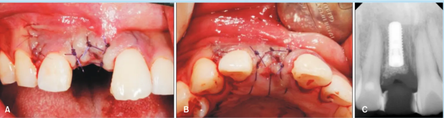
Fig. 7.
(A) Good healing of hard and soft tissue 4 months postoperatively. (B) Subepithelial connective tissue graft was harvested from the palatal donor site. (C) Insertion of a healing abutment and sutured.Jae-Hong Lee et al. : Guided Bone Regeneration Using Type-I Collagen Membrane Cross-Linked by 1-ethyl-3-(3-dimethyl aminopropyl) Carbodiimide in Two Implant Dehiscence Cases. Implantology 2015
A B C
Case Report
A B C
A B
Fig. 10.
Cone-beam computed tomography pre-implant assessment. (A) Axial view. (B) Sagittal view. (C) Coronal view.Jae-Hong Lee et al. : Guided Bone Regeneration Using Type-I Collagen Membrane Cross-Linked by 1-ethyl-3-(3-dimethyl aminopropyl) Carbodiimide in Two Implant Dehiscence Cases. Implantology 2015
Fig. 9.
Preoperative intraoral photo- graphs. (A) Anterior view. (B) Occlu-sal view.Jae-Hong Lee et al. : Guided Bone Regeneration Using Type-I Collagen Membrane Cross-Linked by 1-ethyl-3-(3-dimethyl aminopropyl) Carbodiimide in Two Implant Dehiscence Cases. Implantology 2015
Fig. 8.
Intraoral photographs 6 months after implant placement. (A) Anterior view. (B) Occlusal view.Jae-Hong Lee et al. : Guided Bone Regeneration Using Type-I Collagen Membrane Cross-Linked by 1-ethyl-3-(3-dimethyl aminopropyl) Carbodiimide in Two Implant Dehiscence Cases. Implantology 2015
Case Report perio-test value는 -5였다. 치근단 방사선 사진상 돔 형 태의 골이식부가 방사선 불투과상을 보였으며, 약 5 mm 의 높이를 유지하고 있는 것을 확인할 수 있었다. 1개월 뒤 보철을 진행하였으며, 특별한 합병증의 발생 없이 전 반적으로 양호한 치은의 형태 및 치조제 폭을 유지하였 다(Fig. 13). A B A B C
Fig. 11.
Intraoral photographs of implant placement. (A, B) Correct positioning of the implant.Jae-Hong Lee et al. : Guided Bone Regeneration Using Type-I Collagen Membrane Cross-Linked by 1-ethyl-3-(3-dimethyl aminopropyl) Carbodiimide in Two Implant Dehiscence Cases. Implantology 2015
Fig. 12.
(A) After implant insertion, filling the palatal defect between the alveolar wall and the implant with Bio-Oss (Geistlich Biomaterials) granules. (B) Bone graft materials are covered with a Rapi-Gide (Dalim Tissen) membrane. (C) Postoperative periapical radiograph.Jae-Hong Lee et al. : Guided Bone Regeneration Using Type-I Collagen Membrane Cross-Linked by 1-ethyl-3-(3-dimethyl aminopropyl) Carbodiimide in Two Implant Dehiscence Cases. Implantology 2015
Case Report
III
총괄 및 고찰
첫 번째 증례에서 6 mm의 협측 열개 결손 봉합 후 연 조직 열개 및 차폐막의 노출이 관찰되었으나 특이할 만 한 염증 반응 없이 빠른 치유 양상을 보였으며, 1주일 뒤 ovate pontic type의 임시 치아를 이용하여 자연스러운 치간 유두 형성을 유도할 수 있었다. 3개월 뒤, 이차 수 술 시행 시 차폐막은 모두 흡수되었으며 임플란트 협측 1 mm의 열개 결손만 남아있었다. 일차 수술 당시 연조직 열개 없는 일차 봉합을 이루었다면 더 늦은 차폐막의 흡 수와 골 결손부의 이상적인 회복을 기대할 수 있었을 것 으로 고려된다. 이차 수술을 시행하며 협측 연조직의 증 대를 위하여 상피하 결합조직 이식술을 동반함으로써 만 족스러운 협측 외형을 얻을 수 있었다. 두 번째 증례에서 는 2 mm의 구개측 열개 결손 및 4 mm 높이의 골량이 부 족하여 치조정 접근법을 통하여 6 mm의 높이 증대 및 2 mm의 구개측 열개 결손의 완전한 회복을 얻을 수 있었 다. 일차 봉합 후, 창상 열개 없이 빠른 연조직의 회복과 만족스러운 치조제의 외형을 얻을 수 있었다. 콜라겐은 생체적합성을 지니고 상처 치유 및 혈관화를 촉진시키며, 가교화를 통하여 생체 내 분화 정도를 조절 할 수 있다는 장점을 가지고 있다8,11,12. 가교화되지 않은 콜라겐 차폐막은 2주 후에 약 69%의 흡수가 일어나는 반 면에, 가교화된 콜라겐 차폐막은 4주 후에 28.5%의 흡수 를 보인다고 보고되었다13. 따라서 다양한 화학적 가교화 방법들이 콜라겐의 단백질 분해 활성에 저항을 높이는 방향으로 연구되어 왔다. 특히, 치과영역에서 화학적 가 교화 방법으로 널리 이용되고 있는 glutaraldehyde나 hexamethylene diisocyanate와 같은 bifunctional agents 가 충분한 골생성을 위한 차폐막의 느린 흡수를 목적으 로 널리 사용되고 있다. 하지만 이렇게 가교화된 차폐막 은 oligomeric bridges의 분해과정 중에 독성 물질을 배 출하고 석회화를 유도하는 등의 단점이 존재한다14. 이에 반하여, EDC 가교 결합법은 1 nm 이내의 결합 구 조를 가지고 있기 때문에 트로포콜라겐 분자 사이의 결 합을 강화시키며, 효소 분해 시험에서 EDC로 가교화된 차폐막은 0.625% glutaraldehyde로 가교화된 차폐막에 비하여 높은 저항성을 가지고 있는 것으로 보고되었다15. 또한 이러한 구조의 차폐막은 석회화를 감소시키고 세포 차단 효과 및 효소 저항성을 높여서 골 형성 가능성을 높 A B C
Fig. 13.
Good healing of hard and soft tissue 4 months postoperatively. (A) Anterior view. (B) Occlusal view. (C) Periapical radiograph.Jae-Hong Lee et al. : Guided Bone Regeneration Using Type-I Collagen Membrane Cross-Linked by 1-ethyl-3-(3-dimethyl aminopropyl) Carbodiimide in Two Implant Dehiscence Cases. Implantology 2015
Case Report 이기 때문에 골유도재생술에 장점을 가지고 있는 것으로 보인다. EDC로 가교화된 차폐막은 84~190 μm 크기의 공극과 62%~64%의 다공성을 가지고 있다15. 일반적으로 팽윤이 나 친수성 정도는 가교화될수록 줄어들게 되나, EDC로 가교화된 차폐막은 가교화되지 않은 차폐막과 유사한 정 도의 팽윤성 및 친수성을 보인다. 98%까지의 높은 팽윤 성은 다공성 구조와 관련이 있으며, 차폐막의 임상 적용 시 유리한 혈액의 접촉성과 우수한 조작성을 보인다15. 실제로 본 두 증례에서 골결손부에 적용한 차폐막은 친 수성이 높아 혈액과 자연스럽게 혼합되었고 임상에 적용 시 우수한 조작성을 확보할 수 있었다. 본 연구의 두 증례에서 이차 수술 시, 모두 임상적으로 만족할 만한 골재생량을 확인할 수 있었다. 또한 첫 번째 증례의 경우 차폐막의 노출에도 불구하고 경미한 염증 반응의 발생과 건강한 연조직 형태의 재연은 EDC로 가 교화시킨 콜라겐 차페막의 장점을 확인할 수 있었다. 따 라서 이러한 EDC로 가교화시킨 콜라겐 차폐막의 낮은 세포 독성과 높은 생체 친화성, 그리고 느린 흡수 속도는 임플란트 식립 시 골 결손부의 골재생유도술식에 유용하 게 사용될 수 있을 것으로 고려된다.
IV
결 론
골결손이 존재하는 치조제에서의 임플란트 수복에 있 어서 EDC로 가교화된 제1형 콜라겐 차폐막은 기능적으 로나 심미적으로 유용하게 사용될 수 있다.References
1. Buser D, Dula K, Hirt HP, et al. Lateral ridge augmentation using autografts and barrier membranes: a clinical study with 40 partially edentulous patients. J Oral Maxillofac Surg. 1996; 54: 420-432. 2. Trombelli L, Kim CK, Zimmerman GJ, et al. Retrospective analysis of
factors related to clinical outcome of guided tissue regeneration procedures in intrabony defects. J Clin Periodontol. 1997; 24: 366-371. 3. Ling LJ, Hung SL, Lee CF, et al. The influence of membrane exposure
on the outcomes of guided tissue regeneration: clinical and microbiological aspects. J Periodontal Res. 2003; 38: 57-63.
4. Jung RE, Fenner N, Hämmerle CH, et al. Long-term outcome of implants placed with guided bone regeneration (GBR) using resorbable and non-resorbable membranes after 12-14 years. Clin Oral Implants Res. 2013; 24: 1065-1073.
5. Cortellini P, Pini Prato G, Tonetti MS. Periodontal regeneration of human intrabony defects with bioresorbable membranes. A controlled clinical trial. J Periodontol. 1996; 67: 217-223.
6. Quteish D, Singrao S, Dolby AE. Light and electron microscopic evaluation of biocompatibility, resorption and penetration characteristics of human collagen graft material. J Clin Periodontol. 1991; 18: 305-311. 7. Bunyaratavej P, Wang HL. Collagen membranes: a review. J
Periodontol. 2001; 72: 215-229.
8. Charulatha V, Rajaram A. Influence of different crosslinking treatments on the physical properties of collagen membranes. Biomaterials. 2003; 24: 759-767.
9. Hafemann B, Ghofrani K, Gattner HG, et al. Cross-linking by 1-ethyl-3- (3-dimethylaminopropyl)-carbodiimide (EDC) of a collagen/elastin membrane meant to be used as a dermal substitute: effects on physical, biochemical and biological features in vitro. J Mater Sci Mater Med. 2001; 12: 437-446.
10. Wang HL, Misch C, Neiva RF. "Sandwich" bone augmentation technique: rationale and report of pilot cases. Int J Periodontics Restorative Dent. 2004; 24: 232-245.
11. Ghanaati S. Non-cross-linked porcine-based collagen I-III membranes do not require high vascularization rates for their integration within the implantation bed: a paradigm shift. Acta Biomater. 2012; 8: 3061-3072. 12. Locci P, Calvitti M, Belcastro S, et al. Phenotype expression of gingival
Case Report
Periodontol. 1997; 68: 857-863.
13. Lee EU, Cheryl-Yang, Hwang JW, et al. Early healing processes in guided bone regeneration using cross-linked type-I collagen membrane at rabbit calvarial defect. Biomaterials Research. 2012; 16: 122-128. 14. Simmons DM, Kearney JN. Evaluation of collagen cross-linking
techniques for the stabilization of tissue matrices. Biotechnol Appl Biochem. 1993; 17: 23-29.
15. Park SN, Park JC, Kim HO, et al. Characterization of porous collagen/ hyaluronic acid scaffold modified by 1-ethyl-3-(3-dimethylaminopropyl) carbodiimide cross-linking. Biomaterials. 2002; 23: 1205-1212.