http://dx.doi.org/10.5534/wjmh.2014.32.1.61
Case Report
Received: August 20, 2013; Revised: September 25, 2013; Accepted: October 17, 2013 Correspondence to: Martina Sollini
Nuclear Medicine Unit, Department of Oncology and Advanced Technology, Arcispedale Santa Maria Nuova-IRCCS, v.
le Risorgimento, 80, I-42123 Reggio Emilia, Italy
Tel: +39-0522295053, Fax: +39-0522296153, E-mail: [email protected] Copyright © 2014 Korean Society for Sexual Medicine and Andrology
This is an Open Access article distributed under the terms of the Creative Commons Attribution Non-Commercial License (http://creativecommons.
org/licenses/by-nc/3.0) which permits unrestricted non-commercial use, distribution, and reproduction in any medium, provided the original work is properly cited.
The Role of Imaging in the Diagnosis of Recurrence of Primary Seminal Vesicle Adenocarcinoma
Martina Sollini1, Monica Silvotti2, Massimiliano Casali1, Franco Giovanardi2, Alvise Zadro3, Armando Froio1, Paola Anna Erba4, Annibale Versari1
1Nuclear Medicine Unit, 2Radiology Unit, 3Oncology Unit, Arcispedale Santa Maria Nuova-IRCCS, Reggio Emilia, 4Regional Center of Nuclear Medicine, University of Pisa, Pisa, Italy
Primary seminal vesicle (SV) adenocarcinoma is a rare tumor. A small amount of data about the role of imaging to detect tumor recurrence is available. We report the case of a 58-year-old patient with primary SV clear-cell well-differentiated adenocarcinoma. Clinical and instrumental examinations were negative for the 32 months after treatments when computed tomography scan, [18F]fluoro-D-glucose positron emission tomography/computed tomography and pelvic magnetic resonance imaging showed the appearance of a lesion in the left perineal muscle suspected for recurrence. Patient was symptomless.
Cytology of the suspected lesion confirmed SV adenocarcinoma recurrence. The combined approach, using radiological and nuclear medicine techniques, seems to be effective in the follow-up of SV adenocarcinoma. Technological advances, together with awareness of this rare tumor, have the potential of improving patients outcomes not only by providing earlier detection and accurate staging, but also by detecting recurrence and thereby avoiding delays and therapeutic dilemmas.
Key Words: Magnetic resonance imaging; Multidetector computed tomography; Positron-emission tomography; Seminal vesicles; Urogenital neoplasms
Primary adenocarcinoma of the seminal vesicle (SV) is very rare and poorly understood neoplasm with approx- imately 60 documented cases reported in the literature, in- cluding adenocarcinoma (the most commonly seen malig- nant tumor), cystadenoma, and mesenchymal tumor (the most commonly seen benign tumor) [1]. The prognosis is generally poor, and approximately 95% of the patients die in less than 3 years [2].
The symptoms are generally non-specific, including blad-
der outlet obstruction, hematuria, hematospermia, dysuria, and painful sensation in the pelvis and perineum [3].
Given the rarity of these tumors, there are no estab- lished staging or treatment guidelines. The original diag- nostic criteria of SV tumors were later modified by Benson et al [2]. Radiological imaging plays a crucial role in the as- sessment of SV tumors to confirm the presence of the le- sion and to evaluate the extent of SV involvement.
Usually, they appear as poorly circumscribed solid or cyst-
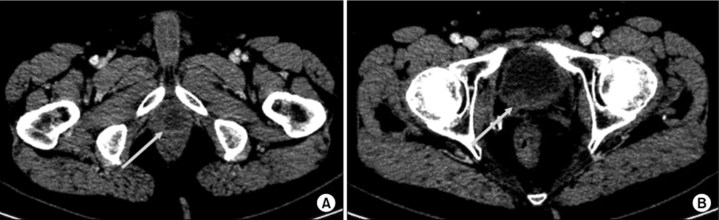
Fig. 1. Computed tomography (CT) images show two lesions suspected for seminal vesicle recurrence. Transaxial CT images show the lesion of the left transverse perineal muscle (arrow in A) associated with a thickening of the posterior wall of the bladder (arrow in B).
ic masses that may be misinterpreted as an abscess or hem- orrhage [1]. Radiology can also help to detect a primary le- sion or any associated congenital genitourinary malforma- tions such as ectopic ureters or renal agenesis [3]. 2-de- oxy-2-([18]F)fluoro-D-glucose positron emission tomog- raphy/computed tomography ([18F]FDG-PET/CT) has been used in the case of SV cancer to stage disease, particularly to exclude distant metastases [3,4] and to assess treatment response [4]. Different from radiological imaging that pro- vides anatomical and morphological data, [18F]FDG-PET/
CT characterizes the biological properties of a tumor, de- picting lesions with enhanced metabolism. Surgical ex- cision, ranging from local excision or vesiculectomy alone to pelvic exenteration depending on the extent of involve- ment of the adjacent organs, is the mainstay of treatment.
Radiotherapy, multiagent chemotherapy, and androgen deprivation therapy may also be beneficial in the adjuvant or palliative setting [5].
CASE REPORT
We present the case of a 58-year-old patient with re- currence of SV clear-cell adenocarcinoma associated with right renal agenesis. Since 2006, the patient had suffered from recurrent hematospermia. Serum tumor markers in- cluding prostate-specific antigen (PSA), neuron-specific enolase, carcinoembryonic antigen (CEA), carbohydrate antigen 19-9 (CA-19.9), alpha-fetoprotein, and chromog- ranin A were negative. Transrectal ultrasound and pelvic magnetic resonance imaging (MRI) diagnosed bilateral
congenital SV cysts. In 2009, a transrectal ultra- sound-guided biopsy of the SV was performed due to the persistence of intermittent hematospermia, resulting negative. Since 2010, the patient had also presented epi- sodes of hematuria. No atypical cells were identified in the urinary cytological examination. Cystoscopy was neg- ative; thus, the transrectal ultrasound-guided biopsy of SV was repeated. In the same examination, a bioptical map- ping of the prostate was also done. The histology results were consistent with SV adenocarcinoma. Staging exami- nations included a contrast-enhanced CT scan, [18F]FDG- PET/CT imaging, and bone scintigraphy. In the CT scan, the primary SV lesion was characterized by a hypodense central area presenting peripheral contrast enhancement.
Further, a left pararectal lymph node (size: 16 mm) was visualized. [18F]FDG-PET/CT confirmed radiopharmaceu- tical uptake in the primary SV lesion and in the pararectal lymph node (SUVmax=7.3 and 4.9, respectively). The bone scan was negative. In May 2010, the patient was sur- gically treated (prostatectomy plus pelvic lymphadenec- tomy plus bilateral vesiculectomy). Definitive histology confirmed primary bilateral SV clear-cell well-differ- entiated adenocarcinoma with a tubulo-papillary growth infiltrating the stroma. In the prostate, normal prostatic tis- sue was not found. The pararectal lymph node resulted metastatic. Immunohistochemistry showed a strong pos- itivity for cytokeratin 7 (CK7), epithelial membrane anti- gen, and p53, and was negative for PSA, carbohydrate an- tigen 125 (CA-125), cytokeratin 20 (CK20), caudal-type homeobox protein 2 (CDX2), and CEA.
Fig. 2. [18F]fluoro-D-glucose positron emission tomography/computed tomography ([18F]FDG-PET/CT) images show the lesion suspected for seminal vesicle recurrence. Maximum intensity projection image shows an area of moderate [18F]FDG uptake under the bladder (arrow in A). Transaxial [18F]FDG-PET/CT images show a central ‘cold’ area and peripheral [18F]FDG uptake at left transverse perineal muscle (arrow in B) as confirmed by images repeated immediately after urination (arrow in C).
Fig. 3. Magnetic resonnace imaging (MRI) images show two lesions suspected for seminal vesicle recurrence. Transaxial MRI images show the lesion of the left transverse perineal muscle characterized by a high signal intensity in T2-weighted (arrow in A) and the thickening of the posterior wall of trigone of the bladder in T1-weighted image (arrow in B).
Thereafter, the patient underwent adjuvant chemo- therapy (cisplatinum plus gemcitabine) and intensity- modulated radiation therapy (60 gray). During the first year of follow-up, the patient was followed up by clinical examination, serum tumor markers, and contrast-enhan- ced CT scan (chest and abdomen) every 3 months. No hematospermia or hematuria occurred. All examinations showed negative results. Thereafter, clinical and radio- logical examinations were performed every 6 months and all of them showed negative results. In January 2013, a contrast-enhanced CT scan showed the appearance of a lesion (size: 24×15×42 mm) in the left transverse peri- neal muscle characterized by a hypodense central area
with peripheral contrast enhancement (Fig. 1). A thicken- ing of the posterior wall of the bladder was also found.
These findings were considered suspicious for tumor re- currence; thus, an [18F]FDG-PET/CT scan was performed.
In the PET/CT, the lesion of the left transverse perineal muscle was characterized by a central ‘cold’ area and pe- ripheral [18F]FDG uptake (SUVmax=4.5), a finding con- firmed by the additional acquisition of the pelvis repeated at the end of the whole-body scan, immediately after uri- nation, performed to better visualize the pelvic region (SUVmax=5.8), as shown in Fig. 2. [18F]FDG-PET/CT did not reveal any other pelvic lesions or distant metastasis. A pelvic MRI was also performed. The lesion of the left trans-
verse perineal muscle presented a high signal intensity in T2-weighted images and a low signal intensity in the T1-weighted images, along with wall contrast enhance- ment after a gadolinium injection, without any cleavage plane with the rectum (Fig. 3). Further, thickening of the posterior wall of the trigone of the bladder was confirmed presenting an internal small hemorrhagic area. The MRI findings were considered suspicious for tumor recurrence although the presence of an abscess could not be ruled out with certainty. The patient did not present any signs or symptoms, and the clinical examination as well as serum tumor markers consistently showed negative results. A transrectal ultrasound-guided cytology of the left trans- verse perineal muscle lesion was performed in February 2013, confirming the recurrence of SV clear-cell ad- enocarcinoma. Thus, a chemotherapy regimen with taxol plus carboplatinum (6 cycles) was started, and the patient is currently in follow-up.
DISCUSSION
We present a case of the usefulness of imaging to detect the recurrence of primary SV clear-cell well-differentiated adenocarcinoma.
Diagnosis of SV adenocarcinoma is difficult and is based on a correlation of clinical, radiological, and histo- logical findings.
Histology is central in this clinical setting, despite the lack of an organ-specific immunophenotype. All reports pub- lished about the immunohistochemistry of SV cancers have revealed that these tumors are negative for PSA. The staining for CK7 has usually been strongly positive and that for CK20, negative. The keratin profile might be helpful in distinguish- ing SV adenocarcinoma from prostate, colorectal, and ur- othelial-type bladder carcinomas [1,5,6]. In our case, CA-125 was negative, similar to other reports, although cas- es positive for CA-125 have also been described [5].
Imaging plays a crucial role in identifying primary SV adenocarcinoma. In the imaging, these tumors appear as a mass behind the bladder with or without a prostatic or ure- teral obstruction or as an infiltrating lesion in the SV with enhancement similar to that of advanced prostate cancer [7]. Further, imaging can identify renal agenesis or dys- genesis that may be associated with both SV cysts and tu-
mors [8]. Our patient, one year before the diagnosis of SV adenocarcinoma, was diagnosed with a bilateral con- genital SV cyst associated with right renal agenesis. SV cysts are commonly associated with renal agenesis or dysgenesis. Incomplete development between the Wolffian duct and the urogenital sinus in males results in an accumulation of secretions and the subsequent for- mation of SV cysts during puberty [9]. Lee et al [8] sug- gested that the cause of large SV cysts is not only the ec- topic ureter opening into the cyst but also a mucin-produc- ing tumor. On the basis of these premises and our case, we suggest a radiological follow-up of patients with SV cyst, particularly when hematospermia and hematuria are pres- ent, in order to eventually detect early SV tumors.
There is little data about the role of imaging in the fol- low-up of SV adenocarcinoma [4]. In our case, the re- currence of SV adenocarcinoma in the left transverse peri- neal muscle presented the same characteristic of primary lesion in both contrast-enhanced CT and [18F]FDG-PET/CT;
thus, tumor recurrence was suspected. Further, the pelvic MRI findings were considered suspicious although the pres- ence of an abscess could not be ruled out with certainty.
Both contrast-enhanced CT scan and pelvic MRI also re- vealed the thickening of the posterior bladder wall, while [18F]FDG-PET/CT did not show any uptake at this level.
Although cytological confirmation of the recurrence of SV adenocarcinoma was obtained only in the left transverse perineal muscle lesion, we cannot exclude the tumor in- volvement of the posterior bladder wall. In fact, with the ex- cretion of [18F]FDG by the kidney into the urine, intense normal [18F]FDG activity is observed in the intrarenal col- lection systems, ureters, and bladder, and the urinary ex- cretion of [18F]FDG continued in well-hydrated patients even 1 hour after [18F]FDG administration [10]. Thus, [18F]FDG-PET/CT may give a false negative result in the de- tection of the recurrence of a bladder tumor. Nevertheless, [18F]FDG-PET/CT allows one to exclude distant metastases.
To summarize, here, we report a case of primary SV clear-cell adenocarcinoma associated with right renal agenesis, recurred after treatments, depicted in both radi- ology and [18F]FDG-PET/CT. Imaging results were very useful in reaching the final diagnosis. In fact, from a clin- ical point of view, the patient did not present any suspi- cious signs or symptoms for the recurrence of SV ad-
enocarcinoma, and both the clinical examination and the tumor markers were yielded negative results. The sim- ilarity between the lesion of the left transverse perineal muscle and the primary SV lesion in both the CT scan and [18F]FDG-PET/CT together with the concordance of the findings of both radiological and nuclear medicine techni- ques led to a strong suspicion of tumor recurrence.
As illustrated by our case, a combined approach using radiology and nuclear medicine techniques seems to be effective in the follow-up of SV adenocarcinoma patients to early diagnose tumor recurrence. Technological ad- vances in imaging and histopathology, together with the awareness of this rare tumor, have the potential of improv- ing the outcomes in these patients not only by providing earlier detection and accurate staging, but also by detect- ing recurrence and thereby avoiding delays and ther- apeutic dilemmas.
REFERENCES
1. Egevad L, Ehrnström R, Håkansson U, Grabe M. Primary seminal vesicle carcinoma detected at transurethral re- section of prostate. Urology 2007;69:778.
2. Benson RC Jr, Clark WR, Farrow GM. Carcinoma of the
seminal vesicle. J Urol 1984;132:483-5.
3. Navallas M, Vargas HA, Akin O, Pandit-Taskar N, Fine SW, Eastham JA, et al. Primary seminal vesicle adenocarcinoma.
Clin Imaging 2011;35:480-2.
4. Kreiner B, Denzinger S, Ganzer R, Fritsche HM, Burger M, Wieland WF, et al. Neuroendocrine carcinoma of the semi- nal vesicles presenting with Lambert Eaton syndrome: a case report. J Med Case Rep 2010;4:320.
5. Oxley JD, Brett MT, Gillatt DA, Burton P. Seminal vesicle carcinoma. Histopathology 1999;34:562-3.
6. Tarján M, Ottlecz I, Tot T. Primary adenocarcinoma of the seminal vesicle. Indian J Urol 2009;25:143-5.
7. Kim B, Kawashima A, Ryu JA, Takahashi N, Hartman RP, King BF Jr. Imaging of the seminal vesicle and vas deferens.
Radiographics 2009;29:1105-21.
8. Lee BH, Seo JW, Han YH, Kim YH, Cha SJ. Primary muci- nous adenocarcinoma of a seminal vesicle cyst associated with ectopic ureter and ipsilateral renal agenesis: a case report. Korean J Radiol 2007;8:258-61.
9. Zaontz MR, Kass EJ. Ectopic ureter opening into seminal vesicle cyst associated with ipsilateral renal agenesis.
Urology 1987;29:523-5.
10. Versari A. Normal findings from different radiopharmaceu- ticals and techniques, with variants and pitfalls. In: Lazzeri E, Signore A, Erba PA, Prandini N, Versari A, D'Errico G, et al, editors. Radionuclide imaging of infection and inflam- mation: a pictorial case-based atlas. 1st ed. New York:
Springer Science Business Media, Inc.; 2013;1-22.