PGHN
Original Article
Amino Acid-Based Formula in Premature Infants with Feeding Intolerance: Comparison of Fecal Calprotectin Level
Hyo-Jeong Jang, Jae Hyun Park, Chun Soo Kim, Sang Lak Lee, and Won Mok Lee*
Departments of Pediatrics and *Laboratory Medicine, Keimyung University School of Medicine, Daegu, Korea
Purpose: We investigated fecal calprotectin (FC) levels in preterm infants with and without feeding intolerance (FI), and compared the FC levels according to the type of feeding.
Methods: The medical records of 67 premature infants were reviewed retrospectively. The fully enteral-fed infants were classified into two groups; the FI group (29 infants) and the control group (31 infants). Seven infants with necrotiz- ing enterocolitis, sepsis, and perinatal asphyxia were excluded. If breast milk (BM) or preterm formula (PF) could not be tolerated by infants with FI, amino acid-based formula (AAF) was tried temporarily. Once FI improved, AAF was discontinued, and BM or PF was resumed. We investigated the FC levels according to the type of feeding.
Results: Significant differences were found in gestational age, birth weight, age when full enteral feeding was ach- ieved, and hospital stay between the FI and control group (p<0.05). The FC levels in the FI group were significantly higher than those in the control group (p<0.05). The FC levels in the AAF-fed infants with FI were significantly lower than those in the BM- or PF-fed infants (p<0.05). The growth velocities (g/d) and z scores were not significantly different between the FI and control group (p>0.05).
Conclusion: The FC levels in AAF-fed infants with FI showed significantly lower than those in the BM- or PF-fed infants with FI. The mitigation of gut inflammation through the decrease of FC levels in AAF-fed infants with FI could be presumed.
Key Words: Fecal calprotectin, Infant formula
Received:February 4, 2017, Revised:December 1, 2017, Accepted:December 29, 2017
Corresponding author: Jae Hyun Park, Department of Pediatrics, Keimyung University Dongsan Medical Center, 56 Dalseong-ro, Jung-gu, Daegu 41931, Korea. Tel: +82-53-250-7524, Fax: +82-53-250-7783, E-mail: [email protected]
Copyright ⓒ 2018 by The Korean Society of Pediatric Gastroenterology, Hepatology and Nutrition
This is an openaccess article distributed under the terms of the Creative Commons Attribution NonCommercial License (http://creativecommons.org/licenses/by-nc/4.0/) which permits unrestricted noncommercial use, distribution, and reproduction in any medium, provided the original work is properly cited.
INTRODUCTION
Enteral feeding advancement in preterm infants is one of the most challenging tasks in postnatal growth [1]. Delayed enteral feeding might be asso- ciated with decreased gastrointestinal (GI) hormone
secretion and intestinal motility [2]. Owing to de- layed enteral nutrition, prolonged use of an in- dwelling catheter for parenteral nutrition is asso- ciated with postnatal infection with an increased risk of mortality, prolonged hospitalization, and ad- verse neurodevelopmental outcome in preterm in-
fants [3,4]. However, many clinical situations with- hold feeding of preterm infants because of the fear of various detrimental GI complications [5]. Feeding intolerance (FI), defined as the inability to digest en- teral feeding associated with increased gastric re- siduals, abdominal distension, and/or emesis, fre- quently develops in preterm infants [1]. The known risk factors for FI are a weak esophageal sphincter, delayed gastric emptying time, intestinal hypo- motility, and abnormal bacterial colonization [6,7].
The immaturity of the GI tract plays a predominant role in preterm infants with FI [8]. Functional and biochemical maturation are established gradually over the last trimester of gestation and are influ- enced by different interacting factors such as age, drug use, diet, and intestinal microflora [9]. This greatly affects the digestive absorptive function of preterm infants. Protein digestion begins with hy- drolysis in the acid environment of the stomach and is processed with multiple proteases [10]. However, limited gastric acid secretion in preterm infants re- sults in decreased activity of enterokinases to hydro- lyze proteins and activate pancreatic protease diges- tive cascade [11]. The aforementioned facts suggest that absorption of hydrolyzed protein could be easier in preterm infants. Mihatsch et al. [12] reported that the use of hydrolyzed protein formulas in preterm in- fants established full enteral feeding more rapidly.
Raimondi et al. [13] reported that a short course of amino acid-based formula (AAF) was a safe and ef- fective rescue strategy for very-low-birth-weight (VLBW) infants with FI.
Calprotectin is an antimicrobial protein found in neutrophils, monocytes, macrophages, and some squamous epithelium cells and known to increase especially in GI inflammation [14,15]. Its presence in stool indicates neutrophil migration to the GI mu- cosa and can suggest the severity of mucosal in- flammation [14,16]. According to a recent systemic review, fecal calprotectin (FC) level was elevated in infants with necrotizing enterocolitis (NEC) and was useful as a noninvasive prognostic marker of NEC [17]. However, high FC levels in healthy infants are related to the physiological response of the gut, such
as intestinal permeability, gut microbiota, and re- sponse to alimentary allergens [18].
We investigated FC levels in preterm infants with and without FI, and compared the FC levels after changing to AAF.
MATERIALS AND METHODS
We retrospectively reviewed the medical records of 67 premature infants born at 29-32 weeks’ ges- tation who were admitted to the neonatal intensive care unit of Keimyung University Dongsan Medical Center between May and September 2016. This study was approved by the Institutional Review Board of Keimyung University Dongsan Medical Center (No. 2017-01-037). Seven infants with peri- natal asphyxia (Apgar score at 5 minutes, ≤5), sep- sis, and NEC were excluded. FI was defined as the in- ability to digest enteral feeding, which presents as a gastric residual volume of >50%, abdominal dis- tension, emesis, or disruption of the patient’s feed- ing plan [7]. Full enteral feeding was defined as en- teral tolerance of at least 120 mL/(kgㆍd) of milk [7].
All enrolled infants were fed within 3 hours of life using either with mother’s breast milk (BM) or with preterm formula (PF) when BM was not available due to mother’s condition. The infants with FI were temporarily fed with AAF (NeocateⓇ; Nutricia, London, UK) instead of BM or PF. Calprotectin was investigated as fecal specimens collected from a fresh diaper which was changed every three hours.
Infants without FI were examined for calprotectin in the feces once on the 7th day of life. In infants with FI, fecal specimens were taken before feeding was changed to AAF. Subsequent FC levels were meas- ured at least 3 days after feeding with AAF. Once the babies with FI showed clinical improvement, AAF feeding was discontinued, and the previous diet, ei- ther BM or PF, was resumed. FC levels were meas- ured using a commercial quantitative enzyme-linked immunosorbent assay kit on the Alegria system (ORGENTEC Diagnostika, Mainz, Germany). The measurable range of FC was from 0 to 1,000 μg/g.
The following variables were analyzed for the peri-
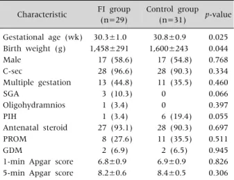
Table 1. Subject Characteristics between FI Group and Control Group
Characteristic FI group (n=29)
Control group (n=31) p-value Gestational age (wk) 30.3±1.0 30.8±0.9 0.025 Birth weight (g) 1,458±291 1,600±243 0.044
Male 17 (58.6) 17 (54.8) 0.768
C-sec 28 (96.6) 28 (90.3) 0.334
Multiple gestation 13 (44.8) 11 (35.5) 0.460
SGA 3 (10.3) 0 0.066
Oligohydramnios 1 (3.4) 0 0.397
PIH 1 (3.4) 6 (19.4) 0.055
Antenatal steroid 27 (93.1) 28 (90.3) 0.697
PROM 8 (27.6) 11 (35.5) 0.511
GDM 2 (6.9) 2 (6.5) 0.945
1-min Apgar score 6.8±0.9 6.9±0.9 0.826 5-min Apgar score 8.2±0.6 8.4±0.5 0.306 Values are presented as mean±standard deviation or number (%).
FI: feeding intolerance, C-sec: cesarean section, SGA: small for gestational age, PIH: pregnancy-induced hypertension, PROM:
premature rupture of membrane, GDM: gestational diabetes mellitus.
Fig. 1. Fecal calprotectin levels by type of feeding in preterm infants with feeding intolerance. BM: breast milk, PF: preterm formula, AAF: acid-based formula.
natal factors: gestational age (GA), birth weight (BW), sex, delivery mode (vaginal delivery or cesar- ean section), multiple gestation, small for GA (SGA), oligohydramnios, pregnancy-induced hypertension (PIH), antenatal steroid, premature rupture of mem- brane, gestational diabetes mellitus (GDM), and Apgar score at 1 and 5 minutes.
The growth velocity and z scores were checked at the time of discharge. The growth velocity was calcu- lated using the following formula: (body weight at discharge [g]−BW [g])/hospital stay[d], g/d). The z score was calculated using the Lambda Mu Sigma method [19]. L determines a nonlinear trans- formation of BW, M stands for the mean of normal distribution, and S stands for its standard deviation (SD). L, M, and S correspond to the following for- mulas: Z=[(X/M)L−1]/LS, where X indicates the measured value of BW, and centile=M (1+LSZ)1/L, where Z is the z score that corresponds to a given percentile. The z score is a measure of the distance in SDs of a sample from the mean [19].
Statistical analysis was conducted using SPSS Statistics ver. 17.0 (SPSS Inc., Chicago, IL, USA).
Descriptive results were reported as the mean±SD.
Statistical analysis between groups were conducted with the Student t-test for independent samples for normally distributed data and with the Mann- Whitney U-test for non-normally distributed data.
The chi-square test was performed for categorical variables, and the Pearson correlation test was used to evaluate the relationship between the selected variable and FC values. For the interpretation of re- sults, p-values of <0.05 were considered significant.
RESULTS
Subject characteristics
The study population included 60 preterm infants (34 males and 26 females), of whom 29 (48.3%) were grouped as the FI group. The mean age at FI onset was 3.7±1.0 days. AAF was fed for a mean duration of 14.2±8.3 days in the FI group. Table 1 shows the comparison between FI and control group. There was a significant difference in the mean GA (30.3±
1.0 weeks vs. 30.8±0.9 weeks, p<0.05) and BW (1,458± 291 g and 1,600±243 g, p<0.05). However, no significant differences were shown in sex, deliv- ery mode, multiple gestation, SGA, oligohydramnios, PIH, antenatal steroid use, GDM, and Apgar score at 1 and 5 minutes between the two groups (p>0.05).
Fig. 2. The postnatal age-related fecal calprotectin levels in preterm infants with feeding intolerance by type of feeding. BM: breast milk, PF: preterm formula, AAF: acid-based formula.
Table 2.Postnatal Outcomes between FI Group and Control Group
FI group (n=29)
Control group (n=31) p-value Time to full enteral
feeding (d)
4.0±0.9 3.2±0.8 0.001
Hospital stay (d) 34.2±13.2 26.4±11.2 0.017 Body weight at
discharge (g)
1,952±171 2,023±205 0.153
Growth velocity (g/d) 9.2±4.0 9.6±5.5 0.763
Z score 0.013±0.007 0.011±0.006 0.287
Values are presented as mean±standard deviation.
FI: feeding intolerance.
FC concentrations
The FC levels in the FI group were significantly higher than in the control group (855±240 μg/g vs.
401±270 μg/g, p<0.05). As shown in Fig. 1, the FC levels in BM- or PF-fed infants with FI were sig- nificantly higher than in AAF-fed infants with FI (898±174 μg/g vs. 273±204 μg/g, p<0.05). At the time of the first FC investigation, there were 3 BM-fed infants (10.3%) and 26 PF-fed infants (89.7%) in the FI group.
Fig. 2 showed the postnatal age-related FC levels in the infants with FI by the type of feeding. There was a negative correlation between FC levels and postnatal age in BM- or PF-fed infants and AAF-fed infants, but not statistically significant (p>0.05).
Postnatal outcomes
Postnatal outcomes were assessed with time to full enteral feeding (d), hospital stay (d), body weight at discharge (g), growth velocity (g/d), and z score. We compared the variables between the FI and control groups (Table 2). Time to full enteral feeding (d) was significantly longer in the FI group than in the control group (4.0±0.9 days vs. 3.2±0.8 days, re- spectively, p<0.05). In addition, hospitalization du- ration was significantly longer in the FI group than in the control group (34.2±13.2 days vs. 26.4±11.2 days, p<0.05). There was no statistically significant difference between the two groups regarding body weight at discharge, growth velocity, and z scores (p>0.05).
DISCUSSION
In our preliminary study, the FC levels in FI group were significantly higher than in non-FI group. And the FC levels in AAF-fed infants with FI were sig- nificantly lower than BM- or PF-fed infants with FI.
No significant differences in postnatal growth pa- rameters were observed between the FI and control groups, which might be associated with the tempo- rary use of AAF.
Previous reports of FC levels in preterm infants in- dicated a wide range of GA in enrolled infants
[12,20]. FC levels depend on the gestational and postnatal ages of preterm infants [21]. In the present study, we selectively investigated FC levels in pre- term infants born at 29-32 weeks’ gestation. A re- search in preterm infants born at <32 weeks’ ges- tation reported a wide range of FC levels and that FC levels correlated positively with the volume of enter- al feeding and the need to interrupt enteral feeding [22]. It is in line with the result of our study that the FI infants showed higher FC levels comparing to the non-FI infants.
The increase in FC level suggests a high level of granulocytes in the gut lumen, which is a hallmark of digestive inflammatory status [20]. Few studies investigated FC levels in preterm infants with FI, and the reference level was investigated a little in term and preterm infants. The studies found higher FC levels in newborns and younger infantsthan in chil- dren and adults, which could suggest greater in- testinal permeability and different immunological status of the gut in newborns. Mussa et al. [16] stud- ied FC levels in preterm infants with and those with- out FI, and suggested a cutoff value for FI screening.
The result of the study showed that the significantly higher level of FC in the FI group even with various types of feeding, which is in line with the result of our study. Some studies researched about the rela- tionship between the types of feeding and FC levels.
Li et al. [20] reported higher FC levels in healthy, ex- clusively BM-fed infants than in non-BM-fed ones in the first 5 months of life. Savino et al. [23] also re- ported a similar result of high FC levels in healthy, exclusively BM-fed infants. However, Rougé et al.
[22] observed that FC levels were lower in infants with human milk than in infants exclusively or pre- dominantly fed with formula. Campeotto et al. [24]
and Yang et al. [25] reported no significant differ- ence in FC levels between the infants fed with BM and those fed with hydrolyzed formula. In the pres- ent study, three infants of FI group were fed with BM and twenty-six infants were fed with PM. Due to those differences, there would be a limit to compare the FC levels between the infants fed with BM and those fed with PF. Further prospective studies will be
needed to investigate the levels of FC in preterm in- fants according to the more specific type of feeding.
However, no study has been conducted yet about the investigation of FC levels according to the change of feeding regimen in preterm infants with FI, which was shown in the present data. Raimondi et al. [13]
compared the infants fed with AAF with the controls fed with PF in terms of time to full enteral feeding, time on parenteral nutrition, time on central venous line, gastric residual volume, and the outcomes on growth at discharge and at 12 months of life. The re- sult and conclusion of the study were that AAF was a safe nutritional alternative for rescuing VLBW in- fants with intrauterine growth restriction with se- vere FI. The mean duration of AAF feeding was 15.4±12.3 days in their study and 14.2±8.3 days in our study. The growth outcome in their study was not impaired in the AAF group, which is in line with the result of our study, even though we did not ex- amine long-term results on growth yet.
So far, little is known about the gut response to en- teral nutrition during the postnatal period in pre- term infants [26]. The immature digestive, absorp- tive, and immunologic functions in preterm infants can lead to nutrient fermentation, bacterial over- growth, and mucosal inflammation, which alto- gether can lead to the intolerance of feeding [26]. In the present study, the lowered level of FC concen- tration after AAF feeding in infants with FI could suggest that the alleviation of the gut inflammation.
AAF is composed of lowered lactose, higher percent- age of medium chain triglycerides, and completely hydrolyzed protein with amino acids [27]. The ami- no acids are essential for the intestinal function with an important role for mucosal blood perfusion, growth, and immunity [28]. The deficiency of the amino acids should result in the impairment of the intestinal function, which can be led to the bacterial overgrowth and mucosal inflammation. According to the previous animal studies, it is known that dur- ing the final weeks of gestation, there is a rapid in- crease in the tissue-specific capacity for glucose up- take, whereas the ability of the mucosa to absorb most amino acids remains low [26,29]. And other re-
searches also revealed that the ability to take up and transfer intact proteins from the epithelium into the circulation is a highly specialized function that de- velops very close to term, which means that the di- gestive function for protein macromolecules is far more impaired in preterm infants [30]. Those results in premature intestine could advocate the use of par- tially or totally hydrolyzed milk diets in a situation of delay in introducing or advancement of enteral nu- trition in preterm infants.
A question can arise about nutritional adequacy in long-term use of AAF and hydrolyzed protein formula. Rigo et al. [31] and Szajewska et al. [32] re- ported about the nutritional insufficiency of hydro- lyzed protein formula for term and preterm infants.
Zuppa et al. [33] also had the same question in their study. In the present study, the patients with FI who switched to AAF for a short termdid not develop any adverse clinical sign or symptom during AAF feed- ing, and it showed improvement in the symptoms and signs of FI with proper body weight gain. Once the clinical presentation of FI improved, AAF feeding was switched to BM or PF. The growth velocity and z score did not show significant differences when compared with those in the control group at the time of discharge.
In summary, in our preliminary study with a small cohort of preterm infants with FI, the FC levels of the BM- or PF-fed infants were significantly higher than those of the AAF-fed infants. No significant differ- ences in postnatal growth parameters were found between the FI and control groups. As an elemental diet is used for cow’s milk protein intolerance or in- flammatory bowel disease, we can hypothesize that FI, an inflammatory status of immature GI tract, can be alleviated by temporary use of elemental diet if BM or other formulas are not suitable. The miti- gation of gut inflammation through the decrease of FC levels in AAF-fed infants with FI could be presumed.
REFERENCES
1. Moore TA, Wilson ME. Feeding intolerance: a concept
analysis. Adv Neonatal Care 2011;11:149-54.
2. Berseth CL, Nordyke CK, Valdes MG, Furlow BL, Go VL. Responses of gastrointestinal peptides and motor activity to milk and water feedings in preterm and term infants. Pediatr Res 1992;31:587-90.
3. Stoll BJ, Hansen N, Fanaroff AA, Wright LL, Carlo WA, Ehrenkranz RA, et al. Late-onset sepsis in very low birth weight neonates: the experience of the NICHD Neonatal Research Network. Pediatrics 2002;110:285-91.
4. Stoll BJ, Hansen NI, Adams-Chapman I, Fanaroff AA, Hintz SR, Vohr B, et al. Neurodevelopmental and growth impairment among extremely low-birth-weight infants with neonatal infection. JAMA 2004;292:2357-65.
5. Hay WW Jr. Aggressive nutrition of the preterm infant.
Curr Pediatr Rep 2013;1:10.1007/s40124-013-0026-4.
6. Mansi Y, Abdelaziz N, Ezzeldin Z, Ibrahim R. Rand- omized controlled trial of a high dose of oral eryth- romycin for the treatment of feeding intolerance in pre- term infants. Neonatology 2011;100:290-4.
7. Indrio F, Riezzo G, Cavallo L, Di Mauro A, Francavilla R. Physiological basis of food intolerance in VLBW. J Matern Fetal Neonatal Med 2011;24 Suppl 1:64-6.
8. Lebenthal E, Lee PC, Heitlinger LA. Impact of develop- ment of the gastrointestinal tract on infant feeding. J Pediatr 1983;102:1-9.
9. Fanaro S. Feeding intolerance in the preterm infant.
Early Hum Dev 2013;89 Suppl 2:S13-20.
10. Hyman PE, Clarke DD, Everett SL, Sonne B, Stewart D, Harada T, et al. Gastric acid secretory function in pre- term infants. J Pediatr 1985;106:467-71.
11. Hyman PE, Feldman EJ, Ament ME, Byrne WJ, Euler AR. Effect of enteral feeding on the maintenance of gas- tric acid secretory function. Gastroenterology 1983;84:
341-5.
12. Mihatsch WA, Franz AR, Högel J, Pohlandt F.
Hydrolyzed protein accelerates feeding advancement in very low birth weight infants. Pediatrics 2002;110:
1199-203.
13. Raimondi F, Spera AM, Sellitto M, Landolfo F, Capasso L. Amino acid-based formula as a rescue strategy in feeding very-low-birth-weight infants with intrauterine growth restriction. J Pediatr Gastroenterol Nutr 2012;
54:608-12.
14. Fagerhol MK, Dale I, Andersson T. A radioimmuno- assay for a granulocyte protein as a marker in studies on the turnover of such cells. Bull Eur Physiopathol Respir 1980;16 Suppl:273-82.
15. Lamb CA, Mansfield JC. Measurement of faecal calpro- tectin and lactoferrin in inflammatory bowel disease.
Frontline Gastroenterol 2011;2:13-8.
16. Mussa R, Khashana A, Kamel N, Elsharqawy SE. Fecal
calprotectin levels in preterm infants with and without feeding intolerance. J Pediatr 2016;92:486-92.
17. Pergialiotis V, Konstantopoulos P, Karampetsou N, Koutaki D, Gkioka E, Perrea DN, et al. Calprotectin lev- els in necrotizing enterocolitis: a systematic review of the literature. Inflamm Res 2016;65:847-52.
18. Kapel N, Campeotto F, Kalach N, Baldassare M, Butel MJ, Dupont C. Faecal calprotectin in term and preterm neonates. J Pediatr Gastroenterol Nutr 2010;51:542-7.
19. Lim JS, Lim SW, Ahn JH, Song BS, Shim KS, Hwang IT. New Korean reference for birth weight by gesta- tional age and sex: data from the Korean Statistical Information Service (2008-2012). Ann Pediatr Endocrinol Metab 2014;19:146-53.
20. Li F, Ma J, Geng S, Wang J, Ren F, Sheng X. Comparison of the different kinds of feeding on the level of fecal calprotectin. Early Hum Dev 2014;90:471-5.
21. Zoppelli L, Güttel C, Bittrich HJ, Andrée C, Wirth S, Jenke A. Fecal calprotectin concentrations in pre- mature infants have a lower limit and show postnatal and gestational age dependence. Neonatology 2012;
102:68-74.
22. Rougé C, Butel MJ, Piloquet H, Ferraris L, Legrand A, Vodovar M, et al. Fecal calprotectin excretion in pre- term infants during the neonatal period. PLoS One 2010;5:e11083.
23. Savino F, Castagno E, Calabrese R, Viola S, Oggero R, Miniero R. High faecal calprotectin levels in healthy, ex- clusively breast-fed infants. Neonatology 2010;97:
299-304.
24. Campeotto F, Kalach N, Lapillonne A, Butel MJ, Dupont C, Kapel N. Time course of faecal calprotectin in preterm newborns during the first month of life. Acta Paediatr 2007;96:1531-3.
25. Yang Q, Smith PB, Goldberg RN, Cotten CM. Dynamic
change of fecal calprotectin in very low birth weight in- fants during the first month of life. Neonatology 2008;94:267-71.
26. Sangild PT. Gut responses to enteral nutrition in pre- term infants and animals. Exp Biol Med (Maywood) 2006;231:1695-711.
27. Kim YJ. Enteral nutrition and its clinical application.
Korean J Pediatr Gastroenterol Nutr 2009;12(Suppl 1):S27-36.
28. Wu G, Jaeger LA, Bazer FW, Rhoads JM. Arginine defi- ciency in preterm infants: biochemical mechanisms and nutritional implications. J Nutr Biochem 2004;15:
442-51.
29. Buddington RK, Elnif J, Puchal-Gardiner AA, Sangild PT. Intestinal apical amino acid absorption during de- velopment of the pig. Am J Physiol Regul Integr Comp Physiol 2001;280:R241-7.
30. Bjornvad CR, Schmidt M, Petersen YM, Jensen SK, Offenberg H, Elnif J, et al. Preterm birth makes the im- mature intestine sensitive to feeding-induced in- testinal atrophy. Am J Physiol Regul Integr Comp Physiol 2005;289:R1212-22.
31. Rigo J, Salle BL, Picaud JC, Putet G, Senterre J.
Nutritional evaluation of protein hydrolysate formulas.
Eur J Clin Nutr 1995;49 Suppl 1:S26-38.
32. Szajewska H, Albrecht P, Stoitiska B, Prochowska A, Gawecka A, Laskowska-Klita T. Extensive and partial protein hydrolysate preterm formulas: the effect on growth rate, protein metabolism indices, and plasma amino acid concentrations. J Pediatr Gastroenterol Nutr 2001;32:303-9.
33. Zuppa AA, Visintini F, Cota F, Maggio L, Romagnoli C, Tortorolo G. Hydrolysed milk in preterm infants: an open problem. Acta Paediatr Suppl 2005;94:84-6.