INTRODUCTION
By definition, sarcomatoid carcinomas are malignant tumors that exhibit histological characteristics of a combination of ma- lignant epithelial and mesenchymal cells. These tumors can oc- cur in various anatomical sites, including lungs, thyroid gland, salivary gland, breast, bladder, prostate, skin and digestive sys- tem (1). Gastrointestinal sarcomatoid carcinomas are unusual tumors that occur most frequently in the stomach, gallbladder, and esophagus (1). These are especially rare in the small intes- tine, with an incidence of only 0.5-0.8 per 100000 population per year (2). To the best of our knowledge, only 26 cases have been reported in the English literature up to date (3-8). More- over, most of the cases are focused on the pathologic reviews.
Therefore, we present a case of sarcomatoid carcinoma arising from small intestinal tract with findings of the multi-detector com- puted tomography (MDCT) scans and reviews of the literature.
CASE REPORT
A 78-year-old man was presented at our hospital with com- plaints of intermittent and aggravated diffuse abdominal pains for three days. His personal history was unremarkable except for the hypertension of one month duration. Physical examina- tion revealed a palpable mass-like lesion in right lower quad- rant. The results of other laboratory investigations, including routine blood tests, liver tests, urine analysis and serum tumor marker (carcinoembryonic antigen) were within normal limits, except for the mild anemia (hemoglobin, 8.7 g/dL). The MDCT of the abdomen was performed for further evaluations on a pal- pable abdominal mass, including unenhanced CT scans and dy- namic contrast-enhanced CT scans with a non-ionic contrast medium (Omnipaque 350, GE Healthcare, Oslo, Norway). The volume of 100 mL contrast medium was injected at a rate of 3 mL/s via an antecubital vein. The dynamic contrast-enhanced
J Korean Soc Radiol 2013;69(4):295-299 http://dx.doi.org/10.3348/jksr.2013.69.4.295
Received June 23, 2013; Accepted August 7, 2013 Corresponding author: Jung-Hee Yoon, MD Department of Radiology, Haeundae Paik Hospital, Inje University College of Medicine, 875 Haeundae-ro, Haeundae-gu, Busan 612-896, Korea.
Tel. 82-51-797-0355 Fax. 82-51-797-0397 E-mail: [email protected]
This is an Open Access article distributed under the terms of the Creative Commons Attribution Non-Commercial License (http://creativecommons.org/licenses/by-nc/3.0) which permits unrestricted non-commercial use, distri- bution, and reproduction in any medium, provided the original work is properly cited.
This work was supported by Grant from Inje University, 2011.
Sarcomatoid carcinomas are rare biphasic tumors composed of mixed malignant epithelial and mesenchymal cells. A few cases for small intestinal sarcomatoid car- cinoma were reported. Moreover, most of the cases are focused on the pathologic review. We experienced a case of monophasic sarcomatoid carcinoma arising in the jejunum in a 78-year-old man. In this case, CT showed fungating mass with central necrosis in the jejunum. We also reviewed literatures on sarcomatoid carcinoma that arises in the small intestine.
Index terms
Sarcomatoid Carcinoma Intestine, Small
Multi-Detector Computed Tomography
Sarcomatoid Carcinoma of the Small Intestine: A Case Report and Review of the Literature
1소장에 생긴 육종성암: 증례 보고 및 문헌고찰1
Kyung A Kang, MD
1, Jung-Hee Yoon, MD
2, Guhyun Kang, MD
31Department of Radiology, Sanggye Paik Hospital, Inje University College of Medicine, Seoul, Korea
2Department of Radiology, Haeundae Paik Hospital, Inje University College of Medicine, Busan, Korea
3Department of Pathology, Sanggye Paik Hospital, Inje University College of Medicine, Seoul, Korea
agnosis was mesenchymal origin tumor such as gastrointestinal stromal tumor (GIST) or lymphoma. A mass with central ne- crosis is an overlapping CT appearance of many small bowel neoplasms, and especially the mesenchymal origin tumors tend to grow exophytically and rarely cause bowel obstructions even though the tumor size is relatively large. In this case, bowel ob- struction was presumed due to seeding nodules rather than the mass itself. Malignant carcinoid tumor and leiomyosarcoma were also considered as a differential diagnosis, even though they are rare, small intestinal mesenchymal tumors. Thus, they may appear as a large intraluminal ulcerating lesion.
Exploratory laparotomy revealed a mass of small bowel locat- ed in jejunum, about 45 cm apart from the duodenojejunal junction with invasion into the serosal surface and the mesen- teric fat. A more proximal segment of jejunum was distorted without the definite palpable mass. A portion of jejunal segment along with adherent loop of proximal jejunum was resected.
Segmental resection revealed a mass located at the jejunum.
The resected specimen showed a large, irregular-shaped, fungat- ing mass with friable and necrotic surface, measuring 4 × 5 cm (Fig. 2). The tumor extended to the subserosa and focally infil- trated the serosa of the adherent, small bowel loop, macroscopi- cally.
Microscopically, the tumor consisted mostly of discohesive po- lygonal giant cells with a few spindle cells. These cells were ar- ranged in sheets or haphazardly. Giant cells have dense eosinophil- ic cytoplasm and pleomorphic nuclei. On immunohistochemistry, tumor cells showed diffused strong positive immunoreactivities for vimentin and focal strong positive reactions for cytokeratin (Fig. 3). The immunoreactivities were negative staining against the other antibodies, such as CD20, CD10 and c-kit. Although abdominal MDCT scan demonstrated a well-defined, round-
shaped, exophytic growing mass with central necrosis, which measured 6 cm in greatest dimension of the mid jejunum. The lesion showed poor contrast enhancements. In addition, signifi- cant dilatation of proximal jejunum with abrupt beaking ap- pearances suggestive of tethering adjacent the mass was demon- strated (Fig. 1). We presumed the small bowel obstruction caused by tumor seeding adjacent proximal jejunum. In this pa- tient, the jejunal loops were located in the lower right quadrant area, probably due to counterclockwise twist of the small bowel mesentery associated with the tethering adjacent the mass.
There was no significant lymphadenopathy. Considering the abovementioned CT features, the patient was diagnosed as ade- nocarcinoma in jejunum. Because the adenocarcinoma is the most common type of primary small intestinal malignancy, the epithelial origin tumor can lead to early bowel obstructions as compared with mesenchymal origin tumor. Our differential di-
Fig. 1. CT images in a 70-year-old man with abdominal pain. Axial pre-contrast (A) and delayed phase (B) images show a well-defined, round shaped fungating mass (solid arrow) with central necrosis. Coronal reconstruction images on delayed phase CT scan (C, D) show focal wall thick- ening suggesting tumor seeding (open arrow in C) and beaking appearance (curved arrow in D) with proximal bowel dilatation.
A B C D
Fig. 2. Photograph of the surgical specimen. The gross specimen shows a fungating necrotic mass in the jejunum.
epithelial-looking and mesenchymal-like cells. Monophasic tu- mors show a predominance of the mesenchymal-like component, with minimal to absent epitheloid area. Because monophagic sar- comatoid carcinoma can be confused with other sarcomas and minimal epitheloid component can be missed, a wide-range panel of immunohistochemical biomarkers should be performed for the differential diagnosis. Most of the tumors show positivity for cytokeratin and vimentin (6). In this case, cytokeratin and vimentin were positive and the carcinomatous nature of the tu- mor was evident only after the immunohistochemistry test.
Clinically, sarcomatoid carcinomas in the small intestine pri- marily affected middle-aged to older patients, with a mean age of 57 years. The male to female ratio was 1.5 : 1 (6). Symptoms may include abdominal pain, intestinal obstruction, palpable abdomi- nal mass, gastrointestinal bleeding, and anemia (5, 8). Sarcoma- toid carcinomas in the lower intestinal tract have aggressive clini- cal courses, often presented with symptoms or signs related to distant metastasis. On the other hand, sarcomatoid carcinomas in upper aerodigestive tract including the esophagus and stomach carcinomatous component was not recognized at microscopic
examination, the carcinomatous nature of the tumor was evident after immunohistochemistry examination, cytokeratin sugges- tive finding of carcinomatous nature of tumor was positive. The histopathological and immunohistochemical transitions be- tween the adenocarcinoma area and the spindle cell area sug- gested that the sarcomatous elements originated from the ade- nocarcinoma during tumor progressions. Therefore, the final diagnosis was sarcomatoid carcinoma. Additional treatment was not given and the patient was lost for follow-ups.
DISCUSSION
Sarcomatoid carcinomas are rare tumors characterized by a mixture of carcinomatous and sarcomatous features. Variable syn- onyms including carcinosarcoma, metaplastic carcinoma, spindle cell carcinoma, and pleomorphic carcinoma have been used.
Histologically, sarcomatoid carcinomas can be classified into biphasic or monophasic (6). Biphasic tumors are composed of
Fig. 3. Pathologic findings of the mass [A. H&E (× 100). B. H&E (× 400). C. Cytokeratin (× 200). D. Vimentin (× 200)]. Microscopically, the tumor consisted mostly of discohesive polygonal giant cells with a few spindle cells (A, B). Tumor cells showed focal strong staining for cytokeratin (C) and diffuse strong staining for vimentin (D).
A B C D
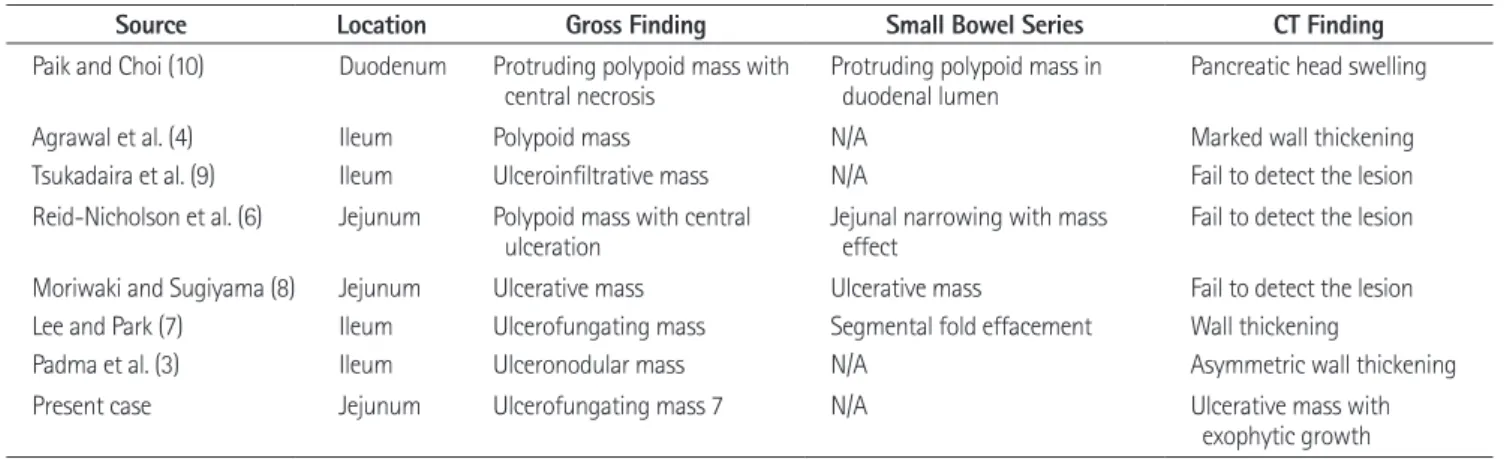
Table 1. Review of Published Cases of Sarcomatoid Carcinoma of the Small Intestine
Source Location Gross Finding Small Bowel Series CT Finding
Paik and Choi (10) Duodenum Protruding polypoid mass with
central necrosis Protruding polypoid mass in
duodenal lumen Pancreatic head swelling
Agrawal et al. (4) Ileum Polypoid mass N/A Marked wall thickening
Tsukadaira et al. (9) Ileum Ulceroinfiltrative mass N/A Fail to detect the lesion Reid-Nicholson et al. (6) Jejunum Polypoid mass with central
ulceration Jejunal narrowing with mass
effect Fail to detect the lesion Moriwaki and Sugiyama (8) Jejunum Ulcerative mass Ulcerative mass Fail to detect the lesion Lee and Park (7) Ileum Ulcerofungating mass Segmental fold effacement Wall thickening
Padma et al. (3) Ileum Ulceronodular mass N/A Asymmetric wall thickening
Present case Jejunum Ulcerofungating mass 7 N/A Ulcerative mass with
exophytic growth Note.-N/A = not applicable
sential diagnostic modalities. Sarcomatoid carcinomas were rare- ly reported in the small intestine, and there were few reported cases which concerned radiologic features and should be differ- entiated. Therefore, data collection on radiologic findings of sar- comatoid carcinomas is important.
Surgical resection remains the mainstay of treatment, as patients show poor responses to chemotherapy and radiation treatment (6). Small intestinal sarcomatoid carcinomas show aggressive be- haviors and have a poor prognosis. The duration of survival is gen- erally only a few months to three years of diagnosis (6, 8). The histological features, tumor stage, and outcome of the reported cases indicate that this neoplasm generally persues a highly ag- gressive and malignant biological course with rapid growth and wide local infiltration which leads to a poor prognosis. The best predictors of outcome in sarcomatoid carcinoma seem to be tu- mor location, size, invasion depth, and the clinical stage of the disease. Radical surgery with adjuvant chemotheraphy, and close follow-up are necessary for the management of this disease.
In conclusion, we present a rare case of sarcomatoid carcino- ma in the small intestine, composed of mixed malignant epithe- lial and mesenchymal cells, with only 26 cases reported to date.
In this case, the CT showed ulcero-fungating mass with necro- sis, which corresponds to the gross morphology. Although it is a nonspecific finding, the sarcomatoid carcinoma must be kept in mind for the differential diagnosis of small intestinal malignan- cies due to its aggressive nature and poor prognosis.
REFERENCES
1. Iezzoni JC, Mills SE. Sarcomatoid carcinomas (carcinosar- comas) of the gastrointestinal tract: a review. Semin Diagn Pathol 1993;10:176-187
2. Robey-Cafferty SS, Silva EG, Cleary KR. Anaplastic and sarcomatoid carcinoma of the small intestine: a clinico- pathologic study. Hum Pathol 1989;20:858-863
3. Padma SK, Permi HS, Patil C, Mathias M. Aggressive mono- phasic sarcomatoid carcinoma of small intestine - a rare case report with review of literature. NUJHS 2012;2:45-47 4. Agrawal S, Trivedi MH, Lukens FJ, Moon C, Ingram EA, Bar- thel JS. Anaplastic and sarcomatoid carcinoma of the small intestine: an unusual tumor. J Clin Gastroenterol 1999;29:99-101
have polypoid growth patterns and can be early diagnosed in their course, and accordingly, are associated with relatively favor- able prognosis. In this case, the patient presented with acute ab- dominal pain and palpable mass was detected through physical examination. We presumed that acute symptom was related to the bowel obstruction which was caused by the tumor seeding.
Although laboratory finding revealed mild anemia, the associa- tion between anemia and the lesion was unclear in this case.
Small intestinal sarcomatoid carcinomas arise most frequently in the jejunum and ileum (2-9). Majority of sarcomatoid carci- nomas in the small intestines showed polypoid or ulcerofungat- ing mass with or without central ulceration, and are frequently necrotic and hemorrhagic, with an average of 7 cm in greatest dimension (range, 3-16 cm) (Table 1) (2-9).
CT findings of small intestinal sarcomatoid carcinoma were described in a few reported cases (Table 1) (1-10). Symmetric or asymmetric wall thickening with fold effacement was noted in three cases (3, 4, 7). CT failed to detect the lesion in three cases (6, 8, 9). Paik and Choi (10) reported duodenal sarcomatoid car- cinoma mimicking focal pancreatic head swelling. Most of the reported cases were not applicable radiologically. In the present case, CT scan showed a predominantly exophytic mass with central necrosis. This imaging finding is consistent with the gross morphology noted here (Fig. 2) and in previous reports (2-10).
It is remarkable how similar the gross specimen is to the CT finding of the mass when compared with the previous reports.
However, this CT finding is remarkably similar to that of the mesenchymal tumor such as GIST, lymphoma, carcinoid or leiomyosarcoma. So, it is not easy to distinguish sarcomatoid carcinoma from mesenchymal tumors radiologically. We pre- sume that it is because monophasic sarcomatoid carcinoma in- cludes minimal epithelial component. Further studies for com- paring CT finding of biphasic and monophasic sarcomatoid carcinoma will be necessary. Small bowel series was done to- gether with the CT in several cases (6-8, 10). The study revealed the intraluminal polypoid mass (6, 8, 10), although CT failed to detect the lesion (6, 8) or mimicked the pancreatic cancer (10).
Thus, it meant that diverse diagnostic tools may be necessary to detect or diagnose the small bowel lesion correctly.
Evaluating primary small intestinal tumors are not easy due to its acceptability. Although immunohistochemistry plays a vi- tal role in the correct diagnosis, CT scans can be one of the es-
Soc 2012;83:321-324
8. Moriwaki Y, Sugiyama M. Severe anemia inducing pre- shock caused by sarcomatoid carcinoma of the small in- testine. Int Surg 2009;94:164-170
9. Tsukadaira A, Koizumi T, Okubo Y, Takashi S, Koide N, Arai K, et al. Small-intestinal sarcomatoid carcinoma with su- perior vena cava syndrome. J Gastroenterol 2002;37:471- 475
10. Paik HJ, Choi YM. A case of carcinosarcoma in duodenum.
J Korean Surg Soc 1991;41:549-553 5. Yucel AF, Kocakusak A, Arikan S, Demirbag N, Tarlaci A,
Batur S. A rare cause of acute abdomen: perforated pri- mary sarcomatoid carcinoma of the small intestine - re- port of a case, with a brief review of the literature. J Can- cer Res Ther 2011;7:348-350
6. Reid-Nicholson M, Idrees M, Perino G, Hytiroglou P. Sarco- matoid carcinoma of the small intestine: a case report and review of the literature. Arch Pathol Lab Med 2004;128:
918-921
7. Lee SE, Park SY. Sarcomatoid carcinoma of the small intes- tine: a rare and highly aggressive tumor. J Korean Surg
소장에 생긴 육종성암: 증례 보고 및 문헌고찰1
강경아
1· 윤정희
2· 강구현
3육종성암은 조직학적으로 상피조직과 간엽조직이 혼재하는 악성종양으로 소장에서는 매우 드문 것으로 알려져 있다. 이 들은 주로 병리학적 증례 형태로 일부 보고되었다. 저자들은 78세 남자의 공장에 생긴 육종성암 1예를 경험하였고 CT에 서는 괴사를 동반한 돌출형 종괴로 보였다. 이에 대한 문헌고찰도 함께 하였다.
1인제대학교 의과대학 상계백병원 영상의학과, 2인제대학교 의과대학 해운대백병원 영상의학과,
3인제대학교 의과대학 상계백병원 병리학과