Introduction
The etiology of osteoporotic vertebral compression frac- ture is multifactorial.23) Osteoporosis is the most common metabolic disorder of the bone and in the case of elderly osteoporotic compression fracture of vertebrae, patients often have risk factors for surgery under general anesthe-
sia.10) If fractured vertebrae are not stabilized early, com- pression may progress, resulting in persistent back or neu- rologic deterioration due to kyphosis. Age-related deformity and secondary medical problems caused by restricted move- ments may additionally develop.19) For the treatment of symptomatic osteoporotic compression fractures, percuta- neous vertebroplasty (PVP) is one of the most effective minimally invasive procedures and has become one of the most rapidly developing techniques in spinal surgery.11)
The goal of PVP is to provide immediate pain relief and bone strengthening in symptomatic vertebral compression fractures.7) Despite some debate, the stabilization of mi- cro-movement, delay of progression of compression, and the thermal and chemical neural damage have been suggest- ed as possible mechanisms of pain relief after PVP.3,5,6,15,21)
There are many studies done on appropriate cement volume in PVP, however, the correlation between the cement volume
Appropriate Cement Volume in Vertebroplasty: A Multivariate Analysis with Short-Term Follow-Up
Hyun Mook Kwon, MD, Sang Pyung Lee, MD, PhD, Jin Wook Baek, MD, and Seong Hwan Kim, MD
Departement of Neurosurgery, Cheju Halla General Hospital, Jeju, Korea
Objective: The optimal threshold of the infusion volume of cement has been a continuous subject in percutaneous verte- broplasty (PVP). This study verifies a causal relationship between the cement volume and the clinical outcome, and sug- gests the parameters of the appropriate volume of cement in PVP.
Methods: This is a retrospective study. One hundred nine patients, who underwent PVP between 2012 and 2015, were in- cluded in the study. Various factors such as patients’ fracture levels, fracture types, fracture body volumes, fracture rates, bone mineral densities, and infused cement volumes were analyzed. Cement infusion ratios were calculated, using total amount of infused cement and fractured body volume. Follow up was done after one-week, one-month and three-months, postoperatively. Changes in the middle body height and the cement leakage levels were monitored and clinical outcomes were evaluated using a visual analogue scale.
Results: Among the variables, the infusion ratio (r=-0.320, p=0.003, Pearson’s correlation) was the only index that showed a significant cause and effect relationship with favorable clinical outcome, except the group with a T-score of higher than -2.5, and the group with a upper thoracic vertebral level. The patients with a cement infusion ratio of 27.8% or more of the fractured body volume had favorable results.
Conclusion: This study showed that high cement infusion ratio revealed favorable outcome in the vertebroplasty of the osteoporotic compression fractures. Infusion ratio of more than 27.8% to osteoporotic compressed vertebrae is optimal for rapid recovery after PVP.
(Korean J Neurotrauma 2016;12(2):128-134) KEY WORDS: Vertebroplasty ㆍInfusion ratio ㆍT-score ㆍBone cements.
Received: July 26, 2016 / Revised: August 25, 2016 Accepted: September 20, 2016
Address for correspondence: Seong Hwan Kim
Departement of Neurosurgery, Cheju Halla General Hospital, 65 Doryeong-ro, Jeju 63127, Korea
Tel: +82-64-740-5036, Fax: +82-64-743-3110 E-mail: mirage97@hanmail.net
cc This is an Open Access article distributed under the terms of Cre- ative Attributions Non-Commercial License (http://creativecommons.
org/licenses/by-nc/3.0/) which permits unrestricted noncommercial use, distribution, and reproduction in any medium, provided the original work is properly cited.
Korean J Neurotrauma 2016;12(2):128-134 https://doi.org/10.13004/kjnt.2016.12.2.128
and clinical outcome has still been under debate.4,13,16,17,22) It has been reported that a higher cement volume improves the results of mechanical restoration of vertebrae by enhanc- ing strength and stiffness in PVP. In a recent cadaver study, PVP increases compression resistant strength and intra- discal pressure when cement volume was infused in at least 15% of the fractured vertebral volume.20) On the other hand, according to another study, cement leakage could be aggra- vated by excessive amount of cement infusion into the frac- tured vertebrae.27)
The purpose of this study is to verify a causal relation- ship between the infused cement volume and the clinical outcome, and suggests the parameters of the appropriate volume of cement in PVP by analyzing cement volume, level of fractured vertebrae, type and grade of fractured ver- tebrae, bone mineral density, and clinical outcome in pa- tients with osteoporotic compression fracture.
Materials and Methods
One hundred nine among two hundred ninety three pa- tients who underwent PVP for thoracolumbar compres- sion fracture in the authors’ hospital, between 2012 and 2015, were retrospectively analyzed. Those who had patho- logic fractures or fractures in more than two vertebral seg- ments were excluded from the study. Three-month follow- up data were analyzed and those who had been lost from the follow up were excluded from this study, as well.
All patients underwent two-weeks of absolute bed rest with conservative treatments prior to PVP except those who were older than 80 years at the time of admission or had medical problems, such as pneumonia. The patients who had -2.5 or lower T-score of any of lumbar or femur in bone mineral density (BMD) received the procedure. The patients were examined for tenderness and motion-induced pain in the fractured site before and after conservative treat- ment and the procedure. computed tomography (CT) and magnetic resonance imaging (MRI) scans were conducted for the definite diagnosis of fractures. With plain X-rays, original body heights of compressed vertebrae were esti- mated by measuring the mean heights of two adjacent ver- tebrae. If the adjacent vertebrae were fractured previously, the nearest vertebrae free from injury were used for estima- tion. Dual-energy X-ray absorptiometry (DXA; Hologic Inc., Waltham, MA, USA) scans were conducted in the fe- mur neck areas as well as lumbar spine to measure the T- score of the BMD. Yet, only the T-scores of the lumbar spines were included for analysis in order to evaluate the correlation between T-scores of the spine and the clinical
results. Moreover, as reference T-scores, the median val- ues were selected, excluding the highest and lowest values among the lumbar T-scores. Thus, some patients were os- teopenic, not osteoporotic, according to the analysis data.
Before the procedures, formal informed consents were taken from all patients and the study was executed in com- pliance with the Helsinki Declaration.
PVP was performed under C-arm monoplane or biplane fluoroscopy. During the procedure, the patient was care- fully monitored with electrocardiogram, sphygmomanom- eter, and pulse oximeter. PVP was performed by three ex- perienced neurosurgeons using the same protocol. Under local anesthesia, the Jamshidi’s needle was inserted into the vertebra through a bilateral pedicle approach. The poly (methyl methacrylate) (PMMA) cement was then infused slowly into one side at a time to prevent cement leakage.
When a cement leakage was detected lateral or anterior to the vertebrae, cement infusion was discontinued temporar- ily and carefully resumed only when no additional leakage was confirmed. When additional leakage was observed, cement infusion was suspended. When leakage into the vertebral foramen was suspected, cement infusion was halted immediately, the patient was examined neurologi- cally, and the extent of canal encroachment was verified with CT scan, postoperatively. The patients started ambu- lation with braces on, six hours after the procedure. Fur- ther compression after the procedure was checked with a standing view of plain spine X-rays.
When cement leakage occurred, the type of the leakage was categorized with an X-ray or CT scan, according to the method by Yeom et al.28) The semi-quantitative visual grading of vertebral fracture was determined by the classi- fication of Genant et al.8) Intervertebral vacuum cleft (IVVC) and preexisting adjacent fractures were identified. The
‘infusion ratio’ was calculated by dividing the infused ce- ment volume by the fractured body volume. The fractured body volume, the compression rate, the compression pro- gression rate, and the amount of cement leakage were mea- sured with X-ray and CT scans using the PiViewSTAR (INFINITT; Infinitt Healthcare, Seoul, Korea) Dicom view- er. Volumetric values were obtained by accumulation of the width of vertebral bodies in 3 mm-thick sliced images, which were calculated and interpreted by one technician.
The pain was scored using the visual analogue scale (VAS) score from zero to ten. Patients were followed up on first week, after one-month, and three-months, postoperatively.
Patients who had a VAS score less than three were classi- fied as ‘favorable outcome’ group whereas three or higher as ‘unfavorable outcome’ group.
Statistical analyses
T-tests, ANOVA, χ2 tests, Fisher’s exact tests, and bivar- iate correlation analyses were conducted on multiple vari- ables such as volumetric data, outcome, age, sex, and the T-score. Simple linear regression analyses were applied to sequent variables. The mean and the mean dispersion of the measured variables were verified, and their 95% con- fidence interval was confirmed. Multiple linear regression analyses were applied to the factors that affect the cement infusion ratio and the volumetric data. The receiver-oper- ating characteristic (ROC) curve and the area under the curve (AUC) were measured to confirm the correlation be- tween the infusion ratio and the clinical outcome of PVP.
The cut-off value was determined based on the maximum AUC value as having high sensitivity and specificity. Sta- tistical significance was accepted when the p-value was less than 0.05 (SPSS statistical software 19.0, SPSS Inc., Chicago, IL, USA).
Results
The patient demographics are summarized on Table 1.
The mean age of the 109 patients included in the study was 74.1±10.3 (range, 44-94), and 86 of them (78.9%) were female. The mean follow-up period was 205 days (range, 92-1,136). The mean BMD was 0.71±0.15 g/cm2 (range, 0.28-1.30), and the mean T-score was -2.86±0.96 (range, 0.74-4.91). The number of patients with a spinal T-score of lower than -2.5 was 74 (67.9%). Fracture level was cate- gorized as upper thoracic (T7-10), thoracolumbar (T11- L1), and lumbar (L2-5). Thoracolumbar injury was pre- dominant above all, 74 cases (67.9%). Based on the fracture classification suggested by Genant et al.8), 40 cases of wedge type (36.7%), 37 cases of crush type (34.0%) and 32 cases of biconcave type (29.7%) were verified. Cement leakage occurred in 71 patients (65.1%). Most cases were of the cor- tical and segmental type, only 2 cases (1.8%) were basiver- TABLE 1. Characteristics of the patient group
Number of patients (n) 109
Gender
Male 23 (21.1%)
Female 86 (78.9%)
Age (range) 74.1±10.3 (44-94)
Follow-up periods (days) 205 (92-1136)
Spine T-score -2.86±0.96 (0.74-4.91)
Fracture level
T7-T10 11 (10.1%)
T11-L1 74 (67.9%)
L2-L5 24 (22.0%)
Fracture type Mild Moderate Severe
Wedge, n=40 (36.7%) 16 18 6
Biconcave, n=32 (29.4%) 14 12 6
Crush, n=37 (34.0%) 17 17 3
Cement leakage (n) 71 (65.1%)
Basivertebral 02 (1.8%)
Cortical 40 (36.7%)
Segmental 25 (23.0%)
Intervertebral discal 04 (3.7%)
None 38 (34.9%)
IVVC 10 (9.2%)
VAS score Total Favorable group Unfavorable group
Pre Op 8.2 8.1 8.3
Post Op
1 week 3.3 2.9 4.5
1 month 3.2 1.6 3.6
3 months 1.4 0.7 3.7
Outcome
Favorable 83 (76.1%)
Unfavorable 26 (23.9%)
VAS: visual analogue scale, IVVC: intravertebral vacuum cleft, Op: operation
tebral type, according to the classification by Yeom et al.28) IVVC exist in 10 patients (9.2%). The mean fracture rate of female patients was 12.58±11.63% (range, 10.1-15.17), whereas that of male patients was 11.88±9.28% (range, 7.9-15.9). The mean infusion ratio of each female and male patient was 24.87±7.51% (range, 23.7-26.5), and 3.18±
5.91% (range, 20.6-25.7). The number of patients who showed favorable outcome was 83 (76.1%) while unfavor- able outcome was 26 (23.9%). Their mean VAS of preop- erative, one-week, one-month and three months postoper- atively were 8.1, 2.9, 1.6, and 0.7, respectively, in the favorable
prognosis group. The values were 8.3, 4.5, 3.6 and 3.7, re- spectively, in the unfavorable prognosis group (Table 1).
Variables of fractured vertebral body volume in favorable outcome group
Among the favorable outcome group, bivariate correlate analyses were conducted on continuous variables such as age, T-score, fracture rate, intervertebral cement volume, as well as the categorical variables such as gender and ver- tebral level. The fractured vertebral body volume had cor- relations with sex, T-score, fracture rate, and fracture lev- el, except for patient’s age (Table 2).
Relationship between infusion ratio and clinical outcome
The mean infusion ratio (%) of all patients was 24.61±
7.40%. The mean infusion ratio of the favorable group was 25.21±7.14%, and that of the unfavorable group was 22.71±7.90% in the first week follow up. The mean differ- ence was 2.50%. Interestingly, the mean infusion ratio of the favorable group after a month postoperatively was 28.13±8.26%, and that of the unfavorable group was 23.02±6.42% showing a mean difference of 5.11%. In ac- cordance with the follow-up period, the infusion ratio of TABLE 2. Variables of fractured vertebra body volume in favor-
able outcome Variables
Fractured body volume Pearson's
correlation Significance (2-tailed)
Sex (M/F) -0.450 0.001
Age -0.115 0.342
Spine T-score 0.287 0.016
Fracture rate -0.389 0.001
Level (T7 to L5) 0.588 0.001
fracture rate=[(original body height-fracture body height)/
original body height]×100. M: male, F: female
TABLE 3. Infusion ratio depending on group and time interval
Groups Infusion ratio
1 week 1 month
Favorable group 25.21±7.14% 28.13±8.26%
Unfavorable group 22.71±7.90% 23.02±6.42%
TABLE 4. Variables affecting favorable outcome Variables
Favorable outcome Pearson's
correlation Significance (2-tailed)
Sex (M/F) -0.030 0.790
Age -0.101 0.363
Spine T-score -0.083 0.456
Fracture rate 0.026 0.817
Infused cement volume -0.115 0.299
Infusion ratio -0.320 0.003
fracture rate=[(original body height-fracture body height)/
original body height]×100. M: male, F: female
TABLE 5. The correlation of infusion ratio according to T-score and fracture level
Spine T-core n VAS (mean) Correlation p-value
> -2.50 41 3.2 -0.172 0.282
<-2.50 68 3.1 -0.406* 0.001
Fracture level
T7-T10 11 4.09 -0.149 0.662
T11-L1 74 2.97 -0.333 0.004
L2-L5 24 3.33 -0.416 0.004
*correlation is significant at the 0.01 level (2-tailed) . VAS: visual analogue scale
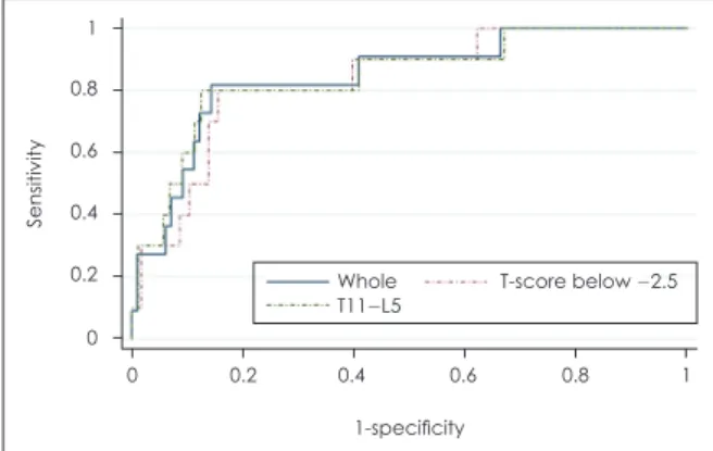
FIGURE 1. The infusion ratios were 27.8% (80.0% sensitivity and 84.5% specificity) and 28.6% (80.0% sensitivity and 87.5%
specificity) in the -2.5 or lower T-score group and the T11-L5 group respectively. An infusion rate of at least 27.8% may be required to obtain favorable outcomes.
1 0.8 0.6 0.4 0.2
0
0 0.2 0.4 0.6 0.8 1 1-specificity
Sensitivity
Whole T-score below -2.5 T11-L5
favorable outcomes had changed (Table 3). Bivariate anal- yses were conducted on the various factors that might af- fect the outcome of PVP in the whole group and the favor- able outcome group. The infusion ratio (r=-0.320, p=0.003, Pearson’s correlation) was the only index that showed a significant cause and effect relationship with favorable clinical outcome (Table 4). The results of the linear regres- sion analyses on the infusion ratio and outcomes were also statistically significant ANOVA (p<0.001). The maximum ROC AUC value of the infusion ratio and the outcome was 0.85 at a 27.83% infusion ratio with sensitivity and speci- ficity 81.8% and 85.7%, respectively (Figure 1).
Correlation between the infusion ratio and outcomes depending on T-score and fractured level
In the spinal T-score comparison, the ‘normal to osteo- porotic’ group, with a T-score of higher than -2.5 did not demonstrate a significant correlation between infusion ra- tio and their outcomes (p=0.282). In the comparison by compressed vertebral levels, no significant correlation be- tween infusion ratio and their outcomes was observed in the upper thoracic level group (p=0.662). Whereas in the comparison between the group with a T-score of lower than -2.5, and the group with a T11 or lower vertebral lev- el, a significant correlation was observed between infu- sion ratio and their outcomes (Table 5). The maximum AUC value in the -2.5 or lower T-score group and the T11- L5 group were 0.83 and 0.84 respectively, with infusion ratio of 27.83% (80.0% sensitivity and 84.5% specificity) and 28.64% (80.0% sensitivity and 87.5% specificity), show- ing no significant differences. Therefore, an infusion ratio of at least 27.83% may be required to obtain favorable out- comes (Figure 1).
The correlation between the infusion ratio and leakage The correlation between infusion ratio of the cement and leakage type was analyzed. According to the leakage criteria suggested by Yeom et al.28), the cortical defect leak- age type was observed in 40 patients (36.8%), the segmen- tal vein leakage type in 25 patients (23.0%), the basivertebral vein leakage type in 2 patients (1.8%), and the intradiscal leakage type in 4 patients (3.7%). Logistic regression anal- yses were conducted on each type of cement leakage, which did not show any significant correlation with the infusion ratio (p=0.152).
Discussion
PVP is a simple, noninvasive, low-risk procedure that
provides immediate and durable pain relief and functional improvement to patients with osteoporotic vertebral com- pression fractures.1,26) Although its simplicity and safety, clinical success of PVP is another issue, influenced by the injected volume of PMMA and BMD.2) Based upon the hy- pothesis that vertebral stability is determined by the cement infusion volume, sufficient cement infusion is crucial in PVP. In this study, the authors focused on verifying an op- timal intervertebral cement volume for favorable outcomes.
There are multiple studies, which investigated the cor- relation between the intravertebral cement volume and the clinical outcome through in vitro, in vivo and experimen- tal studies.9,12-14,16-18,24) However, the consensus has not yet been established, since the optimal cut-off value varies ac- cording to each study. Al-Ali et al.1) and Kaufmann et al.13) asserted that there was no significant correlation between the infused cement volume and clinical outcomes. Howev- er, their mean cement volume ranged from 3.0 to 3.4 mL, which was a small amount, and they analyzed the infused cement volume only, not considering the severity of the fracture. Hence, the scientific evidence has been still in- sufficient to contend how infused cement volume is im- portant to obtain successful outcome in vertebroplasty.
According to a cadaveric study of Liebschner et al.16), the minimal cement infusion ratio to recover the physical strength of compressed vertebrae was 15%. Kim et al.14), on the other hand, suggested 30% of infusion ratio for re- covering the normal strength level of compressed verte- brae through experimental simulation study. Notwithstand- ing the difference of value, many studies assert that cement infusion ratio contributes to the recovery of the mechani- cal strength and stiffness.9,14,16-18) If the optimal amount of cement is determined solely based on each surgeon’s ex- perience, however, there is high possibility of inconsisten- cy since the amount of cement may vary depending on the vertebral level, in addition to the surgeon bias. Thus, it is important to review the patients’ fracture status carefully and establish a solid plan of cement volume amount before the procedure. In order to facilitate the planning, measur- ing optimal cement infusion ratio is necessary as a refer- ence value.
In the most recent in vivo study of Jin et al.12) on osteopo- rotic vertebral fracture, a minimal cement infusion ratio of 11.64% was suggested for reducing pain and minimiz- ing complication developments after the procedure. They advocated a minimal cement infusion ratio to relieve pain, emphasizing the risk of complications as a consequence of massive cement infusion. On the other hand, Nieuwenhui- jse et al.24) suggested that a higher cement infusion ratio of
24% is required to recover physical strength of the com- pressed vertebra and reduce pain. In this study, we sug- gested as high as a 27.8% infusion ratio as an effective ra- tio for favorable outcomes. The ratio was similar to that of Kim et al.14), whose in vitro study determined that a 30%
infusion ratio was effective for strength and stiffness re- covery. In our study, the mean infusion ratio of the favor- able group was 25.21±7.14% in the first week follow up.
After a month, however, the value became 28.13±8.26%, which implies that even the pain was relieved with less than 27.8% of infusion ratio, temporarily, and it ended up aggravating as time goes by. In other words, patients who underwent PVP with less than 27.8% of infusion ratio grad- ually went over from favorable outcome group to unfavor- able outcome group.
Leakage of cement out of the vertebral body may be subclinical, but it also may bring on devastating complica- tions such as pulmonary embolism, neurological, and ad- ditional fractures through the endplate.25,27) In a study of the major complications of cement leakage by Ryu et al.27), the complication of the cement leakage was caused by a high infusion volume. The mean infusion ratio of the leak- age group was slightly higher than that of the non-leakage group. However, in this study, significant correlation be- tween the infusion ratio and cement leakage was not found on the linear regression analysis. The difference of the two studies are that, Ryu et al.27) compared the group using in- jector for cement infusion with the group of manual infu- sion, whereas, all patients underwent manual infusion in our study. Overall frequency of cement leakage was rela- tively higher in this study, however, there was no serious clinical complication such as symptomatic pulmonary embolism or procedure related paralysis. We believe that was because the operators abide by our protocol, strictly;
slow manual cement infusion into one side at a time, tem- porary discontinuance of infusion on a lateral or anterior cement leakage, and cessation of cement infusion on sus- picion of leakage into the vertebral foramen.
In a volumetric analysis on the associated factors, the osteopenia group showed no correlation between the ce- ment infusion ratio and their clinical outcomes, whereas the group with a T-score of lower than -2.5 showed a signif- icant correlation. This result means, in an osteopenic group, paying attention to the avoidance of complications is rec- ommended rather than increasing the infusion ratio. Mean- while, in an osteoporotic group, attainment of the infusion ratio up to 27.8% is recommended. A similar result was found in Graham’s study of the correlation between the BMD and infusion ratio, patients with a low T-score re-
quired the use of a higher infusion volume for favorable outcomes.9) In the upper thoracic level, above the 11th tho- racic spine, there was less correlation between the infu- sion ratio and clinical outcomes, comparing with the tho- racolumbar junction and lumbar area. Thus, patients with fractures in the upper thoracic level should be managed focusing on avoiding complications.
The goal of this study is to seek an optimal infusion ra- tio through tailored methods to improve the clinical out- come. Previously, one study found an optimal infusion ra- tio by the use of a maximum AUC point, the other study found it using a high specificity point.12,24) In our study, a 27.8% infusion ratio was determined using the maximum AUC point of the ROC curve (Figure 1). In regards to op- timal infusion volume, Jin et al.12), recommended an infu- sion ratio of 11.64% which presented sensitivity and speci- ficity of 37% and 93%, whereas 24% by Nieuwenhuijse et al.24), showed sensitivity and specificity of 80.0% and 64%.
Comparing our report with these two reports, our results showed higher reliability, since both sensitivity and speci- ficity was high as 80.0% and 87.5%, respectively.
This study has limitations in that since many patients have short follow-up period, our data does not yet provide definitive conclusions on the efficacy of the procedure.
Another weakness of this study was that VAS score was solely utilized as an indicator of clinical outcome, so that the study had limits in reflecting functional outcome. The correlation between the types and degrees of fracture and the clinical outcome was yet analyzed. Multivariate analy- sis including more variables is required.
Conclusion
This study showed high cement ratio revealed favorable outcome in the vertebroplasty of the osteoporotic compres- sion fractures. Infusion ratio of more than 27.8% to osteo- porotic compressed vertebrae is optimal. In order to obtain better clinical outcome, calculation of the target volume of cement using infusion ratio before vertebroplasty is rec- ommended.
■ The authors have no financial conflicts of interest.
REFERENCES
1) Al-Ali F, Barrow T, Luke K. Vertebroplasty: what is important and what is not. AJNR Am J Neuroradiol 30:1835-1839, 2009 2) Baumann C, Fuchs H, Kiwit J, Westphalen K, Hierholzer J. Com-
plications in percutaneous vertebroplasty associated with punc- ture or cement leakage. Cardiovasc Intervent Radiol 30:161-168, 3) Belkoff SM, Mathis JM, Jasper LE, Deramond H. The biome-2007
chanics of vertebroplasty. The effect of cement volume on me- chanical behavior. Spine (Phila Pa 1976) 26:1537-1541, 2001 4) Boszczyk B. Volume matters: a review of procedural details of
two randomised controlled vertebroplasty trials of 2009. Eur Spine J 19:1837-1840, 2010
5) Dahl OE, Garvik LJ, Lyberg T. Toxic effects of methylmethacry- late monomer on leukocytes and endothelial cells in vitro. Acta Orthop Scand 65:147-153, 1994
6) Deramond H, Wright NT, Belkoff SM. Temperature elevation caused by bone cement polymerization during vertebroplasty.
Bone 25:17S-21S, 1999
7) Galibert P, Deramond H, Rosat P, Le Gars D. Preliminary note on the treatment of vertebral angioma by percutaneous acrylic vertebroplasty. Neurochirurgie 33:166-168, 1987
8) Genant HK, Wu CY, van Kuijk C, Nevitt MC. Vertebral fracture assessment using a semiquantitative technique. J Bone Miner Res 8:1137-1148, 1993
9) Graham J, Ahn C, Hai N, Buch BD. Effect of bone density on vertebral strength and stiffness after percutaneous vertebroplas- ty. Spine (Phila Pa 1976) 32:E505-E511, 2007
10) Harrop JS, Prpa B, Reinhardt MK, Lieberman I. Primary and secondary osteoporosis’ incidence of subsequent vertebral com- pression fractures after kyphoplasty. Spine (Phila Pa 1976) 29:
2120-2125, 2004
11) Jensen ME, Evans AJ, Mathis JM, Kallmes DF, Cloft HJ, Dion JE. Percutaneous polymethylmethacrylate vertebroplasty in the treatment of osteoporotic vertebral body compression fractures:
technical aspects. AJNR Am J Neuroradiol 18:1897-1904, 1997 12) Jin YJ, Yoon SH, Park KW, Chung SK, Kim KJ, Yeom JS, et al.
The volumetric analysis of cement in vertebroplasty: relationship with clinical outcome and complications. Spine (Phila Pa 1976) 36:E761-E772, 2011
13) Kaufmann TJ, Trout AT, Kallmes DF. The effects of cement vol- ume on clinical outcomes of percutaneous vertebroplasty. AJNR Am J Neuroradiol 27:1933-1937, 2006
14) Kim JM, Shin DA, Byun DH, Kim HS, Kim S, Kim HI. Effect of bone cement volume and stiffness on occurrences of adjacent ver- tebral fractures after vertebroplasty. J Korean Neurosurg Soc 52:
435-440, 2012
15) Leeson MC, Lippitt SB. Thermal aspects of the use of polymeth- ylmethacrylate in large metaphyseal defects in bone. A clinical review and laboratory study. Clin Orthop Relat Res:239-245, 1993 16) Liebschner MA, Rosenberg WS, Keaveny TM. Effects of bone
cement volume and distribution on vertebral stiffness after verte- broplasty. Spine (Phila Pa 1976) 26:1547-1554, 2001
17) Luo J, Daines L, Charalambous A, Adams MA, Annesley-Wil- liams DJ, Dolan P. Vertebroplasty: only small cement volumes are required to normalize stress distributions on the vertebral bod- ies. Spine (Phila Pa 1976) 34:2865-2873, 2009
18) Luo J, Skrzypiec DM, Pollintine P, Adams MA, Annesley-Wil- liams DJ, Dolan P. Mechanical efficacy of vertebroplasty: influ- ence of cement type, BMD, fracture severity, and disc degenera- tion. Bone 40:1110-1119, 2007
19) Lyles KW, Gold DT, Shipp KM, Pieper CF, Martinez S, Mulhau- sen PL. Association of osteoporotic vertebral compression frac- tures with impaired functional status. Am J Med 94:595-601, 1993 20) Martinčič D, Brojan M, Kosel F, Štern D, Vrtovec T, Antolič V,
et al. Minimum cement volume for vertebroplasty. Int Orthop 39: 727-733, 2015
21) Mjöberg B, Pettersson H, Rosenqvist R, Rydholm A. Bone ce- ment, thermal injury and the radiolucent zone. Acta Orthop Scand 55:597-600, 1984
22) Molloy S, Riley LH, 3rd, Belkoff SM. Effect of cement volume and placement on mechanical-property restoration resulting from vertebroplasty. AJNR Am J Neuroradiol 26:401-404, 2005 23) Nam DH, Park KH, Kim TW, Chi MP, Kim JO. The effect of
trauma in osteoporotic vertebral compression fractures treated by percutaneous vertebroplasty: a comparison of radiological fea- tures in presence or absence of trauma. J Korean Neurotrauma- tol Soc 7:29-34, 2011
24) Nieuwenhuijse MJ, Bollen L, van Erkel AR, Dijkstra PD. Opti- mal intravertebral cement volume in percutaneous vertebroplasty for painful osteoporotic vertebral compression fractures. Spine (Phila Pa 1976) 37:1747-1755, 2012
25) Nieuwenhuijse MJ, van Rijswijk CS, van Erkel AR, Dijkstra SP.
The intravertebral cleft in painful long-standing osteoporotic ver- tebral compression fractures treated with percutaneous vertebro- plasty: diagnostic assessment and clinical significance. Spine (Phila Pa 1976) 37:974-981, 2012
26) Ploeg WT, Veldhuizen AG, The B, Sietsma MS. Percutaneous ver- tebroplasty as a treatment for osteoporotic vertebral compression fractures: a systematic review. Eur Spine J 15:1749-1758, 2006 27) Ryu KS, Park CK, Kim MC, Kang JK. Dose-dependent epidural
leakage of polymethylmethacrylate after percutaneous vertebro- plasty in patients with osteoporotic vertebral compression frac- tures. J Neurosurg 96:56-61, 2002
28) Yeom JS, Kim WJ, Choy WS, Lee CK, Chang BS, Kang JW. Leak- age of cement in percutaneous transpedicular vertebroplasty for painful osteoporotic compression fractures. J Bone Joint Surg Br 85:83-89, 2003