ABSTRACT
This is a report of a 58-year-old female with Cushing syndrome who underwent posterior lumbar fusion and lost both her vision completely. She was diagnosed with posterior ischemic optic neuropathy. Cushingoid features such as buffalo hump and central obesity might have attributed in triggering posterior ischemic optic neuropathy. When laid prone for surgery, perioperative high abdominal pressure causes venous hypertension leading to increase amount of blood loss. To compensate, infusion of large quantities of intravenous fluids is necessary which leads to hemodilution which decreases ocular perfusion pressure.
Hypercoagulability of Cushing syndrome is also potentially a risk factor of this condition which increases the incidence of venous thromboembolism. For there is no known effective treatment for posterior ischemic optic neuropathy, means to prevent this complication must be strategically reviewed. When performing long spine surgery on patient who has Cushing syndrome or cushingoid features, caution must be taken to avoid this devastating complication.
Keywords: Spinal fusion; Cushing syndrome; Posterior ischemic optic neuropathy;
Intraocular pressure
INTRODUCTION
Blindness after nonocular surgery is a rare and catastrophic complication. One or more small ciliary arteriole occlusions in the optic nerve are responsible for the infarction.13) While the pathogenesis of ischemic optic neuropathy is not fully understood, prone position, anemia, hypotension and venous congestion have been suggested as possible risk factors.6,7,9,14,17,18)
This is a report of a female with iatrogenic Cushing syndrome who lost both her vision after spinal surgery. Informed consent statement was obtained for this study.
CASE REPORT
A 58-year-old female underwent spinal fusion surgery for spinal stenosis and scoliosis to correct coronal imbalance on September 13, 2010. She suffered from bilateral leg weakness Korean J Neurotrauma. 2019 Oct;15(2):221-226
https://doi.org/10.13004/kjnt.2019.15.e39 pISSN 2234-8999·eISSN 2288-2243
Case Report
Received: Sep 11, 2019 Revised: Oct 14, 2019 Accepted: Oct 16, 2019 Address for correspondence:
Seung-Won Choi
Department of Neurosurgery, College of Medicine, Chungnam National University, 282 Munhwa-ro, Jung-gu, Daejeon 35015, Korea.
E-mail: [email protected]
Copyright © 2019 Korean Neurotraumatology Society
This is an Open Access article distributed under the terms of the Creative Commons Attribution Non-Commercial License (https://
creativecommons.org/licenses/by-nc/4.0/) which permits unrestricted non-commercial use, distribution, and reproduction in any medium, provided the original work is properly cited.
ORCID iDs Bumsoo Park
https://orcid.org/0000-0001-7828-4371 Seung-Won Choi
https://orcid.org/0000-0001-8610-887X Conflict of Interest
The authors have no financial conflicts of interest.
Bumsoo Park , Seung-Won Choi , Sanghyun Han, Jin-Young Youm, Jeong-Wook Lim, and Hyon-Jo Kwon
Department of Neurosurgery, College of Medicine, Chungnam National University, Daejeon, Korea
Cushing Syndrome: A Potential Risk of
Bilateral Postoperative Ischemic Optic
Neuropathy after Lumbar Fusion
and aggravated back pain. On magnetic resonance imaging (MRI) scan, multilevel stenosis was found on lumbar spine accompanying scoliosis. To control the symptoms, she went to several hospitals and had frequent steroid injections on her back and knee joints. As a result of numerous injections over a long period of time, she manifested cushingoid features when she came to our institute. She had facial flushing, moon face, buffalo torso, central obesity with thin arms and legs, multiple bruising with thin skin and severe atrophy on her extremities. Her body mass index (BMI) was 22.2 kg/m2. Iatrogenic Cushing syndrome was confirmed after running several tests such as 24-hour urinary free cortisol testing, overnight dexamethasone test and rapid adrenocorticotropic hormone test.
She suffered from weakness which the motor grade for hip flexion were GII. Under the diagnosis of acute bilateral polyradiculopathy of L4-S1 and scoliosis, laminectomy and discectomy of L1/2, L3/4 L4/5, interbody fusion of L3/4 and L4/5 with screw fixation from L1 to L5 was planned (FIGURE 1).
On the day of surgery, the patient was allocated in prone position on a spine table placing her head on a soft foam cushions with cutouts for the eyes, nose and mouth. It took 11 hours to complete the operation which was longer than expected. The total amount of blood loss was approximately 13,000 mL and her hemoglobin and haematocrit fell from 13.2 and 40.4 to the lowest of 5.8 and 17 respectively. Intraoperatively, the lowest systolic blood pressure was 70 mmHg which maintained for 5 hours. For fluid resuscitation, 16,000 mL of crystalloid and 1,000 mL of colloid was infused with 16 packs of packed red blood cell and 6 packs of fresh frozen plasma.
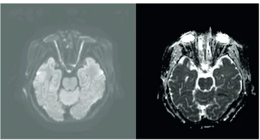
Postoperatively she was sent to intensive care unit but didn't complain anything about her eyes. She said she saw everything bright and thought it was because of the ointment applied on her eyes. Not until the fifth day she complained about her bilateral blindness which brought our attention. On seeing the ophthalmologists, her eyelids were swollen for over 5 days but the intraocular pressure was 18 mmHg on her right and 19 mmHg on her left. She had anisocoric pupil, right being bigger than the left with no response to light. Fundoscopic examination showed flat and pale disc without swelling on both eyes (FIGURE 2). On the orbit MRI, the optic nerve showed high signal intensity on diffuse weighted image, diffusion FIGURE 1. Postoperative X-ray image showing interbody fusion and screw fixation from L1 to L5.
restriction on apparent diffusion coefficient map and mild diffuse peripheral enhancement along intraorbital segment (FIGURE 3 & 4). Low dose steroid was administered after surgery for Cushing syndrome but upon the funduscopic finding which was evident of posterior ischemic optic neuropathy it was surmounted to high-dose.
She had recovered light reflex and a small island of vision in inferotemporal quadrant of her left eye after a month. A year after, she was able to discriminate people from objects with the left eye. However, her right eye remained blind.
DISCUSSION
The incidence of postoperative visual loss after nonocular surgeries account for 0.028% to 0.2%.1) There could be several explanation for postoperative visual loss which should be distinguished from central retinal occlusion and cortical blindness. Among them, posterior ischemic optic neuropathy (PION) is known to be most common.14) Suggestive risk factors of PION include prolonged surgery in the prone position, venous congestion of the head, large blood loss with large fluid resuscitation, hypotension, anemia, use of vasopressors, emboli, Cushing Syndrome Potential Risk of PION
FIGURE 2. Fundoscopic image showing posterior ischemic optic neuropathy. Both eyes have flat and pale disc without swelling.
FIGURE 3. Orbit magnetic resonance imaging showing high signal intensity on the optic nerve on diffuse weighted image and diffusion restriction on apparent diffusion coefficient map.
and susceptible patient vascular anatomy and physiology.6,7,9,14,17,18) Many risk factors fell into the categories in this case mainly due to iatrogenic Cushing syndrome.
Glucocorticoids are frequently used for both diagnostic and therapeutic purposes.5) It has been more than 60 years since the introduction of glucocorticoid therapy as an effective treatment for patients with inflammatory process. Although glucocorticoid therapy has been widely accepted as an essential part of certain clinical settings, long-term administration can suppress the hypothalamic pituitary-adrenal axis, causing secondary adrenocortical insufficiency with surgical or medical stress.12) It is impossible to predict the regimen that will suppress the hypothalamic-pituitary-adrenocortical axis and by that increase the risk of developing adrenal insufficiency.11)
The most common cause of Cushing syndrome is iatrogenic and it is the consequence of long-term glucocorticoid administration using more than 7.5 mg prednisone per day.2) If the amount of nerve blocks and joint injections with steroids are not counted, patients will be susceptible to this syndrome after steroid abuse. The syndrome itself increases the risk of peri- and postoperative complications.
The most common cause of cushingoid features is iatrogenic corticosteroid use which increases circulating corticosteroid levels leading to Cushing syndrome.3) Buffalo torso and central obesity could have adversely affected the patient during surgery.7,16) Prone position itself raises intraocular pressure which rises significantly with time. This could potentially drop the perfusion pressure of the ciliary arterioles to cortical values.8) Buffalo torso aggravated head congestion for making it hard to place the head at the level of heart or higher.
FIGURE 4. Orbit magnetic resonance imaging showing mild diffuse peripheral enhancement along intraorbital segment.
BMI was reported to influence intraabdominal pressure in the prone position more than in the supine position during lumbar spinal surgery. A positive correlation of blood loss and intraabdominal pressure and with BMI in the prone position was reported.10,15) Intraocular pressure also rises if there is direct pressure on the abdomen.8) Placing the patient in prone position causes intraabdominal pressure to increase followed by venous hypertension which leads to intraoperative bleeding. This case eventually resulted in massive bleeding which needed fluid resuscitation.16) Even though central venous pressure is within normal range, infusion of large quantities of fluids in the head-down position may cause venous congestion in the eyes. Increased venous pressure can also cause increased intraocular pressure within the eyes. It is thought to be one of the main factors responsible for triggering posterior ischemic optic neuropathy in susceptible patients. It creates an environment resembling compartment syndrome at the level of the optic canal which obstructs the arterial blood flow to the optic nerve.8)
Hypercoagulability is also an issue of Cushing syndrome which increases the incidence of venous thromboembolism. The risk of developing venous thromboembolism has been reported to be 10 times higher in patients with Cushing syndrome. In addition, the risk is even higher after major spine surgery. Based on many studies, increase levels of procoagulant factors especially VIII, IX, and von Willebrand factor, and impaired fibrinolytic capacity leads to hypercoagulable state in Cushing syndrome.4) Increased production of procoagulant factors with activation of the coagulation cascade shortens activated partial thromboplastin time and an impaired fibrinolytic capacity increases clot lysis time which causes hypercoagulability in Cushing syndrome.19)
CONCLUSION
For there is no known effective treatment for posterior ischemic optic neuropathy, means to prevent this complication must be strategically reviewed.16) When performing long spine surgery on patient who has Cushing syndrome or cushingoid features caution must be taken to avoid this devastating complication.
REFERENCES
1. Baig MN, Lubow M, Immesoete P, Bergese SD, Hamdy EA, Mendel E. Vision loss after spine surgery:
review of the literature and recommendations. Neurosurg Focus 23:E15, 2007 PUBMED | CROSSREF
2. Chabre O. Cushing syndrome: Physiopathology, etiology and principles of therapy. Presse Med 43:376- 392, 2014
PUBMED | CROSSREF
3. Chaudhry HS, Singh G. Cushing syndrome. Treasure Island, FL: StatPearls Publishing, 2019
4. Coelho MC, Santos CV, Vieira Neto L, Gadelha MR. Adverse effects of glucocorticoids: coagulopathy. Eur J Endocrinol 173:M11-M21, 2015
PUBMED | CROSSREF
5. Cota D, Ceroni L, Pasquali R. Steroid therapy and adrenal function. Recenti Prog Med 91:594-600, 2000 PUBMED
6. Dunker S, Hsu HY, Sebag J, Sadun AA. Perioperative risk factors for posterior ischemic optic neuropathy. J Am Coll Surg 194:705-710, 2002
PUBMED | CROSSREF
Cushing Syndrome Potential Risk of PION
7. Epstein NE. Perioperative visual loss following prone spinal surgery: a review. Surg Neurol Int 7 Suppl 13:S347-S360, 2016
PUBMED | CROSSREF
8. Goepfert CE, Ifune C, Tempelhoff R. Ischemic optic neuropathy: are we any further? Curr Opin Anaesthesiol 23:582-587, 2010
PUBMED | CROSSREF
9. Goyal A, Elminawy M, Alvi MA, Long TR, Chen JJ, Bradley E, et al. Ischemic optic neuropathy following spine surgery: Case control analysis and systematic review of the literature. Spine 44:1087-1096, 2019 PUBMED | CROSSREF
10. Han IH, Son DW, Nam KH, Choi BK, Song GS. The effect of body mass index on intra-abdominal pressure and blood loss in lumbar spine surgery. J Korean Neurosurg Soc 51:81-85, 2012 PUBMED | CROSSREF
11. Holland EG, Taylor AT. Glucocorticoids in clinical practice. J Fam Pract 32:512-519, 1991 PUBMED
12. Kajitani M, Tsuzaki K. Steroid. Masui 65:1119-1124, 2016 PUBMED
13. Katz DM, Trobe JD, Cornblath WT, Kline LB. Ischemic optic neuropathy after lumbar spine surgery. Arch Ophthalmol 112:925-931, 1994
PUBMED | CROSSREF
14. Lee LA, Newman NJ, Wagner TA, Dettori JR, Dettori NJ. Postoperative ischemic optic neuropathy. Spine 35 Suppl:S105-S116, 2010
PUBMED | CROSSREF
15. Malhotra A, Gupta V, Abraham M, Punetha P, Bundela Y. Quantifying the amount of bleeding and associated changes in intra-abdominal pressure and mean airway pressure in patients undergoing lumbar fixation surgeries: a comparison of three positioning systems. Asian Spine J 10:199-204, 2016
PUBMED | CROSSREF
16. Roth S, Moss HE. Update on perioperative ischemic optic neuropathy associated with non-ophthalmic surgery. Front Neurol 9:557, 2018
PUBMED | CROSSREF
17. Stevens WR, Glazer PA, Kelley SD, Lietman TM, Bradford DS. Ophthalmic complications after spinal surgery. Spine 22:1319-1324, 1997
PUBMED | CROSSREF
18. Su AW, Lin SC, Larson AN. Perioperative vision loss in spine surgery and other orthopaedic procedures. J Am Acad Orthop Surg 24:702-710, 2016
PUBMED | CROSSREF
19. van der Pas R, Leebeek FW, Hofland LJ, de Herder WW, Feelders RA. Hypercoagulability in Cushing's syndrome: prevalence, pathogenesis and treatment. Clin Endocrinol (Oxf ) 78:481-488, 2013 PUBMED | CROSSREF