ISSN: 2233-601X (Print) ISSN: 2093-6516 (Online)
Received: August 18, 2017, Revised: October 10, 2017, Accepted: October 11, 2017, Published online: April 5, 2018
Corresponding author: Jae Wook Ryu, Department of Thoracic and Cardiovascular Surgery, Dankook University Hospital, Dankook University College of Medicine, 201 Manghyang-ro, Dongnam-gu, Cheonan 31116, Korea
(Tel) 82-41-550-6269 (Fax) 82-41-550-6269 (E-mail) [email protected]
© The Korean Society for Thoracic and Cardiovascular Surgery. 2018. All right reserved.
This is an open access article distributed under the terms of the Creative Commons Attribution Non-Commercial License (http://creativecommons.org/
licenses/by-nc/4.0) which permits unrestricted non-commercial use, distribution, and reproduction in any medium, provided the original work is properly cited.
Clinical Results of Arteriovenous Fistulas Constructed Using Autologous Vessels in End-Stage Renal
Disease Patients on Hemodialysis
Ki Tae Kim, M.D., Jae Wook Ryu, M.D., Pil Won Seo, M.D., Kyoung Min Ryu, M.D.
Department of Thoracic and Cardiovascular Surgery, Dankook University College of Medicine
Background: For hemodialysis patients with end-stage renal disease (ESRD), it is important to construct an efficient vascular access with a superior patency rate. This study investigated the factors influencing the effi- ciency of arteriovenous fistulas (AVFs) constructed using an autologous vessel and evaluated the necessity of ultrasonography as a preoperative tool for AVF construction. Methods: A retrospective analysis was per- formed of 250 patients in whom an AVF was constructed using an autologous vessel due to ESRD at our in- stitution from January 2009 to April 2016. Results: The 1-, 3-, and 5-year patency rates for all subjects were 87.6%, 85.6%, and 84.4%, respectively. The patients who underwent a preoperative evaluation of their ves- sels via ultrasonography had better patency rates than those who did not. Superior patency rates were found in patients under 65 years of age or with an anastomotic vein diameter of 3 mm or more. The 1-year patency rate and the diameter of the anastomotic vein showed a positive relationship. Conclusion:
Ultrasonography is strongly recommended for AVF construction, and efforts should be made to increase the patency rate in patients over 65. Superior clinical results can be expected when an AVF is made using an autologous vessel with an anastomotic vein diameter of at least 3 mm.
Key words: 1. Fistula 2. Hemodialysis
3. End stage renal disease
Introduction
End-stage renal disease (ESRD) involves an irrever- sible decrease in the glomerular filtration rate (GFR).
In ESRD, the GFR decreases to 5%–10% of its normal level, and renal replacement therapy, such as hemo- dialysis or a renal transplant, is needed to remove waste from the body. The ideal treatment for ESRD is a renal transplant. The life expectancy of patients with ESRD has recently increased, and it is diagnosed more frequently, so the prevalence of ESRD is stead- ily increasing. However, the supply of donor kidneys
remains insufficient, so the number of patients re- quiring hemodialysis as renal replacement therapy is increasing [1]. Thus, when performing hemodialysis, an efficient arteriovenous fistula (AVF) that can pro- vide adequate blood flow, is easily accessible, and minimizes complications is needed.
In the early days of hemodialysis treatment, new methods were developed for AVF construction, but those procedures had many problems. In 1966, the Brescia-Cimino fistula method, an anastomosis of the radial artery and cephalic vein, was first introduced by Brescia [2]. This is very similar to the current https://doi.org/10.5090/kjtcs.2018.51.2.122
method for AVF creation [2], and the modified Brescia-Cimino fistula has become the standard for AVF construction. However, hemodialysis using an AVF is accompanied by repetitive punctures, pressure changes, and a sudden decrease in the blood flow rate when on hemodialysis, all of which are detri- mental to maintaining the patency of the AVF.
Most patients in need of an AVF are elderly and in poor medical condition, both overall and with respect to their vasculature, which makes it difficult to con- struct an efficient AVF. Using current techniques, AVFs can be categorized into those using an autolo- gous vessel and those using a prosthetic vessel. It is widely known that AVFs that directly connect autolo- gous vessels have much better results than those that use prosthetic vessels [3]. However, an AVF can- not be constructed using autologous vessels in every patient, and ultrasonography is receiving attention as a way to check for the availability of an autologous vessel. By evaluating the vein and artery for the AVF via preoperative ultrasonography, the most appro- priate surgical method can be selected. This is re- garded as a promising approach for improving the patency rate of AVFs [4].
With these issues in mind, this study retro- spectively analyzed the surgical method, clinical char- acteristics, and postoperative results of 250 patients who underwent AVF construction with an autologous vessel between January 2009 and April 2016 at the Department of Thoracic and Cardiovascular Surgery of the Dankook University Hospital. In this study, we aimed to investigate the effectiveness of ultra- sonography in AVF planning and to identify criteria for using an autologous vessel.
Methods
1) Demographic data
A retrospective analysis was carried out using the medical records of 250 patients who underwent AVF construction using an autologous vessel from January 2009 to April 2016 due to ESRD at the Department of Thoracic and Cardiovascular Surgery of Dankook University Hospital.
The male-to-female ratio of the 250 patients was 161:89, and the average age was 56 years (56±13.5 years). The patients were divided into 2 groups.
Preoperative blood vessel status was evaluated via
ultrasonography in group A (n=126) and using the naked eye in group B (n=124). In each group, the following variables were investigated: the proportion of the elderly population (at least 65 years of age), sex, underlying disease (diabetes or hypertension), complications, the location of the AVF anastomosis, the patency rate of the AVFs (at 1, 3, and 5 years af- ter surgery), and the patency rate according to the diameter of the vein used for the AVF.
Three subgroups were defined in terms of the loca- tion of the venous anastomosis. The first subgroup had an AVF in the snuffbox (group A, 2 cases; group B, 84 cases), the second subgroup had an AVF in the radio- cephalic region (group A, 75 cases; group B, 37 cases), and the third subgroup had an AVF in the brachioce- phalic region (group A, 49 cases; group B, 3 cases).
The complications identified after AVF were (1) lack of AVF blood flow, (2) thrombus, (3) venous hy- pertension, (4) aneurysm, and (5) infection.
2) Blood vessel evaluation using ultrasonography and surgical procedure
In the group in which ultrasonography was used to evaluate the area for the arterial anastomosis (group A), AVF construction was considered in- appropriate when severe calcification of the artery wall or stenosis was observed. In such cases, the proximal artery was checked and if appropriate, anastomosis was performed. When anastomosis was performed in the radial artery, the Allen test was done, and the subject was excluded if peripheral blood flow was insufficient (a positive Allen test).
To evaluate the area of anastomosis in the vein via ultrasonography, the arm was abducted by 60° from an anatomical posture before anesthesia, and a tour- niquet was applied on the brachial shoulder to meas- ure the diameter of the vein. AVF construction was considered to be appropriate in veins with a diame- ter of at least 2 mm with no stenosis in the proximal part that were 6 mm or less deep from the skin surface. However, if the diameter of the vein was smaller than 2 mm but the proximal vein diameter was maintained on ultrasonography, anesthesia of the brachial plexus was carried out, and the vein diame- ter was re-measured. If the diameter expanded to be at least 2 mm, it was used for AVF construction.
In group B, in which preoperative blood vessel sta- tus was evaluated using the naked eye, a tourniquet
Table 1. Clinical characteristics of patients
Characteristic Group A (n=126) Group B (n=124) p-value
Age (yr) 56.33±12.4 56.95±14.5 0.714
Sex 0.571
Male 79 (62.7) 82 (66.1)
Female 47 (37.3) 42 (33.9)
Diabetes mellitus 79 (62.7) 68 (54.8) 0.207
Hypertension 102 (81) 86 (69.4) 0.034
Location <0.001
Brachiocephalic 49 (38.9) 3 (2.4)
Snuff box 2 (1.6), average (3.9 mm) 84 (67.7)
Wrist 75 (59.5), average (3.2 mm) 37 (29.8)
Values are presented as mean±standard deviation or number (%), unless otherwise stated.
was applied on the proximal area. An AVF using an autologous vessel was constructed only when the vessel sufficiently expanded.
The arm used less frequently by the patient was primarily considered as the site for AVF construction.
If the veins in that arm had inappropriate diameters, the opposite arm was evaluated and used if appropriate. Local anesthesia was administered when the surgery was performed in the snuffbox, and an- esthesia of the brachial plexus was carried out in the other cases.
Regarding the surgical procedure, the skin between the anastomotic vein and the artery was cut and exfoliated. Then, 3,000 units of heparin were injected intravenously, the vein was isolated, the proximal part was cut askew, and the vessel was clamped while saline solution was injected for vein expansion.
The artery used for the AVF was isolated using the nearby tissues, and the proximal and dismal areas were then clamped for the anastomosis. After the vein was clamped using a vertical incision, the ar- teries were stitched repetitively using 7-0 poly- propylene, and a 1-sided anastomosis was performed.
Depending on the level of anastomosis on the artery, the diameter of the incision varied. Incisions at the radial artery level had a diameter of 8–10 mm, those at the level of the snuff box had a diameter of 5 mm, and incisions at the brachiocephalic level were smaller than the diameter of the radial artery, which was usually 4 mm, to avoid the steal effect.
3) Statistical analysis
The statistical analysis was conducted using SPSS for Windows ver. 11.0.1 (SPSS Inc., Chicago, IL, USA).
The factors affecting the patency rate of the AVFs were analyzed using a cross-sectional analysis for discrete variables and a logistic regression analysis for continuous variables. A significant correlation was considered to exist when the p-value was <0.05.
The discrete variables included closure of the AVF (patency rate), the presence of diabetes or hyper- tension, the location of surgery (anastomosis area:
i.e., snuff box, radiocephalic region, or brachioce- phalic region). The continuous variables included the size of the anastomotic vein and age. Kaplan-Meier survival analysis was used to analyze the patency rate 1 year, 3 years, and 5 years after surgery.
Results
1) Clinical characteristics
The male-to-female ratio was 79:47 in group A and 82:42 in group B. The number of patients with diabetes was 79 (62.7%) in group A and 68 (54.8%) in group B, and the number of patients with hyper- tension was 102 (81%) in group A and 86 (69.4%) in group B. Regarding the location of the AVF anas- tomoses, 2 (1. 6%) were performed in the snuffbox, 75 (59. 5%) at the radiocephalic region, and 49 (38. 9%) in the brachiocephalic region in group A, while in group B, 84 (67.7%), 37 (29.8%), and 3 (2.4%) AVFs were constructed in the snuffbox, wrist, and brachio- cephalic region, respectively. There were no statisti- cally significant differences between the groups (Table 1).
2) Patency rate of arteriovenous fistulas
The 1-year, 3-year, and 5-year patency rates in all
Fig. 2. Difference in the cumulative patency rate according to preoperative ultrasonography use. Group A, in which ultra- sonography was used, had a 5-year patency rate of 92.9% and group B, in which ultrasonography was not used, had a 5-year pa- tency rate of 75.8%.
Table 2. Patency rate according to the use of ultrasonography Variable No. of
cases
1-year patency (%)
3-year patency (%)
5-year patency (%) Ultrasonography (yes) 126 95.2 92.9 92.9 Ultrasonography (no) 124 79.8 76.6 75.8 p<0.001.
Table 3. Relationship between the venous diameter and early patency (logistic regression analysis)
Method p-value Odds ratio (95%
confidence interval) Venous diameter (total) 0.016 6.139 (1.395–27.020) Venous diameter (wrist) 0.047 5.060 (1.025–24.982) Since a 100% success rate was found for the use of ultrasono- graphy of the brachiocephalic arteries, logistic regression analysis was not performed for that subgroup.
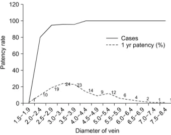
Fig. 1. Overall fistula patency rate. The patency rate decreased to 84.4% at 60 months.
Fig. 3. One-year patency rate according to cephalic vein diameter.
p-value=0.006 (chi-square).
subjects (250 cases) were 87.6%, 85.6%, and 84.4%, respectively. The corresponding rates were 95. 2%, 92.9%, and 92.9% in group A and 79.8%, 76.6%, and 75. 8% in group B, respectively. The patency rates in group A were statistically significantly higher than
those in group B (p<0.001) (Table 2, Figs. 1, 2).
The average diameter of the vein in group A (126 cases) was 3.8±1.3 mm, with 1 case with a diameter of less than 2 mm. In this group, the relationship of the 1-year patency rate to the diameter of the vein and age was analyzed.
A cross-sectional analysis of the 1-year patency rate by vein diameter showed a p-value of 0.006, which was statistically significant. An odds ratio of 6.139 and a p-value of 0.016 were seen in the logis- tic regression analysis, indicating that the diameter and 1-year patency rate had a statistically significant positive relationship (Table 3, Fig. 3).
The 1-year patency rate was compared between the patients in whom the diameter of the vein was at least 3 mm (n=83) and those in whom it was smaller than 3 mm (n=43). The 1-year patency rate in the group with a diameter of at least 3 mm was 98.8%, whereas it was 88.4% in the group with a di- ameter smaller than 3 mm. The patients in whom
Table 4. CE of various factors
Factors Proportion (%) CE p-value
Age >65 yr 27.2 -0.169 0.007
Sex (male) 64.4 -0.010 0.877
Diabetes mellitus 58.8 0.069 0.281
Hypertension 75.2 -0.054 0.398
Cephalic vein diameter
≥3.0 mm
65.9 0.191 0.009
CE, correlation efficiency.
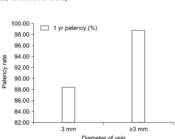
Fig. 4. One-year patency rate according to the vein diameter measured by preoperative ultrasonography. The group with a cephalic vein diameter of less than 3 mm (n=43) had an early pa- tency rate of 88.4%, and those with a cephalic vein diameter of at least 3 mm (n=83) had an early patency rate of 98.8%.
Fig. 5. Difference in the cumulative patency rate according to the vein diameter measured by preoperative ultrasonography.
Patients with a cephalic vein diameter of less than 3 mm group had a 5-year patency rate of 88.4%, those with a diameter of at least 3 mm (n=83) had a 5-year patency rate of 95.2%.
Table 5. Complications
Variable Group A Group B p-value
Inadequate flow 0 2 0.245
Thrombosis 2 2 0.683
Venous hypertension 6 27 0.000
Aneurysm 0 1 0.496
Infection 0 1 0.496
Total 8 33 0.000
the anastomotic vein had a larger diameter had a significantly better patency rate (Table 4, Fig. 4).
The 1-year patency rate was 80.4% in the patients at least 65 years of age and 90. 4% in the patients younger than 65 years of age, showing that the group younger than 65 had better results. This trend was statistically significant, with a p-value of 0.007 in the cross-sectional analysis. Other factors, such as gender, diabetes, and hypertension, had p-values ex- ceeding 0.05, which means that they were not stat- istically significant (Table 4).
In the Kaplan-Meier analysis of the patients with a vein diameter of at least 3 mm and those with a di- ameter smaller than 3 mm, the 1-year, 3-year, and 5-year patency rates in the group with a vein diame- ter of at least 3 mm were 98.8%, 95.2%, and 95.2%, respectively, while the patency rate in the patients with a diameter smaller than 3 mm was 88. 4% at 1 year and remained constant thereafter. The patency
rates were significantly different between these 2 groups (Figs. 4, 5).
3) Postoperative complications
Patients underwent surgery for a second time due to the following postoperative complications: a lack of AVF blood flow (group A, 0 cases; group B, 2 cases), thrombus (group A, 2 cases; group B, 2 cases), venous hypertension (group A, 6 cases; group B, 27 cases), aneurysm (group A, 0 cases; group B, 1 case), and infection (group A, 0 cases; group B, 1 case). A stat- istically significant difference was seen for venous hypertension (p<0.05) (Table 5).
Discussion
According to a study on the status of renal re- placement therapy in Korea published by the Korean Society of Nephrology in 1999, more than half of pa- tients with ESRD undergo hemodialysis [1]. With ad-
vances in medical technology and improvements in economic standards, the average life expectancy of patients with ESRD who are on hemodialysis is increasing. Thus, efficient AVFs must be constructed for ESRD patients on hemodialysis [5]. All ESRD pa- tients want an AVF for long-term use without any adverse effects, but many practical problems may be encountered when an AVF is constructed in patients on hemodialysis, including AVF closure, infection, and aneurysm. Many efforts have been made to solve those problems, but solutions capable of dramatically improving the patency rate of AVFs are still far from becoming reality.
Concerning the historical background of AVF for hemodialysis, Quinton et al. [6] developed an ex- ternal AVF in 1960 using a Teflon tube. However, that AVF could be used only for very short durations due to complications including venous hypertension, infection, thrombus, and aneurysm. Thereafter, Brescia et al. [2] reported on the use of side-to-side anasto- mosis to create an AVF using the radial artery and cephalic vein in 1966, and this method enabled long-term hemodialysis. However, this method also resulted in severe side effects, including congestion, edema, and hot flashes. In 1984, Wedgwood et al. [7]
reported that these side effects could be prevented by side-to-end anastomosis. To date, side-to-end anas- tomosis with an autologous vessel is considered to be the best surgical procedure for AVF construction.
However, since many patients who need long-term hemodialysis have no blood vessels that can be used for AVF, secondary methods of AVF construction for hemodialysis are necessary [8]. One alternative is to construct an AVF using a prosthetic vessel. AVFs with a prosthetic vessel have advantages over AVFs using an autologous vessel in that they allow the choice of a broad range of available blood vessels and are easy to make. Several studies are investigat- ing prosthetic vessels and AVF using Gore-Tex, which has become the standard based on multiple previous studies. However, using a prosthetic vessel has the disadvantages of having a lower patency rate than AVFs using autologous vessels and being prone to secondary complications, including infection and aneurysm. Although studies are investigating ways to improve the patency rate of AVFs constructed using a prosthetic vessel, no clear solution has yet been identified. A realistic strategy for improving the over-
all patency rate of AVFs would be to extend the ap- plication range of autologous vessels. Specifically, the diameter of the anastomotic vein, the direction of flow of the vein, and its structural conditions can be used as criteria for autologous vessel selection to help expand the range of use of this technique [4].
To evaluate the vein to be used for AVF con- struction, a tourniquet has traditionally been applied on the proximal area, and the vein evaluated using the naked eye. However, this method is inherently imprecise in evaluating the diameter and location of the vein, its flow direction, and the presence of stenosis in the proximal area. Therefore, many stud- ies have sought to improve the technique for evaluat- ing candidate veins, and the use of ultrasonography has received particular attention. Ultrasonography can be used to precisely evaluate various factors, in- cluding arterial stenosis and calcification, blood flow, the diameter of the anastomotic vein, stenosis during flow, and the distance between the vein and the skin [4]. However, few studies in Korea have investigated the use or clinical outcomes of ultrasonography in AVF planning, and studies investigating factors asso- ciated with the clinical results of AVFs are still ongoing. In particular, the criterion for the diameter of the anastomotic vein varies across centers, with no clear consensus [4]. As such, this study aimed to quantify the usefulness of using ultrasonography to plan AVF construction, as well as to set an appro- priate criterion for the diameter of the anastomotic vein.
This study investigated several factors that could affect the patency rate of AVFs. Patients younger than 65 exhibited a significantly better patency rate than those 65 years of age or older. However, no statistically significant relationship was found be- tween the patency rate and the prevalence of dia- betes or hypertension.
Ultrasonography use also showed a correlation with the patency rate of the AVFs. Subjects were div- ided into 2 groups. The patients in group A had the size of the vein evaluated using preoperative ultra- sonography, while the patients in group B underwent the traditional approach, in which vein size was eval- uated using the naked eye. The 1-year, 3-year, and 5-year patency rates were then measured. The pa- tency rate of group A was significantly superior to that of group B. We believe that improvement in the
patency rate shown in this study was a result of the patients in group A undergoing preoperative ultra- sonography to evaluate the candidate vein for anas- tomosis, because the most appropriate surgical pro- cedure could then be selected based on the criteria for autologous vessel use. Additionally, an autologous vessel in the brachiocephalic area was used in sig- nificantly more patients in group A, which we believe was the result of the brachiocephalic vein being lo- cated relatively deep, such that it is difficult to see even when a tourniquet is used. However, since ul- trasonography made it easier to evaluate the deep cephalic vein, many patients in group A received an autologous AVF using the cephalic vein. Thus, ultra- sonography is considered essential for evaluating and selecting the most appropriate blood vessel when constructing an AVF using an autologous vessel.
Studies have reported a correlation between the diameter of the vein used for anastomosis and the 1-year patency rate. In 1996, Wong et al. [9] sug- gested that a preoperative diameter of 1.6 mm in the cephalic vein be used as a criterion for anastomosis of the radial artery and cephalic vein, and in 1998, Silva et al. [10] used a diameter of at least 2.5 mm as a criterion. National Kidney Foundation-Dialysis Outcomes Quality Initiative [11] reported in 1997 that the 1-year patency rate was 84% when the size of the anastomosed vein was at least 3 mm with no sign of stenosis. This study showed a significant rela- tionship between the diameter of the anastomotic vein and the 1-year patency rate in group A; in the regression analysis, the odds ratio for 1-year patency in anastomotic veins with a diameter of at least 3 mm was found to be 6. 139, indicating that a larger diameter was associated with a higher 1-year pa- tency rate. A 1-year patency rate of 100% was seen in the 49 patients in whom the brachiocephalic vein was used, and a better 1-year patency rate was seen in the patients in which the anastomotic vein had a diameter of at least 3 mm than in the patients with a diameter of less than 3 mm (98.8% versus 88.4%).
The 5-year patency rate was also significantly superi- or in the former group (95.2% versus 88.4%).
Therefore, the diameter of the vein used for anasto- mosis is closely correlated with the 1-year patency rate, and the patency rate of AVFs can be improved by using autologous vessels with a diameter of at least 3 mm.
Although the size of the anastomotic vein has been established as an important factor for AVF con- struction in several previous studies, a precise crite- rion for the diameter has not yet been determined due to differences in the evaluation method, the in- clusion of a limited number of study subjects, and variation in the characteristics of subjects, including country of origin and ethnicity. In this study, the anastomosis was performed in the snuffbox, wrist, or brachiocephalic region based on the location of the vein, and the average diameter of the veins in each location was measured to be 3.9 mm, 3.2 mm, and 4.8 mm, respectively. Although a diameter of 3 mm was considered to be appropriate for use in an AVF in the subjects in this study, the suitability of using 3 mm as a criterion for vein diameter in each location could be considered separately. Criteria for seg- mented autologous vein use are suggested in this study, although additional studies are needed.
We analyzed the complications after AVF con- struction, and cases of venous hypertension in the proximal part of the anastomosis were significantly more common in group B than in group A, which we think was caused by unidentified stenosis in the proximal vein in group B. In cases with proximal ve- nous hypertension, it is expected that evaluating the vein via ultrasonography rather than with the naked eye could significantly contribute to improvements in the patency rate of AVFs and the prevention of ve- nous hypertension.
In conclusion, this study compared the results of AVF construction using an autologous vessel to those of previous studies. In particular, the patients who had preoperative ultrasonography to evaluate the vein and who underwent surgery that was custom- ized based on the selection criteria showed superior results. Additionally, the patency rate of AVFs was lower in patients 65 years of age or older than in those younger than 65.
Thus, the use of preoperative ultrasonography when constructing an AVF is strongly recommended, and additional efforts should be made to improve the patency rate in patients 65 years of age or older.
Based on the results of this study, a diameter of the anastomotic vein of 3 mm is suggested as a criterion for autologous vessel use. However, additional stud- ies regarding segmented criteria according to the lo- cation of the surgical procedure are required. Surgical
outcomes are expected to be superior when an AVF is constructed using an autologous vein with a diam- eter of at least 3 mm.
Conflict of interest
No potential conflict of interest relevant to this ar- ticle was reported.
Acknowledgments
This study was supported by a Grant of the Samsung Vein Clinic Network (Daejeon, Anyang, Cheongju, Cheonan; Fund no. KTCS04-097).
References
1. Ahn SJ, Choi EJ. Renal replacement therapy in Korea:
Insan Memorial Registry 1997. Korean J Nephrol 1999;18:
1-15.
2. Brescia MJ, Cimino JE, Appel K, Hurwich BJ. Chronic hemo- dialysis using venipuncture and a surgically created arte- riovenous fistula. N Engl J Med 1966;275:1089-92.
3. Kim DS, Kim SW, Kim JC, Cho JH, Kong JH, Park CR.
Clinical analysis of hemodialysis vascular access: compar- ision of autogenous arterioveonus fistula & arteriovenous prosthetic graft. Korean J Thorac Cardiovasc Surg 2011;44:
25-31.
4. Min SK, Ahn JH, Han JJ, Won TH. Factors influencing on early patency rate of autogenous arteriovenous fistula for hemodialysis. Korean J Thorac Cardiovasc Surg 2004;37:
342-8.
5. Yoon YC, Choi BO, Ku BI, et al. Clinical experiences of ar- teriovenous fistula & associated operations for hemodial- ysis in 290 cases. Korean J Thorac Cardiovasc Surg 1993;26:
761-8.
6. Quinton W, Dillard D, Scribner BH. Cannulation of blood vessels for prolonged hemodialysis. Trans Am Soc Artif Intern Organs 1960;6:104-13.
7. Wedgwood KR, Wiggins PA, Guillou PJ. A prospective study of end-to-side vs. side-to-side arteriovenous fistulas for haemodialysis. Br J Surg 1984;71:640-2.
8. Yu JH, Kim JH. Clinical analysis of expanded polytetra- fluoroethylene graft fistula for angioaccess in hemo- dialysis. Korean J Thorac Cardiovasc Surg1996;29:883-8.
9. Wong V, Ward R, Taylor J, Selvakumar S, How TV, Bakran A. Factors associated with early failure of arteriovenous fistulae for haemodialysis access. Eur J Vasc Endovasc Surg 1996;12:207-13.
10. Silva MB Jr, Hobson RW 2nd, Pappas PJ, et al. A strategy for increasing use of autogenous hemodialysis access pro- cedures: impact of preoperative noninvasive evaluation. J Vasc Surg 1998;27:302-7.
11. National Kidney Foundation-Dialysis Outcomes Quality Initiative. NKF-DOQI clinical practice guidelines for vas- cular access. Am J Kidney Dis 1997;30(4 Suppl 3):S150-91.