• 검색 결과가 없습니다.

기능적 자기자극치료 효과

N/A
N/A
Protected

Academic year: 2021

Share "기능적 자기자극치료 효과"

Copied!
6
0
0

로드 중.... (전체 텍스트 보기)

전체 글

(1)

기능적 자기자극치료 효과

The Journal Korean Society of Physical Therapy

조윤우, 김수정

1

, 박해운

2

, 서정민

3

, 황세진

4

, 장성호, 이동규, 안상호

영남대학교 의과대학 재활의학교실,

1

영남대학교 의과학연구소,

2

대구카톨릭대학교 의과대학 재활의학교실,

3

영남대학교 의료공학연구소,

4

한양대학교 의과대학 해부 및 세포생물학교실

The  Effect  of  Direct  Functional  Magnetic  Stimulation  of  the  Lesion  on  Functional  Motor  Recovery  in  Spinal  Cord  Injured  Rat

Yun‐Woo  Cho,  MD,  PhD;  Su‐Jeong  Kim,  PhD

1

;  Hea‐Woon  Park,  MD,  PhD

2

;  Jeong‐Min  Seo,  MS

3

;  Se‐Jin  Hwang,  MD,  PhD

4

;  Sung‐Ho  Jang,  MD;  Dong‐Gyu  Lee;  Sang‐Ho  Ahn,  MD,  PhD

Department  of  Rehabilitation  Medicine,  College  of  Medicine,  Yeungnam  University; 

1

Institute  of  Medical  Science,  Yeungnam  University; 

2

Department  of  Rehabilitation  Medicine,  School  of  Medicine,  Catholic  University  of  Deagu; 

3

Institute  of  Biomedical  Engineering,  Yeungnam  University; 

4

Department  of  Anatomy  and  Cell  Biology,  College  of  Medicine,  Hanyang  University

Purpose:  The  purpose  of  this  study  was  to  determine  the  effect  of  direct  functional  magnetic  stimulation  (FMS)  of  affected  spinal  cord  on  motor  recovery  following  spinal  cord  injury  in  rats.

Methods:  After  a  contusion  injury  at  the  spinal  level  T9  using  an  NYU  Impactor,  functional  magnetic  stimulation  was  delivered  by  a  magnetic  stimulator  through  a  round  prototype  coil  (7  cm  in  diameter).  Stimulation  parameters  were  set  as  follows:  repetition  rate  =  50  Hz  (stimulus  intensity  100%  =  0.18  T),  stimulation  time  =  20  min.  Functional  magnetic  stimulation  was  administered  twice  a  day,  5  days  per  week  for  8  weeks  starting  4  days  after  spinal  cord  injury. 

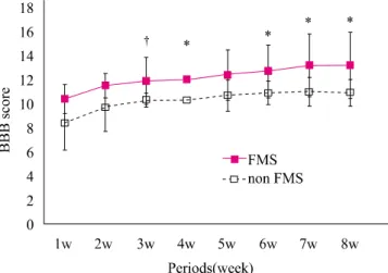
Functional  magnetic  stimulationwas  delivered  directly  to  the  affected  spinal  cord.  Outcomes  of  locomotor  performance  were  assessed  by  the  Basso  Beattie  Bresnahan  (BBB)  locomotor  rating  scale  and  by  an  inclined  plane  test  weekly  for  8  weeks. 

Results: In  the  BBB  test,  hindlimb  motor  function  in  the  Functional  magnetic  stimulation  group  improved  significantly  more  compared  to  the  control  group  at  3,  4,  6,  7,  and  8  weeks  (p<0.05).  In  the  inclined  plane  test,  the  angle  of  the  plane  in  the  functional  magnetic  stimulation  group  increased  significantly  more  compared  to  the  control  group  at  4,  5,  7,  and  8  weeks  (p<0.05).

Conclusion:  Our  results  demonstrate  that  direct  Functional  magnetic  stimulation  of  the  lesional  site  may  have  beneficial  effects  on  motor  improvement  after  spinal  cord  injury. 

Keywords:  Functional  magnetic  stimulation,  Direct  stimulation,  Spinal  cord  injury 논문접수일: 2010년 12월 29일

수정접수일: 2011년 1월 29일 게재승인일: 2011년 2월 7일 교신저자: 안상호, spineahn@ynu.ac.kr

I. 서론

척수손상 후 발생하는 운동기능의 감소는 임상적으로 해결해야

할 과제 중 하나로 운동기능의 회복을 위해 고전적인 물리치료 ,

약물치료 , 전기치료 등이 시행되어 왔고, 근래에는 신경세포 혹

은 지방세포에서 유리된 줄기세포 등을 이용한 세포치료 , 손상

(2)

부위의 이차적 손상 억제 및 축삭의 연장을 위한 향신경성인자 주입치료 , 그리고 원하는 방향으로 세포가 증식되도록 보조하 는 연결다리 제공 등의 다양한 생물학적 , 공학적 치료가 시도되 고 있으나 , 임상적으로 탁월한 변화를 일으킬 정도의 운동기능 향상을 보고한 바는 거의 없다 .

1,2

최근에는 자기자극을 이용한 치료가 보고되고 있다 . 반복적 자기자극은 요실금의 치료 , 척수손상 환자에서 배뇨 조절, 호흡 기능 향상 등을 위해 효과를 보고자 하는 장기부위에서 자극을 주는 방법으로 사용되었고 ,

3,4

두부의 대뇌피질을 자극하는 경두 개 자기자극을 통해 뇌손상 혹은 척수손상 등의 중추신경계 손 상 동물모델에서 운동기능의 회복이 보고되었다 .

5

경두개 자기자극은 척수손상 후 피질척수로 신경세포의 생 존 유도를 증진하여 운동신경 회복을 촉진한다고 보고되었고 ,

6

흰 쥐의 척수손상 모델에서 경두개 자기자극 후 신경생리학적 검사에서도 운동신경유발전위의 진폭 증가가 보고되었다 .

7

하지 만 , 일부 연구에서 경두개 자기자극 후 경련과 같은 의도하지 않은 반응이나,

8

기억, 청력에 미치는 부작용,

9

두통

10

등이 발 생할 수 있다고 보고되었다 . 그리고 Crowe 등

11

은 급성기 척수 손상 고양이에서 자기자극을 경두개를 통하지 않고 중간 흉추 부에 적용한 후 운동기능 회복에 유용하였다고 보고하였다 .

이에 본 연구에서는 대뇌를 자극하지 않고 손상된 척수에 직접 자극하는 기능적 자기자극을 시도하고자 하였고 , 흰 쥐를 이용한 척수손상 모델을 만들어 하지 운동기능회복에 미치는 영향을 알아보고자 하였다 .

II. 연구방법

1. 척수손상 모델

체중 200∼250 g의 암컷 Sprague-Dawley 흰 쥐 40마리를 대 상으로 하였고 , 모든 시술은 영남대학교 실험동물 관리 및 사용 지침에 따랐다. 모든 흰 쥐는 Zoletil (Virbac laboratories, Carros, France, 50 mg/kg)을 복강 내 주입하여 마취를 시행 한 뒤 흉추 부위의 털을 제거하고 povidone iodine용액으로 소 독한 뒤 피부와 근육을 절개하여 흉추 9번 후궁절제술을 실시 하여 아홉 번째 흉수를 노출시켰다. NYU impactor (New York University, New Jersey, 미국)를 이용하여 무게가 10 g 인 추(rod)를 25 mm 높이에서 노출시킨 흉수의 한가운데에 떨어뜨려 척수의 양측이 비슷하게 손상되도록 하였다 .

12

척수좌 상을 유도한 후 각 층별로 근육과 피부를 봉합하였으며 마취에 서 회복될 때까지 37°C 이상으로 체온을 유지하였다. 일정한 중등도의 척수 좌상 모델을 만들기 위하여 떨어지는 속도 및 높이에 대한 결과값을 컴퓨터상으로 측정하였으며 ,

13

압박율

(compression rate) 값이 50∼60% 범위 안에 속하는 쥐들만을 대상으로 하였고 , 나머지는 대상에서 제외하였다.

14

척수손상을 입은 흰 쥐들은 실내온도 24°C 이상의 환경에서 사육되었으며, 자발적 배뇨관리가 가능할 때까지 매일 2회씩 흰 쥐의 복부를 압박하여 배뇨관리를 시행하였다 . 흰 쥐에 대한 각각의 기능적 자기자극 실험 , 행동검사의 시행은 각각 서로 다른 연구자들에 의해 시행되었으며 , 서로간의 정보를 교환하지 않은 상태로 실 험을 진행하였다 .

2. 기능적 자기자극

정해진 범위의 압박율에 해당되지 않은 쥐들은 제외하였으며 , 손상 후 1 주일에 확인한 행동검사 점수가 양측 모두 6∼8점에 해당되는 쥐들만을 대상으로 하였다 . 양측으로 일정하게 척수 손상이 유도된 흰 쥐들을 무작위로 기능적 자기자극 군 (n=5)과 비자극 군 (n=5)으로 나누었으며, 기능적 자기자극을 시행하는 것 외에는 두 군 모두에게 같은 조건을 적용하였다 . 기능적 자 기자극은 BioCon-1000Pro (Mcube Technology Co, 대한민 국 ) 기종의 주문 제작한 반복 자기자극기기를 사용하였고, 70 mm의 원형코일을 사용하였다. 자극 방법으로 최대 자기 자극 은 0.2 Tesla, 370 μsec의 펄스 폭을 가진 최대 50 Hz 주파수, 자극시간 3초, 휴식시간 3초, 총 자극시간 20분이었다. 손상 후 4일째부터 쥐의 척수손상 부위인 흉수 9번 부위에, 활동면이 척수손상부위와 수직을 이루도록 코일을 위치하였다 . 자극을 자극군에만 준 것을 제외하고 , 자극군과 비자극군 모두 같은 조 건으로 기능적 자기자극을 시행하였고 , 자극 강도는 100%, 20 분 /회, 10 Hz (자극 3초-휴식 3초)의 자극을 가하여 하루 2회, 주 5일간, 총 8주 동안 시행하였다.

3. 행동학적 검사

척수 손상 후 기능적 자기자극의 효과를 알아보기 위하여 두 군 모두에서 운동기능을 평가하기 위한 행동검사로서 Basso Beattie Bresnahan 척도 검사(BBB locomotor rating scale)와 경사대 검사 (inclined plane test)를 사용하였다.

14,15

BBB 척도 검사는 매주 1회씩 총 8주간 시행하였고, 부드럽지만 미끄럽지 않은 바닥의 원형 플라스틱 내 (100 cm in the diameter, height of 20 cm)에서 쥐를 걷도록 하였다

경사대 검사는 깊이 3 mm, 10 mm의 간격으로 패여 있는

수평의 홈을 가진 딱딱한 재질의 경사판을 5°씩 증가시키면서

오른쪽 및 왼쪽 신체 면을 위로 향하도록 하여 5초 동안 미끄

러지지 않고 각각의 신체 위치를 잘 유지할 수 있는 최대경사

각도를 측정하여 평균값을 구하였다 .

15

4분 동안 쥐의 움직임을

관찰하여 뒷다리의 움직임 , 체중부하 및 보행양상 등의 운동기

능을 점수화하여 평균값을 구하여 추적 관찰하였다 .

(3)

Figure 2. Locomotor function in the hindlimbs was evaluated by inclined plane method. Compared with non FMS group, FMS group significantly increased the angle of inclination at 4, 5, 7, and 8 weeks. FMS: functional magnetic stimulation, w: week(s). The data represent means. *p<0.05

Figure 1. Locomotor function in the hindlimbs was evaluated by the BBB locomotor scale. FMS group showed significantly improved hindlimb function more than non FMS group (FMS group at 3, 4, 6, 7, 8 weeks). BBB: Basso Beattie Bresnahan, FMS: functional magnetic stimulation, w:

week(s). The data represent mean±SD. *p<0.05,

p<0.01 4. 자료분석

자극군과 비자극근의 비교를 위한 통계적 분석은 SPSS version 12.0을 이용하여 Student’s t test로 통계처리 하였으며 통계적 유의수준은 p값이 0.05 미만인 경우로 하였으며, 측정변수의 값은 평균 ±표준편차로 표시하였다.

III. 결과

1. BBB 척도 검사

척수 손상 후 기능적 운동기능의 회복 측정을 위한 평가항목 중 BBB 척도 검사에서 두 군 모두 손상 후 첫 주부터 8주까지 서서 히 점수가 증가하는 양상을 보였다 . 자극군의 BBB 점수는 비자 극군의 BBB 점수에 비해 의미 있는 향상을 보였고, 척수손상 3 주(p<0.01), 4주(p<0.05), 6주(p<0.05), 7주(p<0.05), 8주 (p<0.05)째 비자극군에 비해 의미 있는 뒷다리의 기능회복을 보 였으며, 이 시기의 BBB 점수는 각각 자극군에서 11.9±1.9, 12.0±1.9, 12.7±2.2, 13.2±2.6, 13.2±2.7, 비자극군에서 10.3±

0.6, 10.3±0.8, 10.9±1.0, 11.0±1.2, 10.9±1.1이었다. 실험의 후 반기에서 자극군은 잘 조화된 뒷다리와 앞다리의 움직임을 보였 고, 일정하게 체중이 부하된 발걸음을 나타내었다(Figure 1).

2. 경사대 검사

경사대 검사에서는 두 군 모두 손상 2주부터 급속한 향상을 보 여 서서히 증가 혹은 유지되는 양상을 보였다 . 자극군에서는 척

수손상 4주(p<0.05), 5주(p<0.05), 7주(p<0.05), 8주(p<0.05) 째 비자극군에 비해 의미 있는 향상을 보였으며 , 이 시기의 경 사각도는 각각 자극군에서 60.6±5.2, 61.3±6.0, 63.8±6.0, 65.0±5.8, 비자극군에서 55.0±2.0, 53.8±2.5, 57.5±2.0, 59.4±3.8이었다. 이때의 쥐의 뒷다리 행동은 자극군의 경우 뒷 다리로 걸을 수 있었지만 앞다리와 가끔씩 조화가 되었고 , 비자 극 군은 때때로 무게를 지탱하면서 걸을 수 있지만 , 앞뒤다리가 조화가 되지 않는 양상을 보여 기능적 자기자극 군이 비자극 군에 비해 뒷다리 운동 기능이 유의하게 회복되는 소견을 나타 내었다 (Figure 2).

IV. 고찰

본 연구에서는 흰 쥐의 척수손상 모델에서 척수손상 후부터 8 주간 손상부위에 기능적 자기자극 치료를 시행하였고 , BBB 척 도검사와 경사대 검사를 이용하여 운동신경기능의 회복을 관찰 하였다 .

자기장은 조직 심부에 전기장을 만들 수 있는 장점이 있다 .

반복적 경두개 자기자극은 대뇌피질부의 선택적 자극이 가능한

비침습적이며 상대적으로 통증이 없는 기술로 Barker 등

16

대뇌기능 평가 및 신경전도 연구를 위해 보고하였다 . 자극기 내

부의 금속 코일을 통해 발생되는 교류전류의 수직으로 박동성

자기장이 형성되며 , 이는 큰 저항 없이 연부조직 및 골을 통과

한 후 충분한 강도의 전류로 바뀌어 신경부위의 탈분극을 유도

(4)

한다 .

17

그리고, 이와 유사한 과정을 통해 자기자극 시 중추신 경계의 장시간 흥분 및 신경전달물질의 변화를 일으키는 것으 로 알려져 있다.

18

반복적 자기자극은 적합한 기준에 따를 경우 상대적으로 부 작용이 매우 적으며, 초기에는 우울증 등의 정신과적 질환에 전 기자극 치료에 비해 덜 침습적인 대체치료 방법으로 사용되었 다.

19

최근엔 척수손상 환자에서 대뇌에 자기자극 후 하지의 경 직감소의 효과를 보고하였고 ,

20,21

척수손상 환자의 통증에도 유 용하다고 보고되었다.

22,23

반복적 자기자극 치료는 만성 경수손 상 환자에서도 적용되었고 , 대뇌피질의 억제 신호 감소를 통해 운동기능이 회복되었다고 보고되었다 .

24

대뇌를 자극하는 경두개 자기자극치료는 비교적 안전한 것 으로 보고되지만 , 일부 연구에서 부작용도 보고되었다. 정상인 및 소수의 간질 환자를 대상으로 경두개 자기자극 시 의도하지 않은 경련이 발생하였고,

25

정상인을 대상으로 대뇌피질 자기자 극 후 기억 , 청력이 감소되었으며,

9

역시 정상인을 대상으로 빠 른 빈도의 대뇌피질 자기자극 후 단기간의 두통이 발생하였 다 .

10

이 같은 단점을 보완하기 위한 이유로 대뇌를 제외한 타 부위를 자극하는 방법과 타 부위 자극 시 그 효과 유무에 대한 의문점으로 인해 손상된 척수를 직접 자극하는 방법을 시행하 고자 하였다 . 따라서 본 연구에서는 흉수 손상 흰 쥐 모델을 만 들었고 , 흉수 손상부위에 직접 자기자극 치료를 시행하였다.

Crowe 등

11

도 급성기 척수손상 고양이에서 8자형 코일을 이용 하여 25 Hz 빈도의 자기장을 중간 흉추부에 적용하였고, 손상 1주 후부터 비자극군에 비해 유의한 향상을 보여 12주 내내 유 의한 향상을 보고하였다 . 본 연구에서는 10 Hz 빈도로 8주간 기능적 자기자극 후 약 3주 후부터 BBB 점수 및 경사대 각도 가 증가되었고 , 이는 8주간 지속되어 코일 모양, 자극 빈도의 차이가 있음에도 불구하고 Crowe 등의 연구와 비슷한 결과를 보였다 .

반복적 자기자극은 다양한 세포에 자극 시 세포막변화 , 세 포증식 , 세포활성 등에 영향을 미치는 것으로 알려져 있으

며,

26-28

세포실험에서 신경성장 인자의 활동성 증가 및 신경돌

기 촉진이 보고되었으며 , 실제 말초신경손상 모델에서 조직학 적 및 신경생리학적 신경재생을 유도하는 것으로 알려져 있 다 .

29-32

Poirrier 등

5

은 척수손상 흰 쥐 모델에서 8주간 10 Hz 의 자기자극 빈도로 치료를 시행하여, BBB 점수의 증가를 보 고하였으며 , 이는 하행적 세로토닌 통로를 통한 central pattern generator 활성화로 인한 것으로 보고하였다. 또 다른 기전으로 는 대뇌 피질척수로가 배부 척수손상 시 손상되는데 반해 피질 하 추체외로는 보존되는 것으로 보고되며,

33

경두개 자기자극 시 대뇌의 배부 및 배부외측 백질의 피질하 추제외로가 활성화 되어 운동기능의 향상이 일어날 수 있다고 보고되었다.

임상적으로 경두개 자기자극 시 적정한 빈도는 알려진 바 없으나 사람에서는 대개 1 Hz 이하의 저빈도는 대뇌피질의 흥 분도를 억제시키고 , 5 Hz 이상의 고빈도는 흥분도를 증가시킨 다고 알려져 있다 .

34,35

이전 연구의 동물실험에서는 주로 10, 20, 25 Hz 등 고빈도의 주파수를 이용하여 자극을 시행하였

고 ,

5,11,36

본 연구에서는 10 Hz의 고빈도 자극을 시행하였다.

본 연구에서는 척수손상부위로의 직접적인 기능적 자기자극 시 척수손상 후 운동기능 회복에 대한 효과를 확인하였으며 , 향후 기능적 자기자극의 기능회복 효과에 대한 조직학적 , 신경생리 학적 , 생화학적 기전에 대한 연구와 경두개 자기자극 및 척수손 상 부위의 직접적 자극의 비교에 대한 추가적 연구가 필요할 것으로 생각한다 .

V. 결론

본 연구에서 저자들은 척수손상부위에 직접적인 기능적 자기자 극치료의 효과를 알아보고자 척수손상 모델 흰 쥐를 이용하여 8주간 기능적 자기자극 치료를 시행하였고, 기능적 자기자극군 에서 비자극군에 비해 BBB 점수 및 경사대 각도가 의미 있는 향상을 관찰하였다 . 이는 기존의 경두개 자기자극 치료 뿐 아니 라 척수 손상부위에 직접 자극하는 기능적 자기자극 치료 역시 척수 손상 환자에게서 운동기능의 회복을 유도하는 새로운 치 료방법으로 사료되는 바이다 .

Author Contributions

Research design: Cho YW, Ahn SH

Acquisition of data: Seo JM, Lee DK, Park HW

Analysis and interpretation of data: Kim SJ, Park HW, Hwang SJ

Drafting of the manuscript: Cho YW, Kim SJ

Administrative, technical, and material support: Hwang SJ, Jang SH

Research supervision: Ahn SH

Acknowledgements

이 논문 (저서)은 2007년도 정부재원(교육인적자원부 학술연구

조성사업비 )으로 한국학술진흥재단의 지원을 받아 연구되었음

(KRF-2007-331-E00175).

(5)

참고문헌

1. Lim PA, Tow AM. Recovery and regeneration after spinal cord injury: a review and summary of recent literature. Ann Acad Med Singapore. 2007;36(1):49-57.

2. Okano H, Kaneko S, Okada S et al. Regeneration-based therapies for spinal cord injuries. Neurochem Int. 2007;

51(2-4):68-73.

3. Lin VW. Spinal cord medicine: principle and practice. New York, Demos, 2003:749-63.

4. Yamanishi T, Yasuda K, Suda S et al. Effect of functional continuous magnetic stimulation on urethral closure in healthy volunteers. Urology. 1999;54(4):652-5.

5. Poirrier AL, Nyssen Y, Scholters F et al. Repetitive transcranical magnetic stimulation improves open field locomotor recovery after low but not high thoracic spinal cord compression-injury in adult rats. J Neurosci Res.

2004;75(2):253-61.

6. Davey NJ, Smith HC, Wells E et al. Responses of thenar muscles to transcranial magnetic stimulation of the motor cortex in incomplete spinal cord injury patients. J Neurol Neurosurg Psychiatry. 1998;65(1):80-7.

7. Park HW, Kim SJ, Seo JM et al. Effects of Functional Magnetic Stimulation on the Functional Recovery in a Rat Model of Spinal Cord Injury. J Korean Acad Rehab Med.

2008;32(6):612-8.

8. Wassermann EM. Risk and safety of repetitive transcranial magnetic stimulation: report and suggested guidelines from the International Workshop on the Safety of Repetitive Transcranial Magnetic Stimulation, June 5-7, Electroencephalogr Clin Neurophysiol. 1998;108(1):1-16.

9. Pascual-Leone A, Houser CM, Reese K et al. Safety of rapid-rate transcranial magnetic stimulation in normal volunteers. Electroencephalogr Clin Neurophysiol. 1993;

89(2):120-30.

10. Epstein CM, Lah JJ, Meador K et al. Optimum stimulus parameters for lateralized suppression of speech with magnetic brain stimulation. Neurology. 1996;47(6):1590-3.

11. Crowe MJ, Sun ZP, Battocletti JH et al. Exposure to pulsed magnetic fields enhances motor recovery in cats after spinal cord injury. Spine (Phila Pa 1976). 2003;28(24):2660-6.

12. Young W. Spinal cord contusion models. Prog Brain Res.

2002;137:231-55.

13. Gruner JA. A monitored contusion model of spinal cord injury in the rat. J Neurotrauma. 1992;9(2):123-6.

14. Basso DM, Beattie MS, Bresnahan JC. Graded histological and locomotor outcomes after spinal cord contusion using the NYU weight-drop device versus transection. Exp Neurol.

1996;139(2):244-56.

15. Rivlin AS, Tator CH. Objective clinical assessment of motor function after experimental spinal cord injury in the rat.

J Neurosurg. 1977;47(4):577-81.

16. Barker AT, Jalinous R, Freeston IL. Non-invasive magnetic stimulation of human motor cortex. Lancet. 1985;

1(8437):1106-7.

17. George MS, Nahas Z, Kozel FA et al. Mechanisms and state of the art of transcranial magnetic stimulation. J ECT.

2002;18(4):170-81.

18. Wassermann EM, Lisanby SH. Therapeutic application of repetitive transcranial magnetic stimulation: a review. Clin Neurophysiol. 2001;112(8):1367-77.

19. Fitzgerald PB, Brown TL, Marston NA et al. Motor cortical excitability and clinical response to rTMS in depression.

J Affect Disord. 2004;82(1):71-6.

20. Kumru H, Murillo N, Samso JV et al. Reduction of spasticity with repetitive transcranial magnetic stimulation in patients with spinal cord injury. Neurorehabil Neural Repair.

2010;24(5):435-41.

21. Mori F, Koch G, Foti C et al. The use of repetitive transcranial magnetic stimulation (rTMS) for the treatment of spasticity.

Prog Brain Res. 2009;175:429-39.

22. Defrin R, Grunhaus L, Zamir D et al. The effect of a series of repetitive transcranial magnetic stimulations of the motor cortex on central pain after spinal cord injury. Arch Phys Med Rehabil. 2007;88(12):1574-80.

23. Kang BS, Shin HI, Bang MS. Effect of repetitive transcranial magnetic stimulation over the hand motor cortical area on central pain after spinal cord injury.Arch Phys Med Rehabil.

2009;90(10):1766-71.

24. Belci M, Catley M, Husain M et al. Magnetic brain stimulation can improve clinical outcome in incomplete spinal cord injured patients. Spinal Cord. 2004;42(7):417-9.

25. Wassermann EM, Cohen LG, Flitman SS et al.Seizures in healthy people with repeated “safe” trains of transcranial magnetic stimuli. Lancet. 1996;347(9004):825-6.

26. Bersani F, Marinelli F, Ognibene A et al. Intramembrane protein distribution in cell cultures is affected by 50 Hz pulsed magnetic fields. Bioelectromagnetics. 1997;18(7):

463-9.

(6)

27. Roman A, Zyss T, Nalepa I. Magnetic field inhibits isolated lymphocytes' proliferative response to mitogen stimulation.

Bioelectromagnetics. 2005;26(3):201-6.

28. Tenuzzo B, Chionna A, Panzarini E et al. Biological effects of 6 mT static magnetic fields: a comparative study in different cell types. Bioelectromagnetics. 2006;27(7):560-77.

29. Greenebaum B, Sutton CH, Vadula MS et al. Effects of pulsed magnetic fields on neurite outgrowth from chick embryo dorsal root ganglia. Bioelectromagnetics. 1996;

17(4):293-302.

30. Longo FM, Yang T, Hamilton S et al. Electromagnetic fields influence NGF activity and levels following sciatic nerve transection. J Neurosci Res. 1999;55(2):230-7.

31. Macias MY, Battocletti JH, Sutton CH et al. Directed and enhanced neurite growth with pulsed magnetic field stimulation. Bioelectromagnetics. 2000;21(4):272-86.

32. Raji AR, Bowden RE. Effects of high-peak pulsed electromagnetic field on the degeneration and regeneration

of the common peroneal nerve in rats. J Bone Joint Surg Br. 1983;65(4):478-92.

33. Simpson RK, Baskin DS. Corticomotor evoked potentials in acute and chronic blunt spinal cord injury in the rat:

correlation with neurological outcome and histological damage. Neurosurgery. 1987;20(1):131-7.

34. Pridmore S, Oberoi G. Transcranial magnetic stimulation applications and potential use in chronic pain: studies in waiting. J Neurol Sci. 2000;182(1):1-4.

35. Romero JR, Anschel D, Sparing R et al. Subthreshold low frequency repetitive transcranial magnetic stimulation selectively decreases facilitation in the motor cortex. Clin Neurophysiol. 2002;113(1):101-7.

36. Shimada Y, Sakuraba T, Matsunaga T et al. Effects of therapeutic magnetic stimulation on acute muscle atrophy in rats after hindlimb suspension. Biomed Res. 2006;

27(1):23-7.

수치

Figure 1. Locomotor function in the hindlimbs was  evaluated by the BBB locomotor scale

참조

관련 문서

영문 : Histomorphometric analysis of bone formation in the bone defects around the implant in adult dogs according to grafts of low or high heat treated autogenous

First, the rhythmic gymnastics exercise program improves the skeletal muscle mass of adult women in office workers and reduces body fat percentage,

Field : Spatial distribution of scalar and vector quantity Electric field and magnetic field.. 3-1

or average level had not only great efficiency in self-directed learning but also high rewarding strategy whereas those who believed themselves in below

High strength, unique magnetic/electrical properties 미세구조 조절: 1) perfection vs imperfection control.. 20. Perfect Crystal is good in many

• To compare this neural activity with MI of a human with spinal cord injury, MN performed a step tracking, ‘center-out’ task using the neural

Mortality and clinical signs in SD rats about single oral administration after 3 weeks with callus extract ..... Hematological values in SD rats administered orally

- The most unstable one: Internal modes with very short wavelengths perpendicular to the magnetic field but long wavelengths parallel to the field.. Classification