大韓獸醫學會誌(2006) 第46卷 第1號 Korean J Vet Res(2006) 46(1) : 75~76
75
Extradural spinal lipoma in a dog
Ji-Hey Lim, Wan Hee Kim, Dae-Yong Kim, Deo-Youn Cho, Oh-kyeong Kweon*
College of Veterinary Medicine, Seoul National University, Seoul 151-742, Korea
(Accepted: January 25, 2006)
Abstract :
A 7-year-old, female pointer dog was referred to the SNU Veterinary Medicine Teaching Hospital for the evaluation of lameness in the pelvic limb of 10 days' duration. After the treatment for 2 weeks (carprofen 2.2 mg/kg, bid), the progressive, symmetric, ambulatory caudal paraparesis was profound. In the spinal myelography, left lateral extradural compression of the spinal cord over the ninth and tenth thoracic vertebral bodies was found. A left hemilaminectomy of the ninth and tenth thoracic vertebrae was done. A 1.5-cm-long, white extradural mass occupied the left side of the spinal canal.
The tumor was identified histologically as lipoma. The 6 weeks after surgery the dog’s complaints were much improved. Continuous evaluation is needed.
Keywords :
dog, extradural, spinal lipoma Extradural tumors account for approximately 50% of spinal tumors in dogs [3, 5, 8]. Only a few extradural and intradural extramedullary spinal lipoma have been reported in dogs [3, 4]. In human beings, 1% of all primary intraspinal tumors are lipomas, and 40% of these intraspinal tumors are extradural with a frequency of 0.8% extradural lipoma described as occurring in the middle and lower thoracic segments [1, 2, 6]. In the present report, extradural lipoma in a Pointer dog was diagnosed after surgical removal.
A 7-year-old, female pointer dog was referred to the SNU Veterinary Medical Teaching Hospital for the evaluation of lameness in the pelvic limb of 10 days’
duration. Before referral, the dog had been treated with carprofen. After the treatment for 2 weeks (carprofen 2.2 mg/kg, bid), the lamness did not improved but progressive, symmetric, ambulatory caudal paraparesis was profound; the dog dragged the dorsum of the left and right tarsus made only feeble attempts to flex the coxofemoral joint and stifles and to advance the hindlimbs. The dog supported much of its weight on the forelimbs. On neurological examination, hyperreflexia of the femoral and sciatic segmental reflexes was evident, as well as lack of conscious proprioception in both hindlimbs. Results of the remainder of the neurologic
examination were normal. Although plain radiography of the thoracic and lumbar parts of the vertebral column, blood and serum biochemical analysis and cistern spinal fluid examination were performed and any abnormalities were not found. However, in the spinal myelography, left dorsolateral extradural compression of the spinal cord over the ninth and tenth thoracic vertebral bodies was found (Fig 1). A left hemilaminectomy of the ninth and tenth thoracic vertebrae was done. A 1.5-cm-long, firm tan extradural mass occupied the left side of the spinal canal, displacing the spinal cord to the right of midline. The mass had a rubbery consistency facilitating complete gross removal, using blunt dissection and suction. The 5 days after surgery, the dog showed decreased hyperreflexia and was discharged. Three weeks after surgery, the clinical sign was improved but still profoundly paretic in both hind limbs.
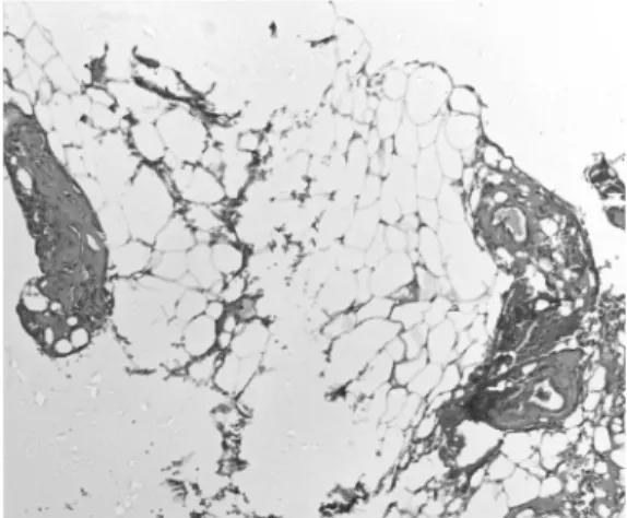
The tumor was identified histologically as lipoma (Fig 2). The tumor was not clearly encapsulated and it proved simple to dissect it from dura and surrounding epidural fat. The tumor was normal adult adipose tissue with sporadic blood vessels, covered in a thin capsule of connective tissue and with fibrous tissue.
Typically, the earliest clinical abnormality associated with extradural spinal tumors in dogs is progressive
*Corresponding author: Oh-Kyeong Kweon
College of Veterinary Medicine, Seoul National University, Seoul 151-742, Korea
[Tel: +82-2-880-1248, Fax: +82-2-888-2866, E-mail: [email protected]]
76 Ji-Hey Lim
et al.and often unrelenting signs of pain as a result of bony destruction and/or nerve root compression [2]. In the present case signs of thoracic pain were not evident, and an area of hyperpathia could not be detected by palpation of the vertebral column. Pain seems to be a less significant symptom particularly with lipomas at thoracic levels [9, 10]. Examination of the CSF in our patient revealed no increased proteins. It has been reported that CSF findings gave on clue for a pre- operative diagnosis of lipoma. [7].
Lipoma may be further subclassified into lipoma, angiolipoma, myxolipoma and embryonic lipoma on the basis of histologic criteria. Subclassification of lipomas may have a relationship with pathophysiology in human beings but little clinical merit because the prognosis after operative removal is very good with very little chance of a recurrence[2]. The tumor in the present case was subclassified as a lipoma; tumor of adult adipose tissue. At 6 weeks, the dog’s complaints were much improved, but paretics in both hindlimbs were still remained. It seems that continuous evaluation is needed.
References
1.
Agnoli AL, Lochner B.Magnetic resonance tomogra- phic findings in spinal lipomas. Radiologe 1988,
28, 284-288.
2.
de Bruine JF, Bongartz EB.Spinal extradural lipomas. Report of two cases. Clin Neurol Neurosurg 1983,
85, 181-190.
3.
De Ley G, Verschooten F, Hoorens J.Extradural lipoma in a young bull. Vet Med Small Anim Clin 1979,
74, 1013-1017.
4.
Funkquist B.Hourglass extradural lipoma in a dog. J Chronic Dis 1961,
138, 302-305.
5.
Levy MS, Kapatkin AS, Patnaik AK, Mauldin GN, Mauldin GE.Spinal tumors in 37 dogs: clinical outcome and long-term survival (1987-1994). J Am Anim Hosp Assoc 1997,
33, 307-312.
6.
Marks SM, Miles JB, Shaw MD.I diopathic spinal extradural lipomas: three cases and review of the literature. Surg Neurol 1985,
23, 153-156.
7.
Meisheri YV, Mehta S, Chattopodhyay K.Acute paraplegia due to an extradural spinal lipoma: case report. Spinal Cord 1996,
34, 633-634.
8.
Prata RG.Diagnosis of spinal cord tumors in the dog.
Vet Clin North Am 1977,
7, 165-185.
9.
Rogers HM, Long DM, Chou SN, French LA.Lipomas of the spinal cord and cauda equina. J Neurosurg 1971,
34, 349-354.
10.
Wright JA, Bell DA, Clayton-Jones DG. The clinical and radiological features associated with spinal tumours in thirty dogs. J Small Anim Pract 1979,
20, 461-472.
Fig. 1.
Spinal cord myelography. Left dorsolateral extradural compression of the spinal cord over the ninth and tenth thoracic vertebral bodies.
Fig. 2.