491
척수손상 쥐에서 인간 배아 줄기세포의 이식이 기능적 회복에 미치는 효과
한림대학교 의과대학 재활의학교실, 1연세대학교 의과대학 재활의학교실 및 재활의학연구소
정광익ㆍ박창일1ㆍ박은숙1
ㆍ신지철1
Effects of Transplantation of Human Embryonic Stem Cells on Functional Recovery in Spinal Cord
Injured Rats
Kwang-Ik Jung, M.D., Chang-il Park, M.D.1, Eun Sook Park, M.D.1 and Ji Cheol Shin, M.D.1
Department of Physical Medicine and Rehabilitation, Hallym University College of Medicine, 1Department and Research Institute of Rehabilitation Medicine, Yonsei University College of Medicine
Objective: To investigate the functional recovery following the transplantation of human embryonic stem (hES) cells into an injured rat spinal cord.
Method: Sprague-Dawley rats were subjected to the spinal cord injury (SCI) using the New York University impactor. The rats were randomly allocated into three groups of 12 rats each, one media-treated and two hES cell-transplanted groups (5×10³/5μl, 2×10⁴/5μl). The hES cells were transplanted 1 week after a SCI.
Results: The hES cells transplanted into the rats were found to promote the hind limb performance 8 weeks after trans-plantation. In the electrophysiological study, the transplanted
rats showed significantly shortened latencies and increased amplitudes of motor and somatosensory evoked potentials, compared to the media-treated rats. In the spinal cord of the hES cell-treated group, the pathological findings including the glial scar formation and degenerative changes were attenuated and the human Tau protein-positive cells were identified in the vicinity of the necrotic cavity and in the white matter.
Conclusion: These results suggest that the transplantation of hES cells might play a role in promoting the functional recovery after a SCI. (J Korean Acad Rehab Med 2008; 32: 491-500)
Key Words: Spinal cord injury, Human embryonic stem cells, Transplantation, Functional recovery
접수일: 2008년 6월 9일, 게재승인일: 2008년 8월 25일 교신저자: 정광익, 경기도 안양시 동안구 평촌동 896번지 431-070, 한림대학교 성심병원 재활의학과 Tel: 031-380-3862, Fax: 031-380-3864 E-mail: [email protected] INTRODUCTION
Recovery from spinal cord injury (SCI) is quite difficult owing to the limited ability of the vertebrate central nervous system to regenerate injured cells, replace damaged myelin sheath and re-establish functional neuronal connections. So many patients with SCI, who are tetraplegics or paraplegics, have difficulty in walking and cannot help being dependent to accomplish the activities of daily living for their remaining years.
Recently, as a therapeutic approach in SCI, stem cells provide a partial solution for the treatment of SCI because they are genetically normal, multipotential and capable of indefinite replication. In 1999, Brustle et al.1 reported that transplantation
in a rat model of a human myelin disease showed that embryonic stem cell-derived glial precursors interacted with host neurons and efficiently myelinated axons in spinal cord. Similarly, Akiyama et al.2 examined the myelin repairing potential of transplanted neural precursor cells derived from the adult human brain from tissue removed during surgery. They also suggested that transplantation of these cells into the demyelinated rat spinal cord resulted in extensive remyelination and the remyelinated axons conducted impulses at near normal conduction velocity. McDonald et al.3 transplanted neural differentiated mouse embryonic stem cells into a rat spinal cord after traumatic injury. They reported that 2∼5 weeks later, transplanted cells survived and differentiated into astrocytes, oligodendrocytes and neurons, furthermore transplanted rats showed hindlimb weight support and partial hindlimb coordi-nation.
Human embryonic stem (hES) cells derived from the inner cell mass of blastocyst-stage embryos are more totipotent.4,5 These hES cells have a remarkable proliferative capacity and stability in a long-term culture6 and can differentiate into various
types of cells including hematopoietic precursors, heart and skeletal muscle, endothelium, and neural cells.4,5 Therefore, hES cells may be a potential source for cell therapy in a central nervous system with different type of injuries.
This study was carried out to investigate whether hES cells could regenerate to replace lost neuronal cells, remyelinate the damaged axons and restore the function of injured rat spinal cord.
MATERIALS & METHODS
1) The establishment of an animal model
Male adult Sprague-Dawley rats (Daehan Biolink Co., Eum-seong, Korea) weighted 300∼350 g at the time of surgery were used in this study. The animals were housed in a facility fully accredited by the Association for Assessment and Accreditation of Laboratory Animal Care (AAALAC). The temperature and humidity were kept constant at 22±2°C and 50±10%, respec-tively. Food and water were available ad libitum. The In-stitutional Review Board in Yonsei University approved all the experimental procedures and the NIH guidelines were followed.
An acute incomplete spinal cord injury was induced using the New York University (NYU) weight-drop device. The rats were anesthetized with 50 mg/kg pentobarbital (Sigma, Saint Louis, USA) given by intraperitoneal injection and a laminec-tomy was performed at the T9 spinal level. The exposed dorsal surface of the cord was then subjected to a weight-drop impact. The moderately contused incomplete SCI models were obtained by dropping a 10 g weight impact rod from a 25 mm height. The contusion impact velocity and compression rate were monitored in order to guarantee the consistency between animals. During their recovery, rectal temperatures were maintained at 37°C by a feedback-regulated heating pad. The postoperative nursing care included a bladder expression twice a day. Prophylactic 1 mg/kg gentamycin sulfate (Sigma, Saint Louis, USA) was regularly administered for a week.
2) Behavioral assessment after SCI
A behavioral test was performed to measure the functional recovery of the hindlimbs of the rats. The open field testing procedure previously described by Basso et al.7 was used. The rats were gently adapted to the open field which was a molded-plastic circular enclosure with a smooth, nonslip floor (90 cm diameter; 21 cm wall height). Once a rat walked continuously in the open field, three investigators conducted the 5 minute testing sessions on each leg. The open field test was performed on all the animals at least once a week from day 1 to 9 weeks after surgery.
3) Culture of hES cells
The hES cells (SNU-hES3: Korea Stem Cell Research Center registered cell line) were maintained in Dulbecco's modified Eagle's medium (DMEM, Life Technologies, Bochum, Germany) supplemented with 20% (v/v) serum replacements (Life Technologies, Bochum, Germany), 100 IU/ml penicillin (Life Technologies, Bochum, Germany), 100 g/ml streptomycin (Life Technologies, Bochum, Germany), 0.1 mM nonessential amino acids (Life Technologies, Bochum, Germany), 0.1 mM mercaptoethanol (Sigma, Saint Louis, USA) and 4 ng/ml basic fibroblast growth factor (bFGF, Dong-A Pharmaceutical Co., Yongin, Korea). The hES cell colonies were cultured on a feeder layer of SIM mouse embryo-derived thioguanine and ouabain resistant (STO) cells pre-treated with mitomycin C (Sigma, Saint Louis, USA). The hES cell colonies were isolated mechanically and dissociated to single cells using trypsin- EDTA 1X (Life Technologies, Bochum, Germany) prior to transplantation.
4) Transplantation of hES cells
After the behavioral test was performed for 1 week after the SCI, the rats were assigned, without bias, into three groups of 12 rats each (one media-treated and two hES cell-transplanted groups) and were anesthetized with halothane (1:2, N2O:O2)
(Sigma, Saint Louis, USA). Using a capillary glass tube, 5μl of cultured hES (5×10³ or 2×10⁴) cells was transplanted into the epicenter of the injury in two transplantation groups. In a preliminary study, 5×10⁴ hES cells were transplanted into injured rat spinal cord. It showed that the transplanted hES cells led to formation of teratomas in many cases. So the amount of the transplanted hES cells in this study should be reduced than 5×10⁴ to prevent tumor formation. The media-treated group received a 5μl culture medium injection into the epicenter of the injury. In order to prevent immune rejection, all rats received 10 mg/kg cyclosporine A (Sigma, Saint Louis, USA) daily from 2 days before the transplant.
5) Electrophysiological study after transplantation of hES cells
(1) Animal preparation: At 8 weeks after transplantation, the electrophysiological evaluation was performed including somatosensory evoked potentials (SSEP) and motor evoked potentials (MEP). The animals were anesthetized with urethan (Sigma, Saint Louis, USA) given by intraperitoneal injection (1.25 g/kg). Each animal was also given 0.8 mg/kg atropine sulfate (Sigma, Saint Louis, USA) to reduce tracheal secretions.
Using a surgical microscope, the right femoral artery and vein were catheterized and the trachea was intubated with tracheostomy. Pancuronium bromide 1 mg/kg (Sigma, Saint Louis, USA) was then perfused through the femoral vein to induce muscle relaxation. The rat was then artificially respirated using a small animal respirator (Model 683, Harvard Instru-ments Inc., Holliston, USA) and expiratory CO2 was maintained
within the physiological range using a capnometer (Model 2200, Traverse Medical Monitors, Saline, USA). Each animal was then placed on a stereotaxic device (Narishige Scientific Instrument Laboratory, Tokyo, Japan) and rectal temperature was main-tained between 36.0 and 37.0°C using a feedback controlled heating blanket (Harvard Instruments Inc., Holliston, USA).
(2) Recording of somatosensory evoked potentials (SSEP): The left sciatic nerve was exposed and isolated. A pair of electrodes, a proximal cathode and a distal anode, was hooked around the nerve. A single square pulse of electrical stimulus was delivered by a stimulus isolator (A365D or A365, World Precision Instruments, New Haven, USA), which was driven by a pulse generator (Pulsemaster A300, World Precision Instru-ments, New Haven, USA). The pulse duration of stimuli was 0.1 msec and the intensity was 6 mA at 1∼4 Hz.
For the SSEP recording, craniectomy was performed in contralateral frontoparietal area. The exact area of craniectomy was 4 mm lateral to sagittal suture and 4 mm posterior to bregma. The special recording electrode (NE-120, Rhodes Medical Instruments, Woodland Hills, USA) was fixed on the sensorimotor cortex at 2 mm lateral to sagittal suture and 2 mm posterior to bregma. This electrode consisted of a round plate, which was 1.4 mm in diameter with a 0.2 mm shaft that was 3 mm in length and protruded from the center of the plate. The electrode was insulated with the exception of the tip of the shaft (0.2 mm) and the bottom surface of the round plate. The pointed tip of the electrode with a 0.5 mm long exposed area was inserted into the cerebral cortex perpendicular to the cortical surface. This enabled gentle contact of the round flat surface with the cortical surface.
(3) Recording of motor evoked potentials (MEP): It was necessary to increase the area of stimulation in the motor cortex area in order to properly monitor the MEP with low stimulus intensity. For this purpose, the same special electrode which was already used for recording of the SSEP in this study was used. In cortical stimulation, the round area of the electrode was the anode and the pointed tip was the cathode. This electrode was designed to keep the cathode and the anode close together in order to prevent current spread. In addition, there was also a large enough surface for activating a large number of
pyramidal neurons with a low intensity electrical current. Furthermore, the direction of current flow was designed to be the same as the projection of the pyramidal cell bodies. The sensorimotor cortex (2 mm lateral and 2 mm posterior to bregma), where the anode was placed, had almost flat geometry due to the rat having no sulci in the frontoparietal cortical area. The electrode was placed using a micromanipulator under microscopic guide, which enabled us to direct the cathode vertical to the cortical surface. A single square pulse (0.1 msec pulse duration and 6 mA intensity) of electrical stimulus was delivered by a stimulus isolator (A365D or A365, World Precision Instruments, New Haven, USA), which was driven by a pulse generator (Pulsemaster A300, World Precision Instru-ments, New Haven, USA).
For MEP recording, laminectomy was performed at L1 spinal level. The shape and specifications of recording elec-trodes were the same as those of the stimulating elecelec-trodes. Following the laminectomy, the electrode was inserted into the contralateral gray matter of the spinal cord near the motor conduction tracts. The pointed tip of the electrode was used as an active electrode and the rounded part of the electrode was used as a reference electrode.
The analog signals of the evoked potential were amplified by AC amplifier (Model RPS 107, Grass Instrument Co., Warwick, USA), filtered (bandpass 1,000∼10,000 Hz), and averaged on an IBM-compatible personal computer system equipped with Spike 2 software (CED, Cambridge, UK). Each SSEP and MEP consisted of an average of 80∼100 single sweep epochs. In order to minimize the effect of anesthetics on the evoked potentials, recording commenced at least 20 minutes after the injection of anesthetics.
The threshold of electrical stimulation was first determined in each experiment. The effect of the stimulation intensity was analyzed on the wave forms and latencies.
6) Histological examination
For the histological examination, the animals were anesthetized with pentobarbital (Sigma, Saint Louis, USA) and perfused transcardially with 150 ml of normal saline followed by 600 ml of 4% paraformaldehyde (Sigma, Saint Louis, USA) in a 0.1 M phosphate buffer 8 weeks after transplantation. The spinal cord was removed and stored in the same fixatives for 2 hours and then processed for paraffin embedding or a cryosection.
For a pathological assessment, the spinal cords were subjected to paraffin embedding and Luxol fast blue-cresyl violet staining. The spinal cord sections were cut to an 8μm
Fig. 1. The Basso, Beattie and Bresnahan (BBB) scores of the spinal cord injured rats before and after transplantation. (A) BBB scores of the left leg. (B) BBB score of the right leg. Arrow (↓) indicates the transplantation point. The asterisks (*) indicate a statistically significant difference between the media-treated group ( ) and the hES cell-trasplanted groups (●, △) using an one-way ANOVA followed by Dunnet's multiple comparison test at each time point (*p<0.05, †p< 0.01).
thickness, deparaffinized and incubated with 1% Luxol fast blue (Chameleon Chemical Reagent, Osaka, Japan) overnight, and differentiated with 0.05% lithium carbonate (Sigma, Saint Louis, USA). The sections were incubated with 0.1% cresyl violet (Sigma, Saint Louis, USA) for 6 minutes and rinsed with 95% ethanol (Sigma, Saint Louis, USA), mounted and observed using an optical microscope (Olympus FV500, Olympus, Tokyo, Japan).
The survival and differentiation of the transplanted hES cells into neuronal components were confirmed using double immunofluorescence for the human Tau protein, which is a class of microtubule associated proteins, and the glial fibrillary acidic protein (GFAP). Double labeling with the microtubule associated protein 2 (MAP2) and synaptophysin was also used to observe the neurons and terminals. The spinal cord tissues were dipped in sucrose for at least 24 hours, and 40μm-thick parasagittal sections were cut with a cryostat (CE, Walldorf, Germany). The sections were permeabilized with 50% ethanol for 30 minutes, blocked with 10% normal donkey serum (Life Technologies, Bochum, Germany) for 30 minutes, and incu-bated overnight in a mixture of the primary antibodies; GFAP (Rabbit anti-cow-GFAP, 1:2000, DAKO, Glostrup, Denmark) and human Tau protein (mouse anti-human Tau protein, 1:100, Abcam, Cambridgeshire, UK) or MAP2 (mouse anti-MAP2, 1:1000, Chemicon, Temecula, USA) and synaptophysin (rabbit anti-synaptophysin, 1:200, Zymed, South San Francisco, USA). The secondary antibodies (FITC- or Cy3-conjugated anti-rabbit or anti-mouse raised in donkey, 1:200, Jackson
Immunoresearch, West Grove, USA) were applied for 3 hours. The sections were coverslipped and examined by confocal microscopy (Olympus FV500, Olympus, Tokyo, Japan). The double fluorescent images were saved in TIFF format, and the contrast and brightness were adjusted using the Adobe Photoshop software (v. 8, Adobe, San Jose, USA). The final plates were composed using Corel Draw (v. 10, Corel Corporation, Ontario, Canada).
One-way analysis of variance (ANOVA), followed by Dunnet's post-hoc multiple comparison test at each time point was conducted to evaluate the data obtained in the behavioral study. An independent t-test was also used to determine statistical differences between the media-treated and the hES cell-transplanted group for the electrophysiological study. A p value less than 0.05 was considered statistically significant.
RESULTS
1) Behavioral assessment
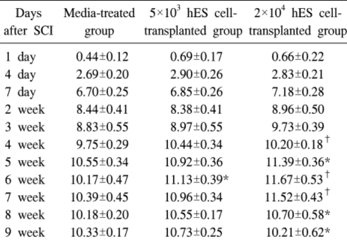
The hind limb locomotor performance was tested in all the rats using the Basso, Beattie and Bresnahan (BBB) open-field scale.7 Prior to transplantation, the BBB tests were performed on all the animals at 1 day, 4 days, and 7 days after a SCI. The animals with a low score and equally malfunctioned hind limbs were selected for the experiments. The media-treated group (n=12) scored very low for both legs 1 day after the injury. The score gradually increased to 9 weeks after the injury (Fig. 1)(Table 1, 2). The 2×10⁴ hES cell-transplanted group
Table 1. BBB Scores of the Left Leg in the SCI Rats Days after SCI Media-treated group 5×10³ hES cell-transplanted group 2×10⁴ hES cell-transplanted group 1 day 4 day 7 day 2 week 3 week 4 week 5 week 6 week 7 week 8 week 9 week 0.44±0.12 2.69±0.20 6.70±0.25 8.44±0.41 8.83±0.55 9.75±0.29 10.55±0.34 10.17±0.47 10.39±0.45 10.18±0.20 10.33±0.17 0.69±0.17 2.90±0.26 6.85±0.26 8.38±0.41 8.97±0.55 10.44±0.34 10.92±0.36 11.13±0.39* 10.96±0.34 10.55±0.17 10.73±0.25 0.66±0.22 2.83±0.21 7.18±0.28 8.96±0.50 9.73±0.39 10.20±0.18† 11.39±0.36* 11.67±0.53† 11.52±0.43† 10.70±0.58* 10.21±0.62* Values were expressed as mean±standard deviation.
BBB: Basso, Beattie and Bresnahan, SCI: Spinal cord injured, hES: Human embryonic stem
*p<0.05, †p<0.01
Table 2. BBB Scores of the Right Leg in the SCI Rats Days after SCI Media-treated group 5×10³ hES cell-transplanted group 2×10⁴ hES cell-transplanted group 1 day 4 day 7 day 2 week 3 week 4 week 5 week 6 week 7 week 8 week 9 week 0.47±0.12 2.85±0.19 6.59±0.26 8.31±0.36 9.19±0.25 9.79±0.30 10.45±0.40 9.94±0.47 10.32±0.44 9.90±0.19 10.22±0.15 0.81±0.18 3.13±0.26 6.53±0.28 8.27±0.40 9.37±0.33 10.44±0.34 10.83±0.32 11.00±0.28 11.04±0.29 10.64±0.17 10.86±0.25 0.74±0.21 3.34±0.25 7.10±0.28 8.58±0.37 9.40±0.65 10.30±0.18 11.43±0.37 11.68±0.51* 11.52±0.50* 10.65±0.52 10.43±0.48 Values were expressed as mean±standard deviation.
BBB: Basso, Beattie and Bresnahan, SCI: Spinal cord injured, hES: Human embryonic stem
*p<0.05
(n=12) showed a significantly improved left (Fig. 1A)(Table 1) and right hind limb (Fig. 1B)(Table 2) performance after the transplant compared with the media-treated group (p<0.05). However, the 5×10³ hES cell-transplanted group (n=12) tended to show an increased hind limb performance after the transplant when compared with the media-treated group, but the difference was not significant except for the left hind limb at 5 weeks after transplantation.
2) Electrophysiological study
At 8 weeks after transplantation, the electrophysiological evaluation, including somatosensory evoked potentials (SSEP) and motor evoked potentials (MEP), was performed in the media-treated group (n=33) and the 2×10⁴ hES cell-transplanted group (n=29). However electrophysiological study was not performed in the 5×10³ hES cell-transplanted group because there was no significant improvement of hind limb performance in the 5×10³ hES cell-transplanted group, compared to the media-treated group, as mentioned above. In the media-treated group, the MEP and the SSEP recordings were detectable in 16 and 28 rats respectively. And in the 2×10⁴ hES cell- transplanted group, the MEP and the SSEP recordings were detectable in 16 and 21 rats respectively too. The responses of the MEP and the SSEP between the media-treated and the hES cell-transplanted group were analyzed using the chi-square test, but the between-group differences were not statistically signi-ficant (p>0.05).
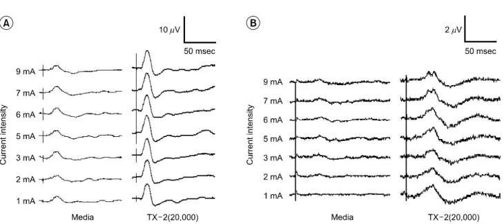
Fig. 2A shows representative wave form of the SSEP, a negative-positive-negative potential (an upward deflection was
designated as negative). The wave of the MEP had the same pattern as the SSEP too (Fig. 2B). However the MEP in the each group consisted of only a few peaks above the detection level of 1μv, on the other hand, the SSEP consisted of distinctive peaks with larger amplitudes than the MEP. The latencies of each evoked potential were measured from the onset of the initial rising phase from the baseline (Initial), the peak of the first negative deflection (N1) and the peak of the first positive deflection (P1). The amplitudes were measured also from the peak of the first negative deflection from the baseline (negative peak amplitude) and the distance between the peak of the first negative and positive deflection (peak to peak amplitude).
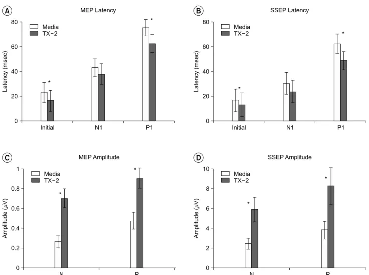
The initial and P1 latency of the MEP and the SSEP in the hES cell-transplanted group were significantly shortened when compared to the media-treated group (p<0.05) (Fig. 3A, B) (Table 3, 4). In addition, the hES cell-transplanted group showed a significant increase in amplitudes of the MEP and the SSEP when compared with the media-treated group (p <0.05) (Fig. 3C, D)(Table 3, 4).
3) Histological examination and immunohistochemistry In the low power view of the spinal cord stained with Luxol fast blue and cresyl violet, the typical pathological findings of the SCI were observed in both the media- and hES cell- transplanted groups at 8 weeks after transplantation. However, the spinal cord of the media-treated group showed more extensive cavity formation, tissue necrosis, gliosis along the rostrocaudal extent and severe demyelination of the white
Table 4. Comparison in Results of the SSEP between the Media- treated and the 2×10⁴ hES Cell-transplanted Group
SSEP Media-treated
group
2×10⁴ hES cell-transplanted group Initial latency (msec)
N1 latency (msec) P1 latency (msec) Negative peak amplitude†
(μv)
Peak to peak amplitude‡ (μv) 16.90±9.10 30.42±8.82 62.44±7.78 2.49±0.54 3.85±1.22 13.18±9.43* 23.85±8.96 48.71±7.23* 5.91±1.22* 8.26±1.83* Values were expressed as mean±standard deviation.
SSEP: Somatosensory evoked potentials, hES: Human embryonic stem, Initial latency: Latency of the onset of the initial rising phase from the baseline, N1 latency: Negative peak latency, latency of the peak of the first negative deflection, P1 latency: Positive peak latency, latency of the peak of the first positive deflection †
: amplitude of the peak of the first negative deflection from the baseline, ‡: amplitude between the peak of the first negative and positive deflection
*p<0.05
Fig. 2. Representative wave forms of evoked potentials recorded in the media-treated group and the human embryonic stem (hES) cell-transplanted group. (A) somatosensory evoked potentials (SSEP), (B) motor evoked potentials (MEP). Media: Media-treated group, TX-2: 2×10⁴ hES cell-transplanted group.
Table 3. Comparison in Results of the MEP between the Media- treated and the 2×10⁴ hES Cell-transplanted Group
MEP Media-treated
group
2×10⁴ hES cell-transplanted group Initial latency (msec)
N1 latency (msec) P1 latency (msec) Negative peak amplitude†
(μv)
Peak to peak amplitude‡ (μv) 22.96±8.03 43.10±7.11 75.25±6.76 0.26±0.06 0.48±0.08 16.34±8.58* 37.58±8.41 62.41±7.21* 0.70±0.10* 0.90±0.10* Values are expressed as mean±standard deviation.
MEP: Motor evoked potentials, hES: Human embryonic stem, Initial latency: Latency of the onset of the initial rising phase from the baseline, N1 latency: Negative peak latency, latency of the peak of the first negative deflection, P1 latency: Positive peak latency, latency of the peak of the first positive deflection †
: amplitude of the peak of the first negative deflection from the baseline, ‡: amplitude between the peak of the first negative and positive deflection
*p<0.05
matter in the ventral funiculus (Fig. 4A, B). In the high power view of the dorsal horns in the vicinity of the necrotic cavity, necrotic cells, glial nuclei and inflammatory cells were predominant in the media-treated group. However, these pathological findings were significantly attenuated and there was no evidence of tumor formation in the hES cell-treated group (Fig. 4C, D). In double labeling immunohistochemistry
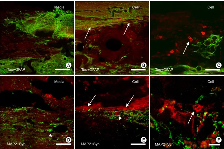
for human Tau and GFAP, the glial scars heavily stained for GFAP were concentrated at the margin of the necrotic cavity. No Tau protein-positive cells were observed in the spinal cords of the media-treated group (Fig. 5A). On the other hand, in the spinal cord of the hES cell-treated group, a few cells positive for the Tau protein were scattered in the necrotic cavity, the spinal gray matter adjacent to the cavity, or in the white matter
Fig. 3. Electrophysiological changes in latencies and amplitudes after spinal cord injury and subsequent transplantation of human embryonic stem (hES) cells. Values are expressed as mean±standard deviation (msec, μV). (A) motor evoked potentials (MEP) latency, (B) somatosensory evoked potentials (SSEP) latency, (C) motor evoked potentials (MEP) amplitudes, (D) somatosensory evoked potentials (SSEP) amplitudes. Media: Media-treated group, TX-2: 2×10⁴ hES cell-transplanted group, Initial: Latency of the onset of the initial rising phase from the baseline, N1: Latency of the peak of the first negative deflection, P1: Latency of the peak of the first positive deflection, N: Amplitude of the peak of the first negative deflection from the baseline, P: Amplitude between the peak of the first negative and positive deflection. The asterisks (*) indicate a statistically significant difference between the media-treated group and the hES cell- transplanted group using a student's t-test (*p<0.05).
(Fig. 5B). These Tau-positive cells were round or ovoid-shaped, and the stain was in the cytoplasm, sparing the nuclei (Fig. 5C). In the double stain for MAP2 and synaptophysin, while most of the neurons were lost and the terminals were clumped in the media-treated rats (Fig. 5D), the anterior horn cells that were positive for MAP2 along with the surrounding terminals in the vicinity of the cavity were spared in the 2×10⁴ hES cell-treated rats (Fig. 5E). In the margin of the cavity in the spinal cord of the hES cell-treated rats, cells with a strong immunoreaction for MAP2 were observed, and these cells were intermixed with varicosities positive for synaptophysin (Fig. 5E, F).
DISCUSSION
Traumatic spinal cord injuries in animals give rise to lesions that are similar to those observed in human SCI. These animals show functional deficits after a SCI resulting from damage to the axons, the loss of neurons and glia, and demyelination.
The results in this study showed that a hES cell transplant significantly improved the locomotion recovery in animals with an acute SCI. In particular, the hind limb performance improved greatly in the 2×10⁴ hES cell-transplanted group, compared with the media-treated group. To our knowledge, this
Fig. 4. Luxol fast blue cresyl violet stain of the spinal cords (parasagittal section) at 8 weeks after the transplantation of human embryonic stem (hES) cells. A and B: Low power view of the spinal cord of the media-treated (Media) and the 2×10⁴ hES cell-transplanted (Cell) rat. Extensive tissue necrosis, glial scar formation (arrows) and demyelination (arrowheads) were observed in the spinal cord of the media-treated group, and these findings were attenuated in the spinal cords of the hES cell-transplanted group. The boxed areas (c, d) are magnified in C and D. C and D: High power view of the areas corresponding to the dorsal horn at the margin of the necrotic cavity. Neuronal necrosis and glial scar formation were predominant in the dorsal horn of the media-treated group (C) but these were not evident in that of the hES cell-transplanted group (D). Scale bars= 500μm.
is the first study to demonstrate the functional recovery after the transplantation of hES cells in spinal cord injured rats. Histologically, the transplantation also attenuated the demyeli-nation and glial scar formation after the SCI. Some cells positive for the human Tau protein were observed 8 weeks after the transplant even though the number was not great. These results suggest that hES cells are effective in the functional recovery after being transplanted in spinal cord injured rats compared with the media-treated rats.
There have been a number of studies that have reported the improvement in the functional recovery after the transplantation in spinal cord injured animals. For example, Zurita and Vaquero8 reported a case of functional recovery in a chronic model using the transplantation of bone marrow stromal cells 3 months after the SCI. They reported that the BBB score showed a significant increase at 4 weeks after the transplant. Similarly, it was previously shown that the hind limb per-formance was significantly improved 28 days after transplanting oligodendrocyte precursor cells.9 In a related study, Groves et al.10 examined the ability of O-2A remyelination in a demyel-inated spinal cord by an X-irradiation. It was shown that in rats, mouse embryonic stem cells differentiated into oligo-dendrocytes, astrocytes and neurons, and the transplantation of mouse embryonic stem cells promoted the functional recovery after transplantation in spinal cord injured rats.3 Therefore, the
transplantation of stem or precursor cells might be effective in the functional recovery after a SCI.
In the present study, a morphological study was performed to examine the survival and differentiation of the transplanted hES cells, which might contribute to the functional and morphological improvement. The results showed that the transplantation of hES cells attenuated glial scar formation and demyelination. This suggests that the functional improvement in this study might be due to the prevention of further degenerative changes or the facilitation of a self regeneration processes rather than to the neuronal differentiation of the transplanted cells. A glial scar is known to be a major cause of functional impairment after a SCI, where a functional deficit is largely due to an interruption in the long ascending and descending tracts rather than to the degeneration of the local neurons.11,12 The inhibition of the glial components, for example, proteoglycans, which implicate neuronal outgrowth, can promote the functional recovery after a SCI.13 The results of double labeling with MAP2 and synaptophysin showed that the neurons and synaptic terminals around the cavity were spared after transplantation. Recently, it was suggested that the host structures may benefit not only by the replacement of lost cells but also from the “chaperone” effect with neuroprotective substances expressed by stem cells.14 On the other hand, cells positive for the human Tau protein were observed in some
Fig. 5. Immunohistochemistry in the spinal cords 8 weeks after transplantation. A∼C: Confocal microscopy of the double immunofluorescence for glial fibrillary acidic protein (GFAP) and human Tau protein (Tau) in the spinal cord of the media-treated (Media) (A) and the 2×10⁴ human embryonic stem (hES) cell-treated (Cell) rats (B and C) 8 week after transplantation. The cells positive for the human Tau proteins were scattered in the cavity and margin of the glial scar of the host spinal cord in the hES cell-treated rats (arrows in B). In the high power view, the Tau protein-positive cells (arrow in C) were round to ovoid in shape. D∼F: Double labeling for microtubule associated protein 2 (MAP2) and synaptophysin (Syn) in the spinal cord of the media-treated (D) and the 2×10⁴ hES cell-treated (E and F) rats. In the media-treated group, the MAP2-positive neurons (arrowheads) were lost and the surrounding synaptic terminals were clumped in the vicinity of the cavity (D), but those were relatively spared in the hES cell-treated rats (E). In this animal, a number of cells at the margin of the cavity were strongly stained for MAP2 (arrows in E and F) and these cells were intermixed with the varicosities stained for synaptophysin (F). Scale bars= 100 μm in A, B, D, and E; 25μm in C; 10μm in F.
animals transplanted with 2×10⁴ hES cells. However, only a small number of Tau-positive cells were observed in these animals, suggesting that the survival rate of the hES cells transplanted in the injured spinal cord was very low.
The possibility that the transplanted cells might also differentiate into neuroglial cells including oligodendrocytes, which play a critical role in remyelination, could not be excluded because the antibodies specific to glial cells of human origin was unavailable. Double labeling with human nuclear antigen (HNA) with other glial markers was attempted, but the antibody for HNA showed nonspecific binding to the de-generating cells (data not shown).
In the electrophysiological evaluation of the present study,
the latencies of motor and somatosensory evoked potentials in the transplanted rats were significantly shortened compared to the media-treated rats. In addition, the hES cell-transplanted group showed increased amplitudes when compared with the media-treated group. In 2001, Akiyama et al.2 examined the myelin repair potential of transplanted neural precursor cells derived from the adult human brain from tissue removed during surgery. They also suggested that transplantation of these cells into the demyelinated rat spinal cord resulted in extensive remyelination and the remyelinated axons conducted impulses at near normal latencies and conduction velocities. Recently, Bambakidis and Miller9 reported that MEP recordings revealed a strong trend towards significant improvement in the latencies
after transplanting oligodendrocyte precursors compared with controls. On the basis of such a prior studies, it seems quite likely that much of the electrophysiological findings in the present study can be explained by activity of the hES cell- derived oligodendrocytes in enhancing myelination or prevention of demyelination, rather than the replacement of lost neuron or axonal regeneration. This is consistent with the histological findings in this study.
Embryonic stem cells are derived directly from early embryos and are pluripotent, indicating that they are individually capable of giving rise to derivatives of each of the three primary germ layers and to germ cells. Embryonic stem cells retain the characteristics of the embryo founder cells, even after a prolonged culture and extensive manipulation.15 Because the cells can be cultured in vitro, there is a greater degree of control over their growth.16 In order to manipulate the cellular behavior, native or artificial factors can also be used. In addition, hES cells have been shown to differentiate into reasonable numbers of neural derivatives.17 Therefore, under these conditions, the transplantation of hES cells may be one of the promising cell replacement therapies for a human SCI. However further studies are necessary to identify the special stain for glial cells of human origin and quantify surviving and differentiated neural cells from the transplanted hES cells by cell counting. And additional long-term studies will be needed not only in animal models but in a human SCI.
CONCLUSION
This study showed that transplantation of hES cells into the injured rat spinal cord improved the hind limb weight support, shortened latencies and increased amplitudes of the evoked potentials and attenuated the glial scar formation and degenerative changes. In addition, some of the transplanted hES cells survived and differentiated into neurons. These results suggest that the transplantation of hES cells plays a role in promoting functional recovery after a SCI, and is one of the important candidates for future cell replacement therapies for a human SCI.
REFERENCES
1) Brustle O, Jones KN, Iearish RD, Karram K, Choudhary K, Wiestler OD, Duncan ID, McKay RD. Embryonic stem cell- derived glial precursor: a source of myelinating transplants. Science 1999; 285: 754-756
2) Akiyama Y, Honmou O, Kato T, Uede T, Hashi K, Kocsis JD. Transplantation of clonal neural precursor cells derived from
adult human brain established functional peripheral myelin in the rat spinal cord. Exp Neurol 2001; 167: 27-39
3) McDonald JW, Liu XZ, Qu Y, Liu S, Mickey SK, Turetsky D, Gottlieb DI, Choi DW. Transplanted embryonic stem cells survive, differentiate and promote recovery in injured rat spinal cord. Nat Med 1999; 5: 1410-1412
4) Thomson JA, Itskovitz-Eldor J, Shapiro SS, Waknitz MA, Swiergiel JJ, Marshall VS, Jones JM. Embryonic stem cell lines derived from human blastocysts. Science 1998; 282: 1145-1147
5) Reubinoff BE, Pera MF, Fong CY, Trouson A, Bongso A. Embryonic stem cell lines from human blastocysts: somatic differentiation in vitro. Nat Biotechnol 2000; 18: 399-404 6) Amit M, Carpenter MK, Inokuma MS, Chiu CP, Harris CP,
Waknitz MA, Itskovitz-Eldor J, Thomson JA. Clonally derived human embryonic stem cell lines maintain pluripotency and proliferative potential for prolonged periods of culture. Dev Biol 2000; 227: 271-278
7) Basso DM, Beattie MS, Bresnahan JC. A sensitive and reliable locomotor rating scale for open field testing in rats. J Neurotrauma 1995; 12: 1-21
8) Zurita M, Vaquero J. Functional recovery in chronic paraplegia after bone marrow stromal cells transplantation. Neuroreport 2004; 15: 1105-1108
9) Bambakidis NC, Miller RH. Transplantation of oligodendrocyte precursors and sonic hedgehog results in improved function and white matter sparing in the spinal cords of adult rats after contusion. Spine J 2004; 4: 16-26
10) Groves AK, Barnett SC, Franklin RJ, Crang AJ, Mayer M, Blakemore WF, Noble M. Repair of demyelinated lesions by transplantation of purified O-2A progenitor cells. Nature 1993; 362: 453-455
11) Reier PJ, Houle JD. The glial scar: its bearing on axonal elongation and transplantation approaches to CNS repair. Adv Neurol 1988; 47: 87-138
12) Silver J, Miller JH. Regeneration beyond the glial scar. Nat Rev Neurosci 2004; 5: 146-156
13) Bradbury EJ, Moon LD, Popat RJ, King VR, Bennett GS, Patel PN, Fawcett JW, McMahon SB. Chondroitinase ABC promotes functional recovery after spinal cord injury. Nature 2002; 416: 636-640
14) Ourednik J, Ourednik V, Lynch WP, Schachner M, Snyder EY. Neural stem cells display an inherent mechanism for rescuing dysfunctional neurons. Nat Biotechnol 2002; 20: 1103-1110 15) Smith A. Embryonic stem cells. In: Marshak DR, Gardner RL,
Gottlieb D, editors. Stem cell biology, 1st ed, New York: Cold Spring Harbor Laboratory Press, 2001, 205-230
16) Kamb A, Ramaswami M, Rao MS. Therapeutic uses of embryonic stem cells. In: Chiu AY, Rao MS, editors. Human embryonic stem cells, 1st ed, Totowa: Humana Press, 2003, 297-322
17) Carpenter MK, Mattson M, Rao MS. Sources of cells for CNS therapy. In: Zigova T, Snyder EY, Sanberg PR, editors. Neural stem cells for brain and spinal cord repair, 1st ed, Totowa: Humana Press, 2003, 3-44