• 검색 결과가 없습니다.

Bisphosphonate-Related Osteonecrosis in a Patient with Florid Cemento-Osseous Dysplasia JOMP

N/A
N/A
Protected

Academic year: 2021

Share "Bisphosphonate-Related Osteonecrosis in a Patient with Florid Cemento-Osseous Dysplasia JOMP"

Copied!
5
0
0

로드 중.... (전체 텍스트 보기)

전체 글

(1)

pISSN 2288-9272 eISSN 2383-8493 J Oral Med Pain 2021;46(2):49-53 https://doi.org/10.14476/jomp.2021.46.2.49

Bisphosphonate-Related Osteonecrosis in a Patient with Florid Cemento-Osseous Dysplasia

Dong-Jun Seo, Seong-Yong Moon, Jae-Seek You, Ji-Su Oh

Department of Oral and Maxillofacial Surgery, College of Dentistry, Chosun University, Gwangju, Korea

Received March 17, 2021 Revised March 31, 2021 Accepted April 1, 2021

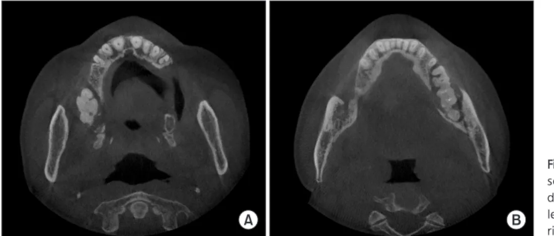
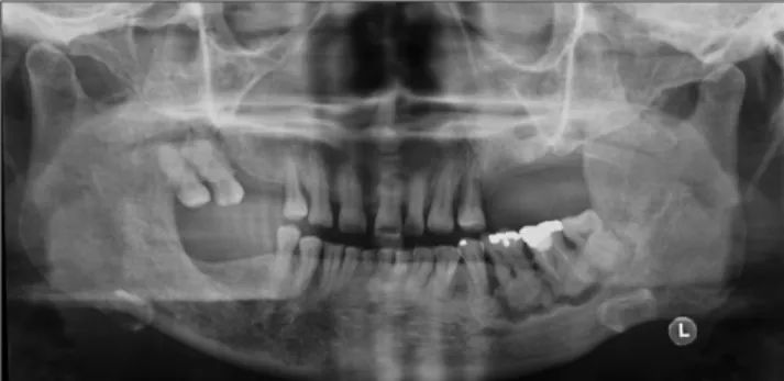
Florid cemento-osseous dysplasia (FCOD) is benign, non-neoplastic lesion characterized by multiple sclerosing masses. Cases of bisphosphonate-related osteonecrosis in FCOD have been rarely reported. we present the patient had multiple osteonecrosis with FCOD in the jaw that occurred after receiving bisphosphonates for eight years due to osteoporosis This report discussed the importance of evaluation of the bone disease in the jaw before bisphos- phonate treatment and periodic follow-up.

Key Words:

Key Words: Bisphosphonate-associated osteonecrosis of the jaw; Jaw; Osteomyelitis;

Osteoporosis

Correspondence to:

Ji-Su Oh

Department of Oral and Maxillofacial Surgery, College of Dentistry, Chosun University, 303 Pilmun-daero, Dong-gu, Gwangju 61452, Korea

Tel: +82-62-220-3813 Fax: +82-62-222-3810 E-mail: [email protected]

https://orcid.org/0000-0002-8369-5025

JOMP

Journal of Oral Medicine and Pain

Copyright

2021 Korean Academy of Orofacial Pain and Oral Medicine. All rights reserved.

CC

This is an open-access article distributed under the terms of the Creative Commons Attribution Non-Commercial License (http://creativecommons.org/licenses/by-nc/4.0/), which permits unrestricted non-commercial use, distribution, and reproduction in any medium, provided the original work is properly cited.

INTRODUCTION

Benign fibro-osseous lesions refer to a group of states characterized by replacing the normal bone with a fi- brous connective tissue that shares the same basic evolu- tive mechanism and becomes gradually mineralized [1].

Among them, cemento-osseous dysplasia (COD) of the jaws originates as a periodontal ligament element and is a fairly common lesion [2], and COD is still a controversial term [3]. According to the latest (4th) edition of the World Health Organization (WHO) classification of head and neck tumors (2017) [3], WHO renames back to the terminology COD, to accept them as odontogenic with an origin in the periodon- tal ligament. There are three categories: periapical COD, fo- cal COD, and florid COD (FCOD) [3].

FCOD is characterized by multiple sclerosing masses only in jaws, which is described as a benign, non-neoplastic le- sion [2]. The word ‘florid’ was introduced to describe the extensive, widespread manifestations in the jaws [4]. The classical FCOD has histologically dense, highly mineralized

characteristics and appears almost acellular cemento-osse- ous tissue [5]. Clinically, these multiple lesions are often as- ymptomatic and can appear as incidental radiological find- ings [6]. So the disease may exist for many years without symptoms [6]. Radiologically, lesions have radiolucent mar- gins with varying degrees of opacity around sclerotic mass [5,6]. The disease has self-limiting characteristics, affects only the alveolar processes, and is independent of teeth [6].

FCOD tends to occur predominantly in middle-aged black women [6]. The disease is prone to secondary infections when exposed to oral pathogens due to the dense sclerotic hypovascular characteristic, which is difficult to manage [7].

So extensive surgical debridement may be required [7].

There are two treatments to manage FCOD. It is recom- mended that asymptomatic patients be observed without surgical intervention. Symptoms usually occur by expos- ing a sclerotic mass to the oral cavity, so biopsy or surgical extraction of teeth in the affected area should be avoided.

No invasive treatment should be performed in asymptom-

atic lesions. If there are no clinical signs, reevaluation with

(2)

panoramic imaging is adequate once every 2 or 3 years.

If new signs or symptoms are observed, examination with dental computed tomography (CT) imaging should be con- sidered. Chronic inflammation and infection appear within dense mineralized tissue, making it more difficult to man- age the symptomatic patients. These infections are known to be difficult to eradicate and often require extensive sur- gical debridement and saucerization. Although antibiotic therapy can be helpful in FCOD, it is often ineffective be- cause the involved area is composed of dense mineralized tissue that is not well perfused [7-9].

Cases of infected FCOD have been rarely reported, es- pecially bisphosphonate-related osteonecrosis in FCOD. In this rare case, we reported the patient with FCOD who pre- sented multiple osteonecrosis that occurred after receiving bisphosphonates for eight years due to osteoporosis. This study was conducted in accordance with the principles of the Declaration of Helsinki, and patient provided written in- formed consent.

CASE REPORT

A 60-year-old female patient was referred with a chief complaint of pain and pus discharge in the left posterior mandible over the past two months. She had diabetes mel- litus, anemia, hypothyroidism, and osteoporosis. The pa- tient was diagnosed asymptomatic FCOD with multiple dif- fuse densely sclerotic lesions confused with roots based on the clinical and radiographic findings before six years (Fig.

1). Further treatments were not planned, periodic follow- up was lost for six years. When the patient returned to the

hospital six years later, the patient presented that she had been taking risedronate monthly for eight years to treat osteoporosis.

Intraoral examination revealed severe gingival swell- ing, exposure of alveolar bone, and yellow, thick purulent discharge.

In cone-beam CT and panoramic radiographs, a wide range of lobulated dense mixed radiopaque-radiolucent le- sions were observed throughout the right maxilla and left mandible (Fig. 2). The lesions included the right first and second molars in the maxilla (Fig. 3A), and continued from the left second premolar to the third molar in the mandible (Fig. 3B). The sclerotic lesions which were not separated and connected like cotton-wool were surrounded by wide osteo- lytic radiolucent lesions. The roots involved in the lesions were confused without resorption. The destruction of the alveolar bone without expansion was extended just above the inferior alveolar canal, but there were fortunately not interruptions of the canal. The alkaline phosphatase level (50 units/L) and white blood cell count (5.14×10

3

/μL) was

Fig. 1.

Fig. 1. Panoramic radiograph showing multiple diffuse densely sclerotic lesions confused with roots before six years.

Fig. 2.

Fig. 2. Axial view computed tomography scan demonstrating a wide range of dense mixed radiopaque-radiolucent lesions without bony expansion in the right maxilla (A) and left mandible (B).

A B

(3)

revealed normal range. Thyroid stimulating hormone was low (0.10 μU/mL) and result of free T4 (1.38 ng/dL) and T3 (94.7 ng/dL) were normal.

The tentative diagnosis of medication-related osteone- crosis of the jaw (MRONJ) complicating FCOD was given.

Based on tentative diagnosis, the drug holiday of bisphos- phonate, the prescription of anti-inflammatory and antibi- otic medication, and the instruction of oral hygiene were planned. The gingival swelling and purulent discharge were subsided after three months. Surgical removal of necrotic bone and curettage was performed with the extraction of the involved teeth under general anesthesia.

On histopathological examination, fibrous connective tis- sue, neutrophils, and granulation tissue are observed around the spherical cementum-like calcification structure (Fig. 4).

Combined with clinical and radiological findings, both the

left mandibular and right maxillary lesions were confirmed as MRONJ associated with infected FCOD. Complete healing was observed 12 months after surgery, and there was no evidence of bone exposure or infection (Fig. 5).

DISCUSSION

Bisphosphonates are administered to malignant neo- plasms, such as multiple myeloma and osteolytic diseases, such as osteoporosis, osteoarthritis. Bisphosphonates sup- press bone resorption by reducing the activity of osteo- clasts, increasing the apoptosis of osteoclasts, and decreas- ing the regulation of matrix metalloproteinases. However, as a result of local sclerosis and inhibition of bone remod- eling, patients with severe osteomyelitis of the jaw re- sulted from its use have been reported [10-13]. The first cases of bisphosphonate-related osteonecrosis of the jaws (BRONJ) were reported in 2003 by Marx [13] and 2004 by Ruggiero et al. [11] Since then, other anticancer (i.e., siroli- mus, bevacizumab) and antiresorptive (denosumab) medi- cations have been implicated in jaw necrosis; as of 2014, Special Committee of the American Association of Oral and Maxillofacial Surgeons (AAOMS) has named this diagnosis as MRONJ. MRONJ can be defined as 1) current or previous treatment with antiangiogenic and/or antiresorptive agents;

2) no history of radiation therapy or no eminent metastatic disease to the jaw; and 3) exposed bone probed through an intraoral or extraoral fistula continuing for more than eight weeks [14].

BRONJ accounts for the largest percentage of MRONJ.

The precise mechanism of BRONJ is not yet unknown.

It seems to be related to the defects in the physiological

Fig. 3.

Fig. 3. Panoramic radiograph revealed sclerotic lesions confused with root not separated and connected to each other surrounded by wide osteolytic radiolucent lesions.

Fig. 4.

Fig. 4. Histopathologic feature showing cementum-like calcification structure around inflammatory cell infiltration (H&E staining, ×200).

Fig. 5.

Fig. 5. Panoramic radiograph showing a normal bone structure in

the right maxilla and left mandible after 1 year of follow-up.

(4)

remodeling or healing of the jaw. Especially, because of the anti-angiogenic properties of bisphosphonates, problems with blood supply to the bone and ischemic changes can occur locally in the affected jaw [11,15].

Dentists should be aware of the key signs and symptoms:

pain, halitosis, exposed necrotic bone, hypoesthesia or par- esthesia, mobility of teeth, and signs of infection such as cellulitis, swelling, fistula, and pus exudation [11,15]. And also, dentists need to know the risk factors that can exac- erbate the development and severity of MRONJ. The pri- mary risk factor for MRONJ is exposure to denosumab or bisphosphonates. Local infection or trauma to the bone or soft tissue (usually surgical trauma or pressure sores) de- velops the condition of MRONJ. In many studies, systemic chronic diseases, the situation of immunocompromise (ie, acquired immune deficiency syndrome), and diseases that reduce the vascularization of the bone seem to affect osteo- myelitis. Smoking, the use of corticosteroids, alcohol abuse, intravenous drugs, radiation, poor oral hygiene, diabetes mellitus, exanthematous fever, anemia, and malignant dis- eases have been considered to be related to increased fre- quency of osteomyelitis [10,11,13,14,16].

When it comes to managing symptoms, MRONJ can be managed effectively, and if precautions are taken, the risk of worsening the condition can be greatly reduced. It is ad- visable for patients, dentists, and physicians to consider prophylactic measures to reduce the risk of MRONJ. If the patients with bone metastases are going to need treatment with denosumab or bisphosphonate, they should fully in- form about the risk of developing MRONJ by physicians.

Also, before the administration of denosumab or bisphos- phonates, the patients should also undergo a clinical and radiological assessment of overall oral hygiene by their dentist. An important key strategy is to keep the patient’s overall oral hygiene good and to have the patient undergo the dental evaluation by dentist every 6 months [11,13-16].

In the event of dental infection such as caries, periodon- titis despite these precautions, timely diagnosis and reso- lution are important in preventing the deterioration of the condition. Extractions should be conducted with the lowest possible trauma level and be performed in a surgical setting to reduce the risk of exacerbating MRONJ. And treatments should be carried out under prophylactic antibiotics and

performed by smoothening of sharp bone and clear closure of the wounds, and then observed until complete soft tissue healing. In stage 1, symptoms and signs of MRONJ can be effectively managed in most patients through a conserva- tive approach. According to the current AAOMS guidelines, debridement is recommended to relieve soft tissue irritation and control infection at stage 2, and resection is recom- mended at stage 3. At later stages, the removal of necrotic bone, sequestrectomy, smoothing of sharp bone margin, and wound closure may be required; some patients may need segmental/continuity resections. Traditionally, surgi- cal treatment is recommended after sequestrum is formed.

However, surgery can be effective in the treatment of MRONJ at any stage. Some physicians recommend stopping the administration of bisphosphonates or denosumab for 2 or 3 months for drug holidays for local treatment and heal- ing after surgery. But there is no clear evidence to support the optimal timing or the effectiveness and safety of this method [11-16].

Lesions such as FCOD are vulnerable to secondary infec- tion because they have hypovascularized and dense sclero- sis properties when exposed to oral pathogens, and can be difficult to manage. Therefore, patients must undergo con- tinuous evaluation and management. Extensive surgical treatment may be necessary if adequate immediate treat- ment is not possible after exposure to the infection. In par- ticular, the surgery of the cases of MRONJ combined with FCOD may be aggressive than the cases of MRONJ alone.

As in this case, in patients with FCOD who have been tak- ing bisphosphonate for a long time, problems with osteo- clasts can degrade the proper healing process for infection and trauma, which can lead to fatal multiple osteomyelitis or osteonecrosis. Therefore, if an early finding of FCOD, the dentist must perform a periodic follow-up and thorough hygiene management in the case of asymptomatic patients, and appropriate treatment should be performed in the case of symptomatic patients. Also, dentists must understand the overall understanding of MRONJ and know how to treat it.

In conclusion, the bone disease in the jaw should be eval-

uated before bisphosphonate is administered. Also, periodic

follow-up and early diagnosis of MRONJ are emphasized in

FCOD patients who are given drugs associated with MRONJ.

(5)

CONFLICT OF INTEREST

No potential conflict of interest relevant to this article was reported.

ORCID

Dong-Jun Seo

https://orcid.org/0000-0002-4301-5881 Seong-Yong Moon

https://orcid.org/0000-0002-7513-4404 Jae-Seek You

https://orcid.org/0000-0001-7638-9583 Ji-Su Oh

https://orcid.org/0000-0002-8369-5025

REFERENCES

1. de Noronha Santos Netto J, Machado Cerri J, Miranda AM, Pires FR. Benign fibro-osseous lesions: clinicopathologic features from 143 cases diagnosed in an oral diagnosis setting. Oral Surg Oral Med Oral Pathol Oral Radiol 2013;115:e56-e65.

2. Coleman H, Altini M, Kieser J, Nissenbaum M. Familial florid cemento-osseous dysplasia--a case report and review of the lit- erature. J Dent Assoc S Afr 1996;51:766-770.

3. El-Mofty SK, Nelson B, Toyosawa S, Wright JM. Cemento-osse- ous dysplasia. In: El-Naggar AK, Chan JKC, Grandis JR, Takata T, Slootweg PJ, eds. WHO classification of head and neck tumours.

4th ed. Lyon: IARC Press; 2017. pp. 254-255.

4. Mangala M, Ramesh DN, Surekha PS, Santosh P. Florid cemento- osseous dysplasia: review and report of two cases. Indian J Dent Res 2006;17:131-134.

5. Ong ST, Siar CH. Florid cemento-osseous dysplasia in a young Chinese man. Case report. Aust Dent J 1997;42:404-408.

6. Melrose RJ, Abrams AM, Mills BG. Florid osseous dysplasia. A clinical-pathologic study of thirty-four cases. Oral Surg Oral Med Oral Pathol 1976;41:62-82.

7. Fenerty S, Shaw W, Verma R, et al. Florid cemento-osseous dys- plasia: review of an uncommon fibro-osseous lesion of the jaw with important clinical implications. Skeletal Radiol 2017;46:581- 590.

8. Beylouni I, Farge P, Mazoyer JF, Coudert JL. Florid cemento- osseous dysplasia: report of a case documented with computed tomography and 3D imaging. Oral Surg Oral Med Oral Pathol Oral Radiol Endod 1998;85:707-711.

9. Waldron CA. Fibro-osseous lesions of the jaws. J Oral Maxillofac Surg 1993;51:828-835.

10. Cavalcante MB, de Oliveira Lima AL, Júnior MA, Santos MB.

Florid cemento-osseous dysplasia simultaneous the chronic sup- purative osteomyelitis in mandible. J Craniofac Surg 2016;27:

2173-2176.

11. Ruggiero SL, Mehrotra B, Rosenberg TJ, Engroff SL. Osteonecrosis of the jaws associated with the use of bisphosphonates: a review of 63 cases. J Oral Maxillofac Surg 2004;62:527-534.

12. Senel FC, Saracoglu Tekin U, Durmus A, Bagis B. Severe osteo- myelitis of the mandible associated with the use of non-nitrogen- containing bisphosphonate (disodium clodronate): report of a case. J Oral Maxillofac Surg 2007;65:562-565.

13. Marx RE. Pamidronate (Aredia) and zoledronate (Zometa) in- duced avascular necrosis of the jaws: a growing epidemic. J Oral Maxillofac Surg 2003;61:1115-1117.

14. Ruggiero SL, Dodson TB, Fantasia J, et al. American Association of Oral and Maxillofacial Surgeons position paper on medication- related osteonecrosis of the jaw--2014 update. J Oral Maxillofac Surg 2014;72:1938-1956.

15. Migliorati CA, Schubert MM, Peterson DE, Seneda LM. Bisphos- phonate-associated osteonecrosis of mandibular and maxillary bone: an emerging oral complication of supportive cancer thera- py. Cancer 2005;104:83-93.

16. Otto S, Pautke C, Van den Wyngaert T, Niepel D, Schiødt M. Med- ication-related osteonecrosis of the jaw: prevention, diagnosis and management in patients with cancer and bone metastases.

Cancer Treat Rev 2018;69:177-187.

수치

Fig. 1. Panoramic radiograph showing multiple diffuse densely  sclerotic lesions confused with roots before six years.
Fig. 3. Panoramic radiograph revealed sclerotic lesions confused  with root not separated and connected to each other surrounded  by wide osteolytic radiolucent lesions

참조

관련 문서

The purposes of the present study are to evaluate the clinical outcomes and radiological outcomes including bone fusion and subsidence that occurred after

In conclusion, as this study found that Internet addiction was significantly related with eating behaviors even after the influences of other variables

Objective: The purpose of this study is to investigate the relationship between employee satisfaction and patient safety and quality improvement after compulsory certification

Diagnosis and Treatment of Oral admidstrated Bisphosphonate-related osteonecrosis of the jaw.. and implantation

This article, in terms of retrospective evaluation, intended to present a suitable alternative for the present with the normative analysis, the comparative

The index is calculated with the latest 5-year auction data of 400 selected Classic, Modern, and Contemporary Chinese painting artists from major auction houses..

SigmaNEST 펀칭 파워팩 제품 관리, 자동 다이나믹™ 배 열 및 펀칭 툴 관리를 갖춘 터렛 펀칭 프로그래밍을 위한 가장 완벽하고 최적화된

The “Asset Allocation” portfolio assumes the following weights: 25% in the S&P 500, 10% in the Russell 2000, 15% in the MSCI EAFE, 5% in the MSCI EME, 25% in the