http://dx.doi.org/10.5090/kjtcs.2014.47.3.211 ISSN: 2233-601X (Print) ISSN: 2093-6516 (Online)
Division of Pediatric Cardiology, Mount Sinai Hospital
Received: September 13, 2013, Revised: December 27, 2013, Accepted: December 31, 2013, Published online: June 5, 2014
Corresponding author: Alexander C. Egbe, Division of Pediatric Cardiology, Mount Sinai Hospital, One Gustave L. Levy place, New York, NY 10029, USA
(Tel) 1-732-581-4858 (Fax) 1-888-719-2864 (E-mail) [email protected]
C
The Korean Society for Thoracic and Cardiovascular Surgery. 2014. All right reserved.
CC
This is an open access article distributed under the terms of the Creative Commons Attribution Non-Commercial License (http://creative- commons.org/licenses/by-nc/3.0) which permits unrestricted non-commercial use, distribution, and reproduction in any medium, provided the original work is properly cited.
Predictors of Intensive Care Unit Morbidity and Midterm Follow-up after Primary Repair of Tetralogy of Fallot
Alexander C. Egbe, M.D., Khanh Nguyen, M.D., Alexander J. C. Mittnacht, M.D., Umesh Joashi, M.D.
Background: Our objectives were to review our institutional early and midterm experience with primary tetralogy of Fallot (TOF) repair, and identify predictors of intensive care unit (ICU) morbidity. Methods: We analyzed perioper- ative and midterm follow-up data for all cases of primary TOF repair from 2001 to 2012. The primary endpoint was early mortality and morbidity, and the secondary endpoint was survival and functional status at follow-up.
Results: Ninety-seven patients underwent primary repair. The median age was 4.9 months (range, 1 to 9 months), and the median weight was 5.3 kg (range, 3.1 to 9.8 kg). There was no early surgical mortality. The incidence of junctional ectopic tachycardia and persistent complete heart block was 2% and 1%, respectively. The median length of ICU stay was 6 days (range, 2 to 21 days), and the median duration of mechanical ventilation was 19 hours (range, 0 to 136 hours). By multiple regression analysis, age and weight were independent predictors of the length of ICU stay, while the surgical era was an independent predictor of the duration of mechanical ventilation. At the 8-year follow-up, freedom from death and re-intervention was 97% and 90%, respectively. Conclusion: Primary TOF repair is a safe procedure with low mortality and morbidity in a medium-sized program with outcomes comparable to national standards. Age and weight at the time of surgery remain significant predictors of morbidity.
Key words: 1. Tetralogy of Fallot 2. Pediatric
3. Outcomes 4. Ventilation 5. Morbidity
INTRODUCTION
Tetralogy of Fallot (TOF) is the most common congenital cyanotic heart disease with an incidence of 3 per 10, 000 live births, and accounts for about 5% to 7% of all congenital heart disease [1]. From the time of the first surgical palliation of TOF by Blalock and Taussig in 1945, surgical manage- ment has evolved to primary corrective repair that can safely be performed in all age groups [2]. The safety of early pri-
mary repair is well documented in the literature with several studies showing that it is a safe procedure even in neonates [3]. As a result, primary repair of TOF is now a routine pro- cedure with a low surgical mortality rate of 0% to 2% [3-7].
Advocates of early primary repair believe that early relief of
right ventricular outflow tract (RVOT) obstruction will pre-
vent right ventricular hypertrophy and dysfunction, as well as
establish unobstructed pulmonary blood flow, which will en-
courage alveologenesis [8-10]. However, the data also clearly
suggest an increased incidence of junctional ectopic tachy- cardia (JET), longer intensive care unit (ICU) and hospital stays, more complicated recovery, and increased need for valve-sacrificing transannular patch (TAP) repairs in patients who undergo primary repair in the neonatal period [4-6,11,12]. Some centers including ours perform neonatal re- pair when clinically indicated for severe RVOT obstruction, cyanosis, and hypercyanotic spells.
In spite of low surgical mortality, ICU morbidity is rela- tively common after primary TOF repair [13]. There is a strong correlation between ICU morbidity and intraoperative factors such as cardiopulmonary bypass (CPB) time, cross clamp, and surgical techniques [3,11,13]. However, our ability to identify patients at risk for significant ICU morbidity based on their preoperative characteristics is limited because of conflicting evidence in the literature [4,5,11,13]. We have hypothesized that certain preoperative demographic and mor- phologic characteristics increase the risk of ICU morbidity af- ter primary TOF repair.
METHODS
1) Patient population
All patients with the diagnosis of TOF with pulmonic stenosis who underwent primary repair between January 2001 and December 2012 at Mount Sinai Hospital, New York, were identified from the hospital database. Our surgical data- base reported that 126 patients underwent TOF repair within the study period. We excluded patients who had a prior Blalock-Taussig shunt, patients who were older than 12 months of age at the time of repair, and patients with asso- ciated anatomic defects such as absent pulmonary valve syn- drome, atrioventricular septal defect, or pulmonary atresia.
Based on our inclusion criteria, we identified 99 patients within this study period who underwent primary TOF repair in infancy. We divided the study period into the early surgi- cal era (January 2001–December 2006) and the late surgical era (January 2007–December 2012) to evaluate the impact of evolving changes in surgical/anesthesia technique and peri-op- erative management in the observed time period. Our study was approved by the hospital’s institutional review board.
The primary objective of this study was to determine over-
all survival and incidence of complications. Our secondary objectives were to identify predictors of ICU morbidity as well as survival and functional status at midterm follow-up.
We defined ICU morbidity as prolonged ICU stay (length of ICU stay ≥7 days) and/or prolonged mechanical ventilation (duration of mechanical ventilation ≥48 hours). For midterm follow-up, we analyzed survival, freedom from re-inter- vention, and freedom from a severe residual lesion at 1 year, 3 years, 5 years, and 8 years postoperatively.
2) Data collection
(1) Preoperative data: Demographic information evaluated included age at the time of surgery, gestational age, gender, weight, history of hypercyanotic spells, baseline systemic sat- uration, history of associated genetic disorders inclusive of Down’s syndrome, and presence of other extracardiac comorbidities.
(2) Echocardiographic data: The pulmonary valve annulus z-score and main pulmonary artery z-score were evaluated as measures of RVOT obstruction.
(3) Operative data: The following were collected and ana- lyzed: CPB time, aortic cross-clamp time, and presence of a hemodynamically significant residual lesion on intraoperative transesophageal echocardiogram (TEE). We defined the re- sidual hemodynamic lesion as greater than trivial residual ventricular septal defect (VSD) or greater than trivial residual pulmonic stenosis (maximum instantaneous gradient of 20 mmHg).
(4) Postoperative intensive care unit data: After the oper- ation, the following were recorded: length of ICU stay, dura- tion of mechanical ventilation, duration of inotropic support, postoperative arrhythmia such as junction ectopic tachycardia (JET), need for extracorporeal membrane oxygenation (ECMO) support, readmission within 30 days of surgery, and death within 30 days of surgery. Hemodynamic data and lab values collected and reviewed include heart rate, central ve- nous pressure, systemic arterial pressure (systolic, diastolic, and mean), serum lactate, blood gases, chemistry, and com- plete blood count.
(5) Midterm follow-up data: We collected and analyzed
data on survival, freedom from re-intervention, and freedom
from a significant residual lesion at 1 year, 3 years, 5 years,
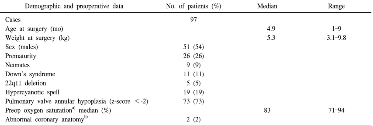
Table 1. Patient demographics
Demographic and preoperative data No. of patients (%) Median Range
Cases
Age at surgery (mo) Weight at surgery (kg) Sex (males)
Prematurity Neonates Down’s syndrome 22q11 deletion Hypercyanotic spell
Pulmonary valve annular hypoplasia (z-score <-2) Preop oxygen saturation
a)median (%)
Abnormal coronary anatomy
b)97
51 (54) 26 (26) 9 (9) 11 (11)
5 (5) 19 (19) 73 (73)
2 (2)
4.9 5.3
83
1–9 3.1–9.8
71–94
a)
Oxygen saturation during pre-surgical evaluation usually about 1 week prior to surgery.
b)
Large conal branch or left anterior descending artery crossing the right ventricular outflow tract.
and 8 years postoperatively. A significant residual lesion was defined as moderate/severe pulmonary regurgitation or tricus- pid regurgitation, moderate/severe right ventricular dilation, or systolic dysfunction.
3) Statistical analysis
Standard descriptive statistical methods were used. Data were described as frequencies and median with ranges as appropriate. To assess the differences between groups, the Mann–Whitney test or Student t-test for continuous data and the chi-square test or Fisher’s exact test for categorical data were used. Multiple logistic regression analysis was per- formed to determine the independent risk factors of prolonged length of ICU stay and prolonged duration of mechanical ventilation. A p-value of <0.05 was considered statistically significant. All analyses were performed using the SAS ver.
7.0 statistical software (SAS Institute Inc., Cary, NC, USA) with default settings except where indicated.
RESULTS
Over the 12-year study period, 99 patients with TOF un- derwent primary repair in infancy in Mount Sinai Hospital.
Two patients had incomplete hemodynamic data and were ex- cluded from the study. Table 1 shows the demographic and preoperative data of the study population. The median age at the time of surgery was 4.9 months (range, 1 to 9 months),
and the median weight was 5.3 kg (range, 3.1 to 9.8 kg).
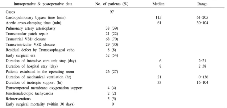
There were 51 males and 46 females making up 53% and 47% of the study population, respectively. Twenty-six patients (26%) were premature (delivery prior to 37 completed weeks of gestation). Nine patients (9%) underwent TOF repair with- in the first month of life. The indication for neonatal surgery was severe hypoxia due to severe RVOT obstruction in all 9 patients. It is Mount Sinai Hospital’s policy to avoid pallia- tive shunts and to perform primary repair irrespective of age whenever possible. Eleven patients (11%) had Down’s syn- drome, five patients (5%) had 22q11 deletion, and 19 patients (19%) had at least one episode of hypercyanotic spells prior to TOF repair. The median pulmonary valve z-score for our population was -3.6 (range, -5.1 to -0.3), and 73 patients (75%) had pulmonary valve annular hypoplasia (pulmonary valve z-score <-2). The median CPB time was 114 minutes (range, 61 to 205 minutes), and the median aortic cross- clamp time was 61 minutes (range, 30 to 104 minutes).
Twenty-one patients underwent TAP repair, and 38 patients
underwent additional pulmonary arterioplasty as part of surgi-
cal relief for RVOT obstruction. The decision to perform
TAP repair versus a valve-sparing procedure was based on
the pulmonary valve z-score (greater or less than -2) or the
decision of the surgeon. The decision to perform pulmonary
arterioplasty was based entirely on the surgeon’s intra-
operative assessment of size on branch pulmonary arteries
(Table 2). Intraoperative TEE showed residual hemodynami-
Table 2. Intraoperative and postoperative data
Intraoperative & postoperative data No. of patients (%) Median Range
Cases
Cardiopulmonary bypass time (min) Aortic cross-clamping time (min) Pulmonary artery arterioplasty Transannular patch repair Transatrial VSD closure Transventricular VSD closure
Residual defect by Transesophageal echo Early surgical era
Duration of intensive care unit stay (day) Duration of hospital stay (day)
Patients extubated in the operating room Duration of mechanical ventilation (hr) Duration of inotropic support (hr)
Extracorporeal membrane oxygenation support Junctionalectopic tachycardia
Reinterventions
Early surgical mortality (within 30 days)
97
38 (39) 21 (22) 68 (70) 29 (30) 8 (8) 52 (54)
26 (27)
4 (4) 2 (2) 5 (5) 0
115 61
6 8
21 33
61–205 30–104
2–21 2–38
0–136 16–104
VSD, ventricular septal defect.
cally insignificant lesions in eight patients (8%). These re- sidual lesions were a trivial/small residual VSD patch leak in 5 patients, mild residual RVOT obstruction in 2 patients, and small VSD patch leak plus mild RVOT obstruction in 1 pa- tient, none of which required a repeat bypass run. Five pa- tients (5%) underwent reintervention before hospital discharge. Four of these reinterventions were re-exploration for bleeding. One patient underwent placement of a perma- nent epicardial pacemaker for persistent complete heart block.
The median length of ICU stay was 6 days (range, 2 to 21 days), and the median duration of mechanical ventilation was 19 hours (range, 0 to 136 hours). Four patients (4%) required ECMO support for hemodynamic instability. The clinical courses of these 4 patients were as follows: patient #1 (age, 3 months; weight, 5.8 kg) and #2 (age, 2 months; weight, 4.1 kg) were placed on veinarterial extracorporeal-ECMO support on the first postoperative night because of hemodynamic in- stability due to intractable JET. In both cases, JET resolved with amiodarone (patient #1) and amiodarone with procaina- mide (patient #2). Both patients were successfully decannu- lated on postoperative day 2 (patient #1) and postoperative day 3 (patient #2). Patient #3 underwent neonatal TOF repair (age, 4 days; weight, 3.9 kg). The patient also underwent
TAP repair with extensive pulmonary artery plasty and was placed on ECMO in the operating room (OR) because of in- ability to wean the patient from CPB. Intraoperative TEE did not show any significant anatomic residual lesion, and the pa- tient has normal coronary artery anatomy. The patient was decannulated on postoperative day 2. Patient #4 (weight, 9.4 kg) was an 11-month-old infant with severe RVOT ob- struction and restrictive RV physiology who had marginal cardiac output coming off bypass and had cardiac arrest with- in 6 hours of arrival in the ICU. The patient was immedi- ately ‘crashed’ onto ECMO support. Atrial communication was created percutaneously in the catheterization lab on post- operative day 3 because of persistent right ventricular dia- stolic dysfunction. The patient was successfully decannulated on postoperative day 6. All of the patients survived to hospi- tal discharge, and there was no early surgical mortality (Table 2).
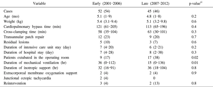
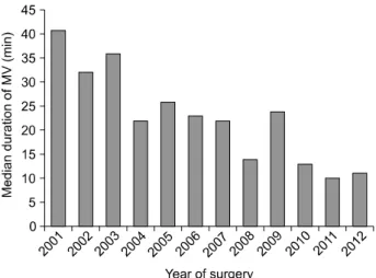
Comparing surgical eras, we found that there was a statisti-
cally significant decrease in the duration of mechanical ven-
tilation from the early surgical era (2001–2006) to the late
surgical era (2007–2012) (p-value=0.01). The median duration
of ventilation in the patients who were not extubated in the
OR decreased from 36 hours (range, 0 to 112 hours) in the
Table 3. Data by surgical era
Variable Early (2001–2006) Late (2007–2012) p-value
a)Cases Age (mo) Weight (kg)
Cardiopulmonary bypass time (min) Cross-clamping time (min)
Transannular patch repair Residual lesions
Duration of intensive care unit stay (day) Duration of hospital stay (day)
Patients extubated in the operating room Duration of mechanical ventilation (hr) Duration of inotropic support (hr)
Extracorporeal membrane oxygenation support Junctional ectopic tachycardia
Reintervention
52 (54) 5.1 (1–9) 5.4 (3.1–9.4) 121 (61–205) 58 (35–104) 12 (23) 5 (10) 7 (4–20) 7 (4–28) 9 (17) 36 (0–112) 32 (16–91) 2 (4) 2 (4) 3 (4)
45 (46) 4.8 (1–8) 5.1 (3.2–9.8) 113 (65–196) 63 (30–101) 9 (20) 3 (7) 6 (2–21) 8 (2–38) 17 (38) 15 (0–136) 36 (18–104) 2 (4) 0 2 (13)
0.2 0.6 0.4 0.3 0.7 0.6 0.2 0.3 0.02 0.01 0.8 0.9
0.8 Values are presented as number (%) or median (range).
a)
Statistical significance in multivariate analysis.
early surgical era to 13 hours (range, 0 to 136 hours) in the late era. Apart from the change in the duration of mechanical ventilation, there was no significant difference in the length of ICU stay, patient demographics, surgical technique, intra- operative factors (CPB time and aortic cross-clamping time), and postoperative complications between the two surgical eras (Table 3).
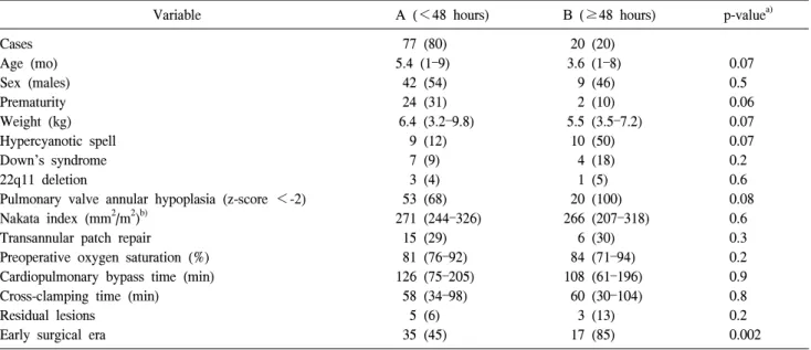
Based on our definition of prolonged ICU stay (length of ICU stay ≥7 days), 22 patients (23%) had prolonged ICU stay. These patients with prolonged ICU stay underwent sur- gical repair at a younger age (median, 3.8 months; range, 1 to 7 months) as compared to patients with ICU stay <7 days (median, 5.1 months; range, 1 to 9 months) (p-value=0.001).
Also, there was a significant difference in the weight of pa- tients with prolonged ICU stay (median, 4.8 kg; range, 3.1 to 7.5 kg) as compared to patients with the length of ICU stay
<7 days (median, 6.2 kg; range, 3.2 to 9.8 kg) (p-val- ue=0.001). A majority of the patients (73%) with prolonged ICU stay underwent TOF repair in the early surgical era as compared to 48% of the patients with an ICU stay duration of <7 days, p-value=0.02. Additionally, there was higher in- cidence of pulmonary valve annular hypoplasia in the patients with prolonged ICU stay than in those with normal ICU stay duration (100% vs. 68%, p-value=0.002). However, on multi-
variate analysis, only age and weight at the time of surgery emerged as statistically significant predictors of prolonged ICU stay as shown in Table 4.
Table 5 compares the preoperative and intraoperative char-
acteristics of patients with prolonged duration of mechanical
ventilation (≥48 hours) to patients with a normal duration of
mechanical ventilation (<48 hours). Patients with a pro-
longed duration of mechanical ventilation underwent surgical
repair at a younger age (median, 3.6 months; range, 1 to 8
months) as compared to patients with a duration of mechan-
ical ventilation of <48 hours (median, 5.4 months; range, 1
to 9 months) (p-value=0.001). Also, there was a significant
difference in the weight of patients with a prolonged duration
of mechanical ventilation (median, 5.5 kg; range, 3.5 to 7.2
kg) as compared to patients with a duration of mechanical
ventilation of <48 hours (median, 6.4 kg; range, 3.2 to 9.8
kg) (p-value=0.001). Eighty-five percent of the patients
(n=17) with a prolonged duration of mechanical ventilation
underwent TOF repair in the early surgical era as compared
to 45% (n=35) of the patients with a duration of mechanical
ventilation of <48 hours (p-value=0.002). Additionally, there
was a higher incidence of pulmonary valve annular hypo-
plasia in the patients with a prolonged duration of mechanical
ventilation than in those with a duration of mechanical ven-
Table 4. Length of intensive care unit stay
Variable A (<7 days) B (≥7 days) p-value
a)Cases Age (mo) Sex (males) Prematurity Weight (kg) Hypercyanotic spell Down’s syndrome 22q11 deletion
Pulmonary valve annular hypoplasia (z-score <-2) Nakata index (mm
2/m
2)
b)Transannular patch repair
Preoperative oxygen saturation (%) Patients extubated in the operating room Cardiopulmonary bypass time (min) Cross-clamp time (min)
Residual lesions Early surgical era
75 (77) 5.1 (1–9) 41 (54) 21 (28)
6.2 (3.2–9.8) 10 (13) 7 (9) 3 (4) 51 (68) 284 (251–326) 16 (21) 85 (71–94) 21 (28) 118 (75–205) 58 (34–98) 5 (6) 36 (48)
22 (23) 3.8 (1–7) 10 (45) 5 (22)
4.8 (3.1–7.5) 9 (40) 4 (18) 1 (5) 22 (100) 262 (207–314) 5 (23) 82 (74–91) 5 (23) 114 (61–175) 60 (30–104) 3 (13) 16 (73)
0.001 0.4 0.6 0.001 0.07 0.3 0.6 0.07 0.1 0.2 0.4 0.6 0.6 0.8 0.3 0.09 Values are presented as number (%) or median (range).
a)
Statistical significance in multivariate analysis.
b)
Nakata index data were only available in 76 patients (78% of our cohort).
Table 5. Duration of mechanical ventilation
Variable A (<48 hours) B (≥48 hours) p-value
a)Cases Age (mo) Sex (males) Prematurity Weight (kg) Hypercyanotic spell Down’s syndrome 22q11 deletion
Pulmonary valve annular hypoplasia (z-score <-2) Nakata index (mm
2/m
2)
b)Transannular patch repair
Preoperative oxygen saturation (%) Cardiopulmonary bypass time (min) Cross-clamping time (min)
Residual lesions Early surgical era
77 (80) 5.4 (1–9) 42 (54) 24 (31)
6.4 (3.2–9.8) 9 (12) 7 (9) 3 (4) 53 (68) 271 (244–326) 15 (29) 81 (76–92) 126 (75–205) 58 (34–98) 5 (6) 35 (45)
20 (20) 3.6 (1–8) 9 (46) 2 (10)
5.5 (3.5–7.2) 10 (50) 4 (18) 1 (5) 20 (100) 266 (207–318) 6 (30) 84 (71–94) 108 (61–196) 60 (30–104) 3 (13) 17 (85)
0.07 0.5 0.06 0.07 0.07 0.2 0.6 0.08 0.6 0.3 0.2 0.9 0.8 0.2 0.002 Values are presented as number (%) or median (range).
a)
Statistical significance in multivariate analysis.
b)