Introduction
Chronic expanding EDH cases have been infrequently reported. It has been hypothesized that this expansion is due to rebleeding of chronic epidural hematomas in their dural capsules. We report a case of a chronic expanding epidural hematoma with increased intracranial pressure symptoms and an onsetof approximately 12 days after head trauma.
Case Report
A 12-year-old boy fell to the ground during a soccer game.
Immediately, he presented at our department complaining of headacheand nausea. His medical history was unremarkable. He denied a previous episode of seizure.
His neurologic examination showed Glasgow Coma Scale score of 15 and no neurologic deficit, and his laboratory findings were within normal limits. He underwent urgent brain computed tomography(CT) (Fig. 1-A), and an abnormal extracerebral space containing heterogenous
intermediate and high density materials, was observed in both posterior parietal convexities. Attenuation coefficient measures of 60 to 70 Hounsfield units suggest hemorrhage.
However, theses materials was adherent to the inner membrane crossing the midline falx. This initial brain CT scan indicated a relapsing subacute and chronic epidural process with partial calcification within chronic hematoma.
We asked the patient about his prior head truma, and he recalled an episode about 4 years ago, when he fell down stairs. His symptoms (headache and nausea) gradually subsided (Fig. 1-B). A magnetic resonance(MR) image taken at post trauma 12 days showed subacute stage EDH in the left posterior parietal eonvexity with extension to the right side. An MR venogram showed patent sagittal sinuses and minimal displacement of venous structures by EDH.
However he was relatively tolerable at that time.
On the 13th day, a follow up brain CT examination with contrast enhancement showed significant interval changes compared with the initial examination (Fig. 1-C). Fluid density was loculated in posterior parietal convexity in abiconvex configuration. Review of the MR image suggested the fluid to be a methemoglobin stage hemorrhage. MRI also revealed adjacent lateral ventricle compression, and an enhanced dural margin was noted
고신대학교 의과대학 학술지 제 권 제 호24 2 Kosin Medical Journal
Vol. 24. No. 2, pp. 261 264, 2009∼
- -
Ju-Ho Jeong ・ Jin-Wook Kim
Department of Neurosurgery, Kosin University College of Medicine, Busan, Korea
――― Abstract ――――――――――――――――――――――――――――――――――――――――
The authors report a case of chronic expanding epidural hematoma (EDH). A 12-year-old boy presented with headache, nausea, and vomiting 12 days after headtrauma. Chronic epidural hematoma was diagnosed and craniotomy was performed at post trauma 14 days. The authors comfirmed the pathologic findings of the hematoma membrane, which appeared to play a key role in hematoma expansion, much like that of chronic subdural hematoma. Delayed expansion of chronic EDH should be borne in mind when planningconservative management.
―――――――――――――――――――――――――――――――――――――――――――――――――
Key words : Chronic epidural hematoma, expanding, membrane
교신저자 김 진 욱:
주소 : 602-702 부산광역시 서구 암남동34번지 고신대학교 의과대학 신경외과학교실 TEL : 051-990-6465, FAX : 051-990-3042 E-mail: [email protected]
고신대학교 의과대학 학술지 제 권 호24 2 , 2009
following contrast injection. Contralateral extension across the midline was also noted at the vertex.
Craniotomy was performed on the 14th day, and a dark brown colored fluid was noted in the epidural space (Fig.
2). In addition, a dark red blood clot with a fibrous membrane was attached to the dural surface at the left frontoparietal convexity (Fig. 3). However, there was no active bleeding focus.
Abnormal calcification, and fresh and old red blood cells were evident in hematoxylin and eosin (H & E) stain, which suggested repeated hemorrhage (Fig. 4).In addition, multiple capillary formations at membranous surfaceof the hematoma was evident in immunohistochemistry for factor VIII (Fig. 5).
He was discharged without any neurologic deficit at 8 days post craniotomy.
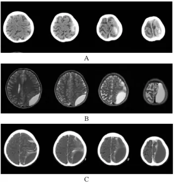
A
B
C
Fig. 1. Serial Brain CT and MR images of 12-year-old male patient. A: Initial brain CT showed high density materials suggesting hemorrhage. This initial brain CT showed a relapsing subacute and chronic epidural process with partial calcification within chronic hematoma. B: T2 weighted MR image taken at 12 days after head trauma showing subacute stage epidural hematomas at the left posterior parietal convexity. C: On the 13th day, contrast enhanced CT showed significant interval changes as compared with the initial examination. Fluid density was loculated in the posterior parietal convexity in a biconvex configuration.
Fig. 2. Operative views. The upper portion of the photograph showed the left frontoparietal convexity. The lower portion of the photograph is near the sagittal sinus. A dark brown colored fluid was found in the epidural space. A dark red blood clot with a fibrous membrane was found attached to the dural surface on the frontoparietal convexity. No active bleeding focus was evident.
Fig. 3. Gross feature of the specimen obtained from the epidural surface. The specimen appeared to be composed of old blood clots with a membranous surface.
Fig. 4. An abnormal calcification(blue arrow), and fresh and old red blood cells were evident suggesting repeated hemorrhage. The presence of calcification in the initial CT scan indicated an old hemorrhage (H & E stain, × 100).
Chronic Expanding Epidural Hematoma
Fig. 5. Immunohistochemistry for factor VIII showed multiple capillary formations at the membranous surface of the specimen, suggesting that the hematoma membrane played a key role in hematoma expansion (Immunohistochemistry, × 100).
Discussion
No meaningful consensus has been achieved regarding the definition of ‘chronic’ epidural hematoma with respect to from acute trauma to surgery.1) This interval was 14 days in our patient, which matches the definition of Iwakuma and Brunngraber, who examined EDH histologically and defined chronic EDH based on a diagnosis at more than 13 days after head injury based on a finding of capsule ossification.2) Other investigators have regarded 3, 4, or 7 days post-injury as ‘chronic’.3~5)
The differentiation of chronic EDH and delayed EDH is not straightforward but is important.1, 6)Delayed EDH is a rare entity,7) and has been defined as a hematoma "not apparent in CT scans (or by angiography) performed immediately after craniocerebral injury, but which manifests radiologically at subsequent examination".6) The prognosis of delayed EDH is poor, compared with those of acute or chronic EDH, and surgical treatment is almost always required.6)
Because the hematoma membrane resembles the membranes formed during chronic subdural hematoma, these two lesion types may share a hematoma resorption mechanism.8, 9) However, it is surprising that few authors have emphasizedthe role played by the hematoma membrane in
hematom expansion in chronic subdural hematoma, because this has been suggested on several occasions to be a possibility in epidural hematoma.10-14)
In the present case, abnormal calcification and fresh and old red blood cells were demonstrated in H & E sections, which suggested repeat hemorrhage. Furthermore, the presence of calcification indicates remote onset hemorrhage, and in our patient, was observed in initial brain CT scans.
Furthermore, factor VIII stained sections demonstrated multiple capillary formations at the membranous surface of the hematoma, indicating that the hematoma membrane played a key role in the expansion of the hematoma.
Conclusion
We report a case of chronic expanding EDH, and caution that delayed expansion of a chronic EDH should be borne in mind when planning conservative management.
국문초록
저자들은 만성 팽창성 경막상 혈종을 례 치험하였기에1
문헌고찰과 함께 보고 하고자 한다. 12살 소년이 두부 외 상을 받은지12일 후에 두통 오심 구토를 주소로 내원하,
였다 만성 경막하 혈종으로 진단되었고 두부 외상을 받.
은지14일째 개두술을 통한 혈종제거 수술을 받았다 병, . 리학적 검사상 혈종의 팽창에 핵심적인 역할을 하는 혈
종막을 확인하였다 이런 혈종막은 만성 경막하 혈종에.
서 자주 발견되는 것이다 결론적으로 만성 경막상 혈종. ,
을 보존적으로 치료하는 경우에는 반드시 만성 경막상 혈종의 팽창 가능성을 확인해야 한다.
중심 단어 만성 경막상 혈종 팽창성 막: , ,
References
1) Tatagiba M, Sepehrnia A, el Azm M, Samii M: Chronic epidural hematoma-report on eight cases and review of the
고신대학교 의과대학 학술지 제 권 호24 2 , 2009
literature. Surg Neurol 32 : 453-458, 1989
2) Iwakuma T, Brunngraber CV:Chronic extradural hematomas. A study of 21 cases. J Neurosurg 38: 488-93, 1973
3) Bullock R, Van Dellen JR:Chronic extradural hematoma. Surg Neurol 18: 300-302, 1982
4) Clevel M, Onzain I, Gutierrez F. Chronic epidural hematomas.
Acta Neurochir 66: 71-81, 1982
5) Fiume D, Interligi M, Nardi P, Parziale G, Savino S.
L'ematoma epidurale cronico. Studio di 53 casi operati. Riv Neurol 55: 350-356, 1985
6) Milo R, Razon N, Schiffer J, Delayed epidural hematoma. A review. Acta Neurochir (Wien) 84 : 13-23, 1987
7) Rivas JJ, Lobato RD, Sarabia R, Cordob s F, Cabrera A,é Gomez P., Extradural hematoma: analysis of factors influencing the courses of 161 patients. Neurosurgery 23:
44-51, 1988
8) Masuzawa H, Sato J, Kamitani H, Miyake H, Ishiko T, Yoshimasu N. Membrane formation in epidural and subperiosteal hematomas. Relationship with the neomembrane of chronic subdural hematomas. Neurol Med Chir (Tokyo) 22 : 995-1001, 1982
9) Zuccarello M, Fiore DL, Pardatscher K, Trincia G, Andrioli GC. Chronic extradural haematomas. Acta Neurochir (Wien) 67 : 57-66, 1983
10) Klingler M, Scheidegger S. Organisationsvorg nge beiä epiduraler Bluntung. Zugleich ein Beitrag zur Frange der Membranbildung beim chronischen subduralen H matom.ä Acta Neurochir (Wien) 19 : 39-50, 1968
11) Pang D, Horton JA, Herron JM, Wilberger JE, Vries JK.
Nonsurgical management of extradural hematoma. J Trauma 27 : 579-580, 1983
12) Pozzati E, Staffa G, Nuzzo G, Frank F. Late recurrence of bleeding in a chronic extradural hematoma. J Trauma 27:
579-580, 1987
13) Watanabe T, Nakahara K, Miki Y, Shibui S, Takakura K, Nomura K. Chronic expanding epidural haematoma. Case report. Acta Neurochir (Wien) 132 : 150 153, 1995– 14) Kanamori M, Seki K, Kitahara M. A case of chronic
expanding epidural hematoma. No Shinkei Geka 250: 1137–
1142, 1997