CASE REPORT
대한화상학회지 제 23 권 제 2 호
64
Journal of Korean Burn Society Vol. 23, No. 2, 64-67, 2020 https://doi.org/10.47955/jkbs.20.0014
자궁 근종에 대한 HIFU 치료 후 발생한 하부 복부의 심부 화상
유성훈ㆍ김동철
분당제생병원 성형외과, 화상재건센터
Deep Burn Injuries on the Lower Abdomen after HIFU Treatment for Uterine Myoma
Sung Hoon Yu, M.D. and Dong Chul Kim, M.D., Ph.D.
Department of Plastic and Reconstructive Surgery, Advanced Burn Reconstruction Center, Bundang Jesaeng Hospital, Seongnam, Korea
High-intensity focused ultrasound (HIFU) has been regarded as a non-invasive uterine-preserving treatment for women with uterine myoma. Numerous studies have reported that it is a relatively safe and effective treatment for uterine myoma.
However, severe complications, such as deep thermal burn injuries, bowel perforation, and bladder injury, were reported on rare occasions. We report a case of a 4th degree burn on the lower abdomen after HIFU treatment for uterine myoma.
Physicians must consider the possibility of deep thermal burn injuries when managing uterine myoma with HIFU. (J Korean
Burn Soc 2020;23:64-67)
Key Words: HIFU, Burns, Uterine myoma
Received: 2020. 11. 4, Accepted: 2020. 11. 16
Corresponding author: Dong Chul Kim, Department of Plastic and Reconstructive Surgery, Advanced Burn Reconstruction Center, Bundang Jesaeng General Hospital, 20 Seohyeon- ro 180, Seongnam 13590, Korea
Tel: 82-31-779-0281, Fax: 82-31-779-5032 E-mail: [email protected]
INTRODUCTION
High-intensity focused ultrasound (HIFU) is a non-in- vasive ultrasound technique that focuses and transmits ultrasound beams through solid body tissues resulting in bio-effects that can destroy and coagulate deeper tissues by thermal effects and cavitation [1]. The possibility of HIFU as a local thermal ablation technique was first in- troduced in the 1940s [2]. HIFU treatment is also offered as a potential therapeutic option to women of child- bearing age with symptomatic myoma. Treatment of my- oma using HIFU is an organ-sparing, thermoablative pro- cedure applied transcutaneously. In this procedure, the myoma, under continuous MRI monitoring, is gradually heated in small, focused stages, until temperatures greater than 55
oC are reached to achieve complete denaturation
of the localized tumor. The high energy concentration in a very small, ellipsoid focus to create discrete areas of tis- sue necrosis preserves both adjacent tissue and the struc- tures between the energy source and the target tissue [3].
HIFU has been thought to be a generally safe modality for solid tumors. However, the heat diffusion out of the target can cause problems by damaging adjacent tissues.
We report a case of a 4th degree burn on the lower abdomen after HIFU treatment for uterine myoma. The aim of this case report is to inform other physicians of an uncommon severe thermal complication from HIFU treatment.
CASE REPORT
A 46-year-old female patient visited a local gynecology
hospital due to multiple (5.6×4.0 cm, 1.3×1.3 cm, 1.0×1.0 cm)
uterine myoma, and received HIFU (JC200D, CHONGQING
HAIFU MEDICAL TECHNOLOGY CO., LTD, China)
treatment on May 28, 2019. 6 days after the HIFU proce-
dure, she visited our clinic with a 6×4 cm sized deep der-
mal burn on her lower abdomen including the upper por-
tion of the suprapubic area (Fig. 1). Burn dressing was
Sung Hoon Yu and Dong Chul Kim:Deep Burn Injuries after HIFU for Uterine Myoma
65
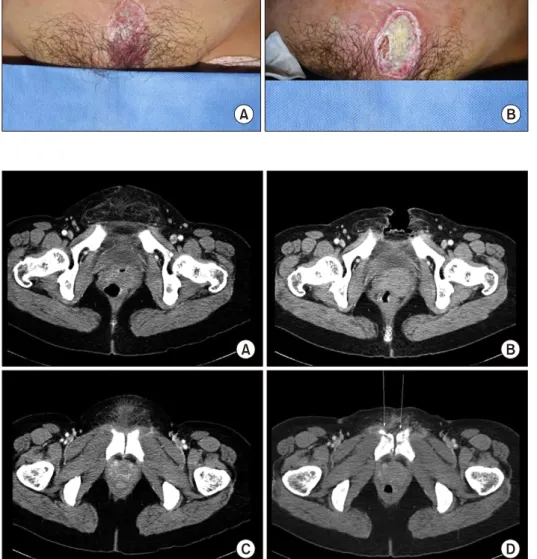
Fig. 1. (A) Gross photographs show-
ed 6×4 cm sized, sharply demar- cated, progressive thick eschars on lower abdomen including upper portion of suprapubic area on post- HIFU day 6, (B) and post-HIFU week 3.Fig. 2. (A) The APCT on 4 weeks
postburn showed diffuse fatty infil- tration at the lower abdominal wall.(B) The APCT on 7 weeks postburn showed newly appeared abdominal defects about 6×4 cm in size on the lower abdomen involving skin, sub- cutaneous fat and a portion of both lower rectus abdominis muscles and left pectineus muscle. (C) The APCT on 4 weeks postburn showed normal pubic symphysis, but infla- mmatory edema within the adjacent soft tissues. (D) The APCT on 7 weeks postburn showed newly developed septic arthritis in the pubic sym- physis and osteomyelitis in both adja- cent pubic bones.
done once every two days. On 4 weeks postburn, the ab- dominopelvic computed tomography (APCT) showed dif- fuse fatty infiltration in the lower abdominal wall. On 7 weeks postburn, the APCT showed an approximately 6×4 cm sized abdominal wall defect in the lower abdomen in- volving skin, subcutaneous fat, and a portion of both low- er rectus abdominis muscles and left pectineus muscle.
The size of uterine myoma had not decreased (Fig. 2). On 8 weeks postburn, the magnetic resonance imaging (MRI) showed septic arthritis in the pubic symphysis with small abscess formation and osteomyelitis with a pathologic fracture. On 8 weeks postburn, surgical debridement of the infected pubic bone was done. Then daily copious sal- ine irrigation was done using broad spectrum antibiotics, and vacuum assisted closure (VAC) dressing with V.A.C.
Ⓡ(Kinetic Concepts, Inc., USA) was applied every third day. After two months of VAC dressing, the wound showed improvement (Fig. 3). On 15 weeks postburn, a local advancement skin flap was elevated and transferred to cover the exposed pubic bone and abdominal skin defect. At 23 days postoperatively, the abdominal full- thickness skin defects were well reconstructed without complications (Fig. 4).
DISCUSSION
Common complications reported after HIFU therapy
include superficial skin burns, skin disruptions, subcuta-
neous edema, lower abdominal pain, sciatic pain, and
vaginal discharge with small amounts of vaginal bleeding.
66
대한화상학회지 Vol. 23, No. 2, 2020Fig. 3. (A) On 8 weeks postburn,
the MRI showed septic arthritis in the pubic symphysis with small abscess formation and osteomyelitis with a pathologic fracture. (B) On 7 weeks postburn, gross photographs showed about 5×4 cm sized abdo- minal skin defect. (C) On 15 weeks postburn, after the surgical debride- ment of infected pubic bone, VAC dressing was applied on every third day. (D) On 16 weeks postburn, gross photograph showing the wound was much more improved.Fig. 4. (A, B) On 15 weeks post-
burn, the fasciocutaneous advance- ment flap was elevated and trans- ferred to cover the exposed pubic bone and abdominal skin defect.(C) At 23 days postoperatively, the lower abdominal skin defects caused by HIFU were well healed without complications. (D) 18 weeks after HIFU, the ultrasonography showed the size of uterine myoma had not decreased since previously studied.
In addition, severe complications, such as deep dermal burns requiring surgical repair, bowel perforation, blad- der injuries, and deep vein thrombosis, were also re- ported [4].
In general instructions in the US beam path, the pa- tients with myoma close to the sacral bone surface, with extensive cutaneous scars, or with bowel interposition
should be strictly excluded from HIFU treatment. Also,
the presence of concomitant severe adenomyosis, pe-
dunculated subserosal myoma with a pedicle less than
50% of total myoma diameter, or multiple myomas count-
ing more than five in numbers or over 10 cm in size
should also be considered as factors limiting treatment
using magnetic resonance-guided HIFU therapy. However,
Sung Hoon Yu and Dong Chul Kim:Deep Burn Injuries after HIFU for Uterine Myoma
67
there is lack of organized criteria about indications or contraindications of US-guided HIFU [5]. In recent years, numerous studies have been conducted about the factors that contribute to HIFU treatment complications. The re- flection of ultrasound caused by bones can deposit ultra- sonic energy in skin and subcutaneous layers, resulting in skin burns, especially when targeting tumors in the up- per abdomen and lower abdomen with HIFU. Also, ab- sorption of ultrasonic energy by HIFU can damage tissues in the HIFU path and hurt the adjacent tissues. The differ- ence in acoustic impedance can result in different dis- tributions of energy into tissues and cause damage to the tissue in the interface [6]. Canney et al. (2010) demon- strated that localized heating by shockwaves at the HIFU focus can raise tissue temperatures to 100
oC in a few mil- liseconds, resulting in the formation of a boiling vapor bubble at the HIFU focus [7]. This bubble then grows to a millimeter in size, and may destroy adjacent tissue be- cause of shear stresses produced around the oscillating bubble [8]. Some studies have been conducted about the mechanism of boiling histotripsy to help predict the size of a mechanically induced lesion. Pahk et al. (2017) sug- gested that the overall size of a lesion generated by boil- ing histotripsy is dependent on the spatial extent of the heated region at the HIFU focus and the backscattered ul- trasound wave by the original vapor bubble [9]. Future work needs to be focused on predicting the size of a me- chanically induced lesion and the factors that contribute to complications in HIFU treatment.
In our case, a burn injury occurred on the lower abdo- men near the pubic symphysis. Septic arthritis in the pu- bic symphysis and osteomyelitis in both adjacent pubic bones were also observed. These complications were sus- pected to be as a result of ultrasound reflection. Many studies have reported that most skin burns after HIFU are 1st and 2nd degree. However, even if it looks like a su- perficial skin burn, physicians must pay attention to pro- gressive deep burn injuries when examining patients with skin burns after HIFU treatment.
Currently, HIFU has been regarded as a non-surgical, minimally invasive therapeutic option for uterine myoma.
However, severe complications such as deep burn injuries at the HIFU site, have been reported [10]. For the safe ap- plication of HIFU in treating uterine myoma, the safety
verification of HIFU instruments and performing the pro- cedures by experienced practitioners according to stand- ard criteria and guideline are emphasized.
CONFLICTS OF INETEREST
The authors have no financial disclosures or conflicts of interest to declare.
REFERENCES
1. Burgess SE, Iwamoto T, Coleman DJ, Lizzi FL, Driller J, Rosado A. Histologic changes in porcine eyes treated with high-intensity focused ultrasound. Ann Ophthalmol 1987;19:
133-8.
2. Lynn JG, Zwenmer RL, Chick AJ. A new method for genera- tion and use of focused ultrasound in experimental biology.
J Gen Physiol 1942;26:179-93.
3. Griffiths A, ter Haar G, Rivens I, Giussani D, Lees C. High- intensity focused ultrasound in obstetrics and gynecology: the birth of a new era of noninvasive surgery? Ultraschall in Med 2012;33:E8-15.
4. Zhang L, Zhang W, Orsi F, Chen W, Wang Z. Ultrasound guided high intensity focused ultrasound for the treatment of gynaecological diseases: a review of safety and efficacy. Int J Hyperthermia 2015;31:280-4.
5. Froling V, Kroncke TJ, Schreiter NF, Scheurig-Muenkler C, Collettini F, Hamm B, et al. Technical eligibility for treatment of magnetic resonance-guided focused ultrasound surgery.
Cardiovasc Intervent Radiol 2014;37:445-50.
6. Jian-Jun Li, Guo-Liang Xu, Mo-Fa Gu, Guang-Yu Luo, Zhang Rong, Pei-Hong Wu, et al. Complications of high intensity focused ultrasound in patients with recurrent and metastatic abdominal tumors. World J Gastroenterol 2007;13:2747-51.
7. Canney MS, Khokhlova VA, Bessonova OV, Bailey MR, Crum LA. Shock-induced heating and millisecond boiling in gels and tissue due to high intensity focused ultrasound. Ultra- sound Med Biol 2010a;36:250-67.
8. Khokhlova TD, Canney MS, Khokhlova VA, Sapozhnikov OA, Crum LA, Bailey MR. Controlled tissue emulsification produced by high intensity focused ultrasound shock waves and milli- second boiling. J Acoust Soc Am 2011;130:3498-510.
9. Ki Joo Pahk, Pierre Gélat, David Sinden, Dipok Kumar Dhar, Nader Saffari. Numerical and experimental study of mechanisms involved in boiling histotripsy. Ultrasound in Med. & Biol.
2017;43:2848-61.
10. Sang Hyup Hong, Gil-Sun Hong, Choong Wook Lee, Gi Hong Kim. Complication following ultrasound-guided high-intensity focused ultrasound for the treatment of uterine adenomyosis:
case report of CT imaging features. J Korean Soc Radiol 2019;80:579-84.