Received on August 12, 2013. Revised on August 23, 2013. Accepted on August 23, 2013.
CC This is an open access article distributed under the terms of the Creative Commons Attribution Non-Commercial License (http://creativecommons.org/licenses/by-nc/3.0) which permits unrestricted non-commercial use, distribu- tion, and reproduction in any medium, provided the original work is properly cited.
*Corresponding Author. Sang-Heon Cho, Department of Internal Medicine, Seoul National University College of Medicine, 28, Yongon-dong, Chongno-gu, Seoul, Korea. Tel: 82-2-760-2971; Fax: 82-2-764-3954; E-mail: [email protected] Kim YK and Jeon SG are now at the Division of Molecular and Life Sciences, Pohang University of Science and Technology, Pohang, Korea.
Kim YY is now at the Department of Internal Medicine, National Medical Center, Seoul, Korea.
Keywords: Allergy, Asthma, Rodent, House dust mite, Der f 2
Abbreviations: rDer f 2, recombinant Der f 2; FP, Freund’s adjuvants with pertussis toxin; CD, cluster of differentiation;
TSLP, thymic stromal lymphopoietin
Influence of the Adjuvants and Genetic Background on the Asthma Model Using Recombinant Der f 2 in Mice
Yoon-Seok Chang1,2,3, Yoon-Keun Kim1,2, Seong Gyu Jeon2, Sae-Hoon Kim1,2,3, Sun-Sin Kim1,2,4, Heung-Woo Park1,2, Kyung-Up Min1,2, You-Young Kim1,2 and Sang-Heon Cho1,2,4*
1Department of Internal Medicine, Seoul National University College of Medicine, Seoul 110-799, 2Institute of Allergy and Clinical Immunology, Seoul National University Medical Research Center, Seoul 110-799, 3Department of Internal Medicine, Seoul National University Bundang Hospital, Seongnam 463-707, 4Department of Internal Medicine, Seoul National University Gangnam Healtcare Center, Seoul 135-984, Korea
Der f 2 is the group 2 major allergen of a house dust mite (Dermatophagoides farinae) and its function has been re- cently suggested. To determine the optimal condition of sen- sitization to recombinant Der f 2 (rDer f 2) in murine model of asthma, we compared the effectiveness with different ad- juvants in BALB/c and C57BL/6 mice. Mice from both strains sensitized with rDer f 2 by intraperitoneal injection or subcutaneous injection on days 1 and 14. The dosage was 20μg. Freund’s adjuvants with pertussis toxin (FP) or alum alone were used as adjuvants. On days 28, 29, and 30, mice were challenged intranasally with 0.1% rDer f 2. We eval- uated airway hyperresponsivenss, eosinophil proportion in lung lavage, airway inflammation, and serum allergen specif- ic antibody responses. Naive mice were used as controls.
Airway hyperresponsiveness was increased in C57BL/6 with FP, and BALB/c with alum (PC200: 13.5±6.3, 13.2±6.7 vs.
>50 mg/ml, p<0.05). The eosinophil proportion was in- creased in all groups; C57BL/6 with FP, BALB/c with FP, C57BL/6 with alum, BALB/c with alum (24.8±3.6, 20.3±
10.3, 11.0±6.9, 5.7±2.8, vs. 0.0±0.0%, p<0.05). The se- rum allergen specific IgE levels were increased in C57BL/6 with FP or alum (OD: 0.8±1.4, 1.1±0.8, vs. 0.0±0.0).
C57BL/6 mice were better responders to rDer f 2 and as for
adjuvants, Freund’s adjuvant with pertussis toxin was better.
[Immune Network 2013;13(6):295-300]
INTRODUCTION
Allergic diseases such as asthma, allergic rhinitis, and atopic dermatitis are increasing worldwide including in Korea. The prevalence has been doubled or tripled for recent decades (1,2). House dust mite is the most common inhalant allergen that causes asthma, allergic rhinitis, and even atopic dermati- tis (3). There are two species of house dust mites, Dermato- phagoides farina and Dermatophagoides pteronyssinus, in Korea. D. farinae is the predominant species (65.3%) fol- lowed by D. pteronyssinus (20.6%) (4). House dust mites have various proteins that could IgE-mediated immune re- sponses and molecules with adjuvant-like characteristics or their affinity to adjuvant (4). Among the major allergens, group 1 and group 2 allergens constitute 40∼60% of the total allergenicity in house dust mites (5).
Group 1 major allergens, Der f 1 and Der p 1, have proteo- lytic properties as cysteine protease; 1) they can cause dis-
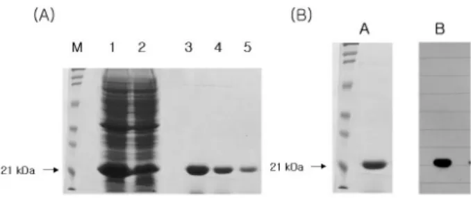
Figure 1. Molecular cloning, functional expression and purification of recombinant Der f 2. (A) 12% SDS-PAGE analysis of the expressed Der f 2 allergen. M; molecular size marker, Lane 1, 2; homogenates from JM 109 transformed of pET-15b+Der f 2 cDNA in induced condions, Lane 3-5; purified recombinant Der f 2 allergens. (B) 12 % SDS-PAGE and Western blot analysis of the expressed Der f 2 allergen. A;
coomassie blue staining of SDS-PAGE gel. B; immunoblot with pooled patients sera sensitized against Dermatophagoides farinae.
ruption of the epithelium, allowing access of allergens to anti- gen-presenting cells; 2) Der p 1 can cleave immunomo- dulators such as CD23 (low-affinity IgE receptor) and CD25 (α-subunit of IL-2 receptor); 3) they can elicit inflammatory reactions by activating protease-activated receptor-2; 4) they can release inflammatory cytokines independent of PAR-2 ac- tivation; 5) Possibly Der p 1 has been shown to activated and recruit basophils to the draining lymph nodes and stimulate production of Th2-inducing cytokines such as IL-4 and thymic stromal lymphopoietin (TSLP) (4).
Group 2 major allergens, Der f 2 and Der p 2, have lip- id-binding properties and show almost 90% sequence homol- ogy which made them highly cross-reactive (4). However, the function of group 2 major allergens, especially Der f 2 has not been well elucidated. Only a few studies used recombi- nant Der f 2 (rDer f 2) to evaluate its function (4,6-8). Reports on murine asthma model using rDer f 2 are few (9,10). There was no report on the influence of adjuvants and genetic back- ground on the asthma model using rDer f 2 in mice. This study showed which strain and adjuvants were the best com- binations in the development of murine asthma model using rDer f 2.
MATERIALS AND METHODS Animals
Female C57BL/6 and BALB/c mice aged 8∼10 weeks were used in this study. The mice were purchased from Dae Han Biolink (Choongbuk, Korea) and kept in specific pathogen free conditions in the preclinical center of the Clinical Re- search Institute of Seoul National University Hospital (Seoul, Korea). None of the mice were exposed to rDer f 2 before the experiment. The study was approved by the appropriate committees on animal experimentation at our institution and performed according to the guide for the care and use of lab- oratory animals.
Molecular cloning, functional expression and purifi- cation of recombinant Der f 2
cDNA cloning, over-expression and purification of Der f 2 was performed as described previously (11,12). In brief, am- plification of Der f 2 gene with the exception of the signal peptide was carried out in 50 ul volume by using Ex.Taq polymerase. Thirty cycles of PCR were performed on the cDNA of Dermatophagoides farinae using the upstream pri- mer 5’-CAAGTCGATGTTAAAGATTG-3’ and the downstream
primer 5’-TTAATCACGGATTTTACCATGG-3’ with denaturing for 45 s at 94oC, annealing for 45 s at 55oC, and extension for 1 min at 94oC. The PCR product was inserted into pET-15b vector for over-expression, and then transformed in- to E.coli JM109 (DE3). Single positive colony was grown overnight at 37oC in 2X-YT media (16 g of Bacto-tryptone, 10 g of yeast extract, 5 g of NaCl) supplemented with 100 mg/ml ampicillin. The next day, 990 ml of 2X-YT media sup- plemented with 100 mg/ml ampicillin was inoculated with 10 ml of the overnight culture and grown at 30oC with shaking at 260 rpm until O.D600=1.0. We induced gene expression by adding isopropylthio-b-D-galactoside to a final concentration of 0.1 mM, and the cultures were grown at 28oC overnight with gentle agitation. The cells were pelleted at 1,500 g and washed twice in phosphate-buffered saline. The cell pellet was then resuspended in 50 ml of a 10 mM potassium phos- phate buffer (pH 7.0), 1 mM 2-mercaptoethanol, 1 mM PMSF, and 1% (v/v) Triton X-100, and then it was sonicated while being chilled in an ice bath. We removed cell debris by cen- trifugation at 15,000 g for 30 min, and the supernatant were purified by using the His-bind purification kits (Novagen, Darmstadt, Germany) as explained in the manufacturer’s manual. The purified recombinant proteins were confirmed by SDS-PAGE and immunoblot analysis (Fig. 1).
Protocols for sensitization and rDer f-specific intra- nasal challenge
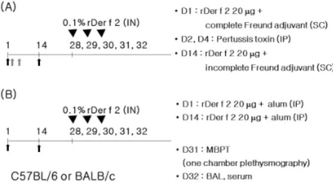
The protocols of sensitization and intranasal challenge were designed according to those previously recommended with some modifications (13,14). Twenty μg of rDer f 2 was in- jected for sensitization and boosting using Freund’s adjuvants
Figure 2. Experimental design. Twenty μg of recombinant Der f 2 was injected for sensitization and boosting using Freund adjuvants with pertussis toxin (A) or alum (B) as adjuvants. On day 28, intranasal (IN) challenge was performed with 50μl of 0.1% recombinant Der f 2 and for consecutive 3 days.
with pertussis toxin (Freund/Pertussis) or alum as adjuvants.
For alum group, 4 mg of alum hydroxide (alum, Sigma, St. Louis, USA) was dissolved in 200μl of phosphate buf- fered saline (PBS). Mice were injected intraperitoneally with rDer f 2 and alum on days 1 and 14.
For Freund/Pertussis group, 100μl of complete Freund’s adjuvant (Sigma, St. Louis, USA) was injected subcutaneously at the time of sensitization on day 1. On days 2 and 4, 300 ng of Pertussis toxin (Sigma, St. Louis, USA) was injected intraperitoneally. On day 14, 100μl of Incomplete Freund’s adjuvant (Sigma, St. Louis, USA) was injected subcutaneously with rDer f 2.
On day 28, intranasal challenge was performed with 50μl of 0.1% rDer f 2 and for consecutive 3 days after light anes- thesia with ether.
We evaluated airway hyperresponsivenss (day 31), eosino- phil proportion in lung lavage, airway inflammation, and se- rum allergen specific antibody responses (day 32). Naive mice were used as controls. The mice were divided into 6 groups according to the protocols (6 mice for each ex- perimental group, 4 mice for control groups, Fig. 2).
Evaluation of asthma phenotypes
Airway hyperresponsiveness: Twenty-four hours after the final intranasal challenge with rDer f 2, airway hyper- responsiveness was assessed by methacholine-induced air- flow obstruction using single chamber whole body ple- thysmography (Allmedicus, Anyang, Korea) as previously de- scribed (15). Increases in enhanced pause (Penh) were meas- ured as an index of airway resistance {Penh=[(Te/RT−1)×
(PEF/PIF)], where Penh=enhanced pause, Te=expiratory time (s), RT=relaxation time (s), PEF=peak expiratory flow (ml/s), PIF=peak inspiratory flow (ml/s)}. Increasing doses of meth- acholine (ranging from 2.5 to 50 mg/ml; Sigma, St. Louis, USA) were administered by nebulization for 3 minutes, and the values of Penh were calculated over the subsequent 3 minutes. During the experiment, the activity of the mice and the barometric plethysmograph flow tracings were monitored.
For the quantification of the dose-response to methacholine, the linear regression of Penh on log was calculated for in- dividual mice. The log dose corresponding to an increase in Penh of 200%, respectively, was determined, and the average log doses of the different groups were compared. The results are presented as PC200, which is the concentration of meth- acholine required to increase the baseline Penh by 200%.
Inflammatory cells in bronchoalveolar lavage: Forty- eight hours after the final intranasal challenge with rDer f 2, mice tracheae were cannulated and the lungs were lavaged five times with 0.4 ml aliquots of pyrogen-free saline. After Diff-quikR staining (Dade Behring AG, Dudingen, Switzer- land) of lung lavage cells in a cytospin preparation, two in- vestigators blindly counted more than 300 inflammatory cells under a light microscope and classified them as macrophages, lymphocytes, neutrophils, and eosinophils (16,17).
Serum anti-rDer f 2 specific IgE: Forty-eight hours after the final intranasal challenge with rDer f 2, blood samples were obtained from the mice via the inferior vena cava. Serum anti- rDer f 2 specific IgE was measured by ELISA as previously described with some modifications (18). Briefly, microtiter plates (Nunc, Roskilde, Denmark) were coated overnight with 10μg/well of rDer f 2 in a 50 mM carbonate buffer (pH 9.6) at 4oC. Nonspecific binding was blocked with 2% bovine se- rum albumin for 1 hour at 20oC. After incubation of test sera for 2 hours, the plates were incubated with horse radish per- oxidase-labeled goat anti-mouse IgE (Pharmingen, San Diego, USA) for 1 hour at 20oC. The reaction was developed with a tetramethylbenzidine (Sigma, St. Louis, USA) substrate and then stopped by adding 2 N H2SO4. Subsequently, the optical density was measured at 490 nm. The antibody titers of the samples were related to pooled standards that were generated in the laboratory and expressed as arbitrary units (AU) ac- cording to each O.D. value.
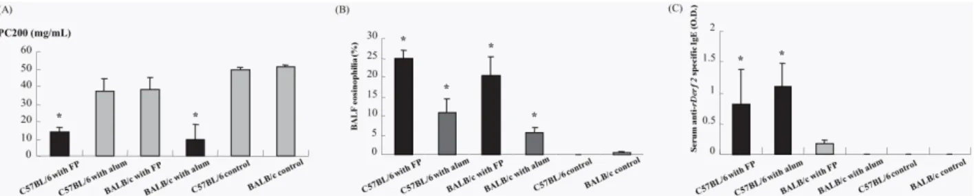
Figure 3. Evaluation of asthma phenotypes. (A) Airway hyperresponsiveness measured using whole body plethysmograph, (B) airway eosinophilia from bronchioalveolar lavage fluid, (C) Serum anti-recombinant Der f 2 (rDer f2) specific IgE measured by ELISA. Each bar indicates the mean (±SEM) value. *p<0.05, compared with the control groups. BALF, bronchoalveolar lavage fluid.
Statistical analysis
Statistical analysis was performed using the Kruskal-Wallis and the Mann-Whitney tests. Statistical significance was ac- cepted at p<0.05. Analysis was performed using SPSS 9.0.
Values for all measurements are expressed as the means±the standard error of the mean.
RESULTS AND DISCUSSION
The airway responsiveness was increased in the groups of C57BL/6 with Freund/Pertussis and BALB/c with alum and the control groups (p<0.05). No difference of PC200 was found between them (p>0.05). The PC200 of C57BL/6 with alum, BALB/c with Freund/Pertussis were not significantly dif- ferent from those of the control groups (p>0.05) (Fig. 3A).
The percentages of BALF eosinophil were higher in all ex- perimentally sensitized groups compared with those of the control groups (p<0.05). The BALF eosinophil levels in the groups of C57BL/6 or BALB/c with Freund/Pertussis were higher than in the groups of C57BL/6 or BALB/c with alum (Fig. 3B).
The serum anti-rDer f 2 specific IgE levels were higher in C57BL/6 with Freund/Pertussis or alum than in the control groups (p<0.05). There was no significant difference in the serum anti-rDer f 2 levels between the C57BL/6 with Freund/Pertussis or alum (p>0.05) (Fig. 3C).
We evaluated the influence of the adjuvants and genetic background on the murine model of asthma using rDer f 2, especially evaluating the important features of asthma - air- way hyperresponsivenss, eosinophilic airway inflammation and serum allergen-specific IgE levels, which has not been reported so far. This study showed that C57BL/6 mice with Freund adjuvant and pertussis toxin were the best model among the experimental groups. In terms of airway hyper-
sensitivity, C57BL/6 with Freund/Pertussis and BALB/c with alum were good combinations. Freund and pertussis toxin was more effective adjuvants for inducing airway eosinophilia in both strains although alum also induced airway eosinophilia.
C57BL/6 mice were ideal for inducing serum anti-rDer f 2-specif- ic IgE using Freund/Pertussis or alum.
House dust mite is the most common inhalant allergen in the world as well as in Korea (3). Allergens from mite groups 1, 3, 6, and 9 were identified as cysteine protease, trypsin, chymotrypsin, and collagenolytic serine protease, respectively (4). Group 2, 13, and 14 allergens are associated with lip- id-binding activity (4). Der f 2 as well as Der P 2 was lo- calized in the midgut and fecal pellets of the mite (19,20).
The exact function of Der f 2 has not been known yet. The group 2 major mite allergens, Der f 2 and Der p 2, are 14 kD protein and show structural homology with MD-2, the lip- opolysaccharide (LPS)-binding component of the Toll-like re- ceptor (TLR) 4 signaling complex (4). It has been reported that Der f 2 binds to LPS in a molar ratio of 1 : 1 and that LPS binds Der f 2 between the two large beta-sheets, similar to its binding to MD-2, the LPS-binding component of the in- nate immunity receptor TLR4 (21). There is interesting reports that Der f 2 showed stronger IgE reactivity than Der f 1 in Korean mite-allergy patients (22,23). rDer f 2 produced in E.
coli is recognized by 90∼100% of serum IgE from Korean D. farinae-sensitized subjects (4). Recently it has been re- ported that Der f 2 induced interleukin-13 expression by acti- vating the PI3K/Akt pathway and by phospholipase D1 through activating transcription factor-2 activation in human bronchial epithelial cells (24,25).
There has been several murine models using Der f 2 but only few researchers evaluated only limited aspects of allergic reactions in mice using rDer f 2 (7,9,10). However, this study shows unique data on the influence of the adjuvants and ge-
netic background on the murine model of asthma using re- combinant Der f 2 evaluating the important features of asthma.
C57BL/6 mice is a black with H2b allele which has been reported to be a good responder to house dust mite extract allergen but a poor responder to ovalbumin (26,27). BALB/c mice, a white with H2d allele, have been reported to be a good responder to ovalbumin but a poor responder to house dust mite extract allergen (28). As shown in this study, C57BL/6 mice showed generally good response to rDer f 2 with Freund/Pertussis but not with alum. In terms of airway hyperresponsiveness, BALB/c with alum showed a better re- sult than C57BL/6 with alum. It is interesting that BALB/c with alum showed increased airway hyperresponsiveness with less eosinophilic airway inflammation than C57BL/6 with alum. It is well known that airway hyperresponsiveness and airway inflammation are independent pathways: There are several hypotheses on that (17). It was also interesting that BALB/c with alum showed airway hyperresponsiveness in the almost absence of rDer f 2-specific IgE. As presented in Fig.
3C, there was a small increase in rDer f 2-specific IgE, which could mean that BALB/c with alum was enough to induce local rDer f2-specific IgE production after intranasal challenge but was not enough to induce systemic rDer f2-specific IgE production to be detected from serum.
In conclusion, the influence of the adjuvants and genetic background were important factors on the murine model of asthma using rDer f 2. The optimal condition for the asthma model using rDer f 2 in mouse was using C57BL/c mice treat- ed with Freund adjuvant with pertussis toxin.
ACKNOWLEDGEMENTS
This study was supported by a research grant from the Ministry of Health and Welfare (HMP-00-CH-06-0006), and by a grant of Seoul National University Bundang Hospital (02- 2006-024), Korea.
CONFLICTS OF INTEREST
The authors have no financial conflict of interest.
REFERENCES
1. Cho, S. H., Y. K. Kim, Y. S. Chang, S. S. Kim, K. U. Min, and Y. Y. Kim. 2006. Asthma insights and reality in Korea.
Korean J. Med. 70: 69-77.
2. Kim, Y. Y. 2010. Past, present, and future of allergy in Korea.
Allergy Asthma. Immunol. Res. 2: 155-164.
3. Kim, T. B., K. M. Kim, S. H. Kim, H. R. Kang, Y. S. Chang, C. W. Kim, J. W. Bahn, Y. K. Kim, H. T. Kang, S. H. Cho, H. S. Park, J. M. Lee, I. S. Choi, K. U. Min, C. S. Hong, N. S. Kim, and Y. Y. Kim. 2003. Sensitization rates for in- halant allergens in Korea; a multi-center study. J. Asthma.
Allergy Clin. Immunol. 23: 483-493.
4. Jeong, K. Y., J. W. Park, and C. S. Hong. 2012. House dust mite allergy in Korea: The most important inhalant allergen in current and future. Allergy Asthma. Immunol. Res. 4:
313-325.
5. Jeong, K. Y., S. Y. Choi, J. H. Lee, I. Y. Lee, T. S. Yong, J. S. Lee, C. S. Hong, and J. W. Park. 2012. Standardization of House Dust Mite Extracts in Korea. Allergy Asthma.
Immunol. Res. 4: 346-350.
6. Wu, C. C., E. C. Liao, M. F. Lee, and J. J. Tsai. 2009.
Augmentation of regulatory T cells in allergic individuals by recombinant Der f 2 peptide with fungal immunomodulatory peptide fve. Ann. Allergy Asthma. Immunol. 102: 216-222.
7. Kikuchi, Y., T. Takai, M. Ota, T. Kato, K. Takeda, K. Mitsuishi, S. Ikeda, K. Okumura, and H. Ogawa. 2006. Application of immunoreaction enhancer solutions to an enzyme-linked im- munosorbent assay for antigen-specific IgE in mice immu- nized with recombinant major mite allergens or ovalbumin.
Int. Arch. Allergy Immunol. 141: 322-330.
8. Jin, H. S., T. S. Yong, J. W. Park, C. S. Hong, and S. H.
Oh. 2003. Immune reactivity of recombinant group 2 aller- gens of house dust mite, Dermatophagoides pteronyssinus, and Dermatophagoides farinae. J. Investig. Allergol. Clin.
Immunol. 13: 36-42.
9. Yasue, M., T. Yokota, Y. Kajiwara, M. Suko, and H.
Okudaira. 1997. Inhibition of airway inflammation in rDer f 2-sensitized mice by oral administration of recombinant der f 2. Cell Immunol. 181: 30-37.
10. Yasue, M., T. Yokota, M. Fukada, T. Takai, M. Suko, H.
Okudaira, and Y. Okumura. 1998 Hyposensitization to aller- gic reaction in rDer f 2-sensitized mice by the intranasal ad- ministration of a mutant of rDer f 2, C8/119S. Clin. Exp.
Immunol. 113: 1-9.
11. Jeon, S. G., J. H. Bahn, J. S. Jang, S. H. Jang, B. R. Lee, K. S. Lee, J. Park, T. C. Kang, M. H. Won, H. B. Kim, O.
S. Kwon, S. W. Cho, and S. Y. Choi. 2001. Molecular cloning and functional expression of bovine brain GABA trans- aminase. Mol. Cells 12: 91-96.
12. Jeon, S. G., J. H. Bahn, J. S. Jang, J. Park, O. S. Kwon, S.
W. Cho, and S. Y. Choi. 2000. Human brain GABA trans- aminase tissue distribution and molecular expression. Eur. J.
Biochem. 267: 5601-5607.
13. Yu, C. K., B. C. Yang, S. C. Lee, J. Y. Wang, T. R. Hsiue, and H. Y. Lei. 1997. Dermatophagoides-farinae-induced pul- monary eosinophilic inflammation in mice. Int. Arch. Allergy Immunol. 112: 73-82.
14. Chang, Y. S., Y. K. Kim, J. W. Bahn, S. H. Kim, H. W. Park, T. B. Kim, S. H. Cho, K. U. Min, and Y. Y. Kim. 2005.
Comparison of asthma phenotypes using different sensitizing protocols in mice. Korean J. Intern. Med. 20: 152-158.
15. Chang, Y. S., Y. K. Kim, S. H. Kim, H. W. Park, K. U. Min, Y. Y. Kim, and S. H. Cho. 2013. Murine subcutaneous im- munotherapy models with beneficial immunological and physiological effects. Asia Pac. Allergy 3: 50-58.
16. Park, Y., Y. S. Chang, S. W. Lee, S. Y. Cho, Y. K. Kim, K. U. Min, Y. Y. Kim, S. H. Cho, and Y. C. Sung. 2001.
The enhanced effect of a hexameric deoxyriboguanosine run conjugation to CpG oligodeoxynucleotides on the protection against allergic asthma. J. Allergy Clin. Immunol. 108: 570- 576.
17. Chang, Y. S., Y. K. Kim, T. B. Kim, H. R. Kang, S. S. Kim, J. W. Bahn, K. U. Min, Y. Y. Kim, and S. H. Cho. 2004.
Airway inflammation and allergen specific IgE production may persist longer than airway hyperresponsiveness in mice.
J. Korean Med. Sci. 19: 69-73.
18. Chang, Y. S., Y. K. Kim, J. W. Bahn, S. H. Kim, H. W. Park, T. B. Kim, S. H. Cho, K. U. Min, and Y. Y. Kim. 2005.
Comparison of asthma phenotypes using different sensitizing protocols in mice. Korean J. Intern. Med. 20: 152-158.
19. Park, G. M., S. M. Lee, I. Y. Lee, H. I. Ree, K. S. Kim, C.
S. Hong, and T. S. Yong. 2000. Localization of a major aller- gen, Der p 2, in the gut and faecal pellets of Dermatophago- ides pteronyssinus. Clin. Exp. Allergy 30: 1293-1297.
20. Jeong, K. Y., I. Y. Lee, H. I. Ree, C. S. Hong, and T. S.
Yong. 2002. Localization of Der f 2 in the gut and fecal pel- lets of Dermatophagoides farinae. Allergy 57: 729-731.
21. Ichikawa, S., T. Takai, T. Yashiki, S. Takahashi, K. Okumura, H. Ogawa, D. Kohda, and H. Hatanaka. 2009. Lipopolysa- ccharide binding of the mite allergen Der f 2. Genes Cells 14: 1055- 1065
22. Hong, C. S., J. W. Park, and D. H. Nahm. 1994. Measure- ment of IgE and IgG subclass antibodies to whole body anti-
gen and two major allergens (Der fI & Der fII) of Dermato- phagoides farinae in normal subjects and asthmatics. Yonsei Med. J. 35: 453-463
23. Nahm, D. H., J. W. Park, C. S. Hong, S. Y. Lee, and K.
Y. Lee. 1995. Specific IgE antibodies to D. farinae whole body extract, Der f I and Der f II in child age groups. Pediatr.
Allergy Respir. Dis. 5: 117-124
24. Ro, E. J., P. H. Cha, H. Y. Kim, Y. H. Cho, J. W. Park, J. S. Han, and K. Y. Choi. 2013. House dust mite allergen Der f 2 induces interleukin-13 expression by activating the PI3K/Akt pathway. Immunol. Res. 56: 181-188.
25. Park, S. Y., J. H. Cho, D. Y. Oh, J. W. Park, M. J. Ahn, J. S. Han, and J. W. Oh. 2009. House dust mite allergen Der f 2-induced phospholipase D1 activation is critical for the pro- duction of interleukin-13 through activating transcription fac- tor-2 activation in human bronchial epithelial cells. J. Biol.
Chem. 284: 20099-20110.
26. O'Brien, R., M. A. Ooi, A. H. Clarke, and W. R. Thomas.
1996. Immunologic responses following respiratory sensitiza- tion to house dust mite allergens in mice. Immunol. Cell Biol.
74: 174-179.
27. Lee, Y. L., C. L. Fu, Y. L. Ye, and B. L. Chiang. 1999.
Administration of IL-12 prevent mite Der p 1 allergen-IgE anti- body production and airway eosinophil infiltration in an ani- mal model of airway inflmmation. Scand. J. Immunol. 49:
229-236.
28. Zhang, Y., W. J. Lamm, R. K. Albert, E. Y. Chi, W. R.
Henderson, Jr., and D. B. Lewis. 1997. Influence of the route of allergen administration and genetic background on the murine allergic pulmonary response. Am. J. Respir. Crit. Care Med. 155: 661-669.