High-resolution MR Imaging of Carotid Atherosclerotic Plaques
Wonseon Shin, Sung Mok Kim, Yeon Hyeon Choe
Department of Radiology, Samsung Medical Center, Sungkyunkwan University School of Medicine, Seoul, Korea
High-resolution carotid MRI allows visualization of carotid atherosclerotic plaque characteristics. MRI serves as a nonin- vasive option for the detection of active plaque inflammation and intraplaque hemorrhage. Significant gains in signal-to- noise ratio and contrast-to-noise ratio can be obtained for carotid atheroma imaging at 3T compared with 1.5T.
Normalized wall index or wall area on MRI has shown its efficacy in monitoring the response after medical therapy. T(2)*
quantification in carotid plaques before and after the administration of ultrasmall superparamagnetic iron oxide particles shows difference in response to treatment according to drug doses. In conclusion, high-resolution MRI is useful in the diagnosis and monitoring of carotid atherosclerotic plaques prone to transient ischemic attack and stroke.
Index words : Atheroslcerosis∙Carotid artery∙Magnetic resonance imaging (MRI)
Carotid artery is a major source of cerebral embolism. The detection of unstable carotid plaques is important to prevent future cerebrovascular events, because vulnerable plaques may rupture and cause sudden arterial occlusion or distal embolization.
Carotid MRI is considered as a tool for individualized management of carotid atherosclerotic disease to prevent stroke (1).
MRI capabilities in plaque characterization MRI has been shown to accurately identify the fibrous cap, lipid-rich necrotic core, intraplaque hemorrhage, neovasculature and vascular wall inflam- mation (Figs. 1-3). MRI is a histologically validated technique for prospective testing of the vulnerable plaque hypothesis (2). High signal intensity (SI) in the
plaque may suggest a focal area of fresh hemorrhage on time-of-flight (TOF) images (3). New contrast agents and targeted molecular imaging open a window for MRI detection of thrombus and assessment of atherosclerotic activity and plaque vulnerability.
MRI techniques
Significant gains in signal-to-noise ratio (SNR) can be obtained for carotid atheroma imaging at 3T compared with 1.5T. There was also a trend towards increased contrast-to-noise ratio (CNR) (4). Excellent agreement was seen between contrast-enhanced MR angiography and T2-weighted SPACE sequence (dark blood-MRI using a 3-dimensional turbo spin echo with variable flip angles sequence) and between observers for assessment of lumen diameter, lumen area, vessel wall area, and degree of North American Symptomatic Carotid Endarterectomy Trial (NASCET) stenosis (r > 0.80, p < 0.001) (5).
Carotid intima-media thickness (IMT) is not adequately reproducible when tracking the progression of carotid atherosclerosis. The variability of ultrasound measurements of carotid IMT are likely to be reduced by further development of automatic calculation of this index by magnetic resonance imaging (6). Vessel INTRODUCTION
�Received; July 30, 2012�Accepted; August 20, 2012
Corresponding author : Yeon Hyeon Choe, M.D., Department of Radiology, Samsung Medical Center, Sungkyunkwan University School of Medicine, 50 Ilwon-dong, Gangnam-gu, Seoul 135-710, Korea.
Tel. 82-2-3410-2509, Fax. 82-2-3410-2559 E-mail : [email protected]
Review Article
wall imaging could quantify atherosclerotic plaque measurements more reliably with an improved blood suppression technique (7). Accurate co-registration between ultrasonography and other modalities is feasible with a global positioning system (GPS)-like technology, which has significant clinical and research applicability (8). The advantage of MRI over ultrasound is that the measurement variability is smaller, enabling smaller sample sizes and potentially shorter study duration in cardiovascular prevention trials (9).
Horie et al. (10) investigated whether a difference in dynamic enhancement pattern in plaque components could be useful to assess plaque stability with multide- tector CT angiography. With MRI, a higher HU was negatively correlated with signal intensity on T1- weighted imaging (r = -0.56, p<0.0001). Histology confirmed that HU was positively correlated with fibrous tissue (r = 0.67, p=0.001) and negatively correlated with a lipid-rich necrotic core with hemorrhage (r = -0.70, p<0.001). Moreover, less neovascularization and inflammation was found in plaques with a higher HU. Delayed-phase images of dynamic CT provide information regarding the dynamic change in contrast media from the early arterial phase. An increase in attenuation (HU) from the early phase on multidetector CT angiography
indicates plaque stability with more fibrous tissue and a less lipid-rich necrotic core, intraplaque hemorrhage, and neovascularization. According to Tartari et al.
(11), ongoing contrast-enhanced (CE)-MR angiography with a neurovascular coil for the simultaneous detection of unstable plaques is feasible. Plaque assessment was performed starting with a diffusion- weighted sequence and followed by a fat-suppressed T1-weighted sequence; after contrast-enhanced MR angiography (CE-MRA), all patients were evaluated with a T1-weighted 3-dimensional high-resolution sequence.
Kwee et al. (12) sought to compare 18F-fluoro-2- deoxyglucose positron emission tomography (18F-FDG PET), CT, and MRI of carotid plaque assessment.
Overall, correlations between 18F-FDG PET and CT/MRI findings are weak. Correlations between CT and MRI measurements are moderate to strong, but there is considerable variation in absolute differences.
Patterson et al. (13) investigated T(2)* quantification in carotid plaques before and after the administration of ultrasmall superparamagnetic iron oxide particles (USPIOs) in a cohort of patients receiving statin therapy. In the patient study, there was a highly signifi- cant difference in post-USPIO T(2)* measurements in plaques between the low- and high-dose statin groups (10 mg and 80 mg of atorvastatin). Quantitative T(2)*
a b
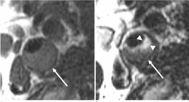
Fig. 1. Lipid-rich plaque. T1-weighted image (a) shows slightly increased signal intensity in the plaque (arrow). T2-weighted image shows a portion of bright signal intensity (arrowheads in the plaque which suggests the presence of fibrosis. The darker signal intensity in the plaque (arrow) implicates lipid-rich components.
(qT(2)*) measurements provide an alternative method of quantifying USPIO uptake. These results also demonstrate that changes in USPIO uptake can be measured using post-USPIO imaging only.
Clinical evidences
According to Virani et al. (14), extent of carotid atherosclerosis was associated with atherogenic choles- terol and lipoproteins in 1670 participants of Atherosclerosis Risk in Communities study.
Atherogenic/anti-atherogenic cholesterol or particle ratios were associated with presence of a detectable lipid-rich core.
In a study of patients with atherosclerotic disease with 4 years of follow-up, only severe or bilateral carotid stenosis, and not moderate carotid stenosis and increased carotid intima-media thickness, were associ- ated with progression of brain atrophy (15). According to Ota et al. (16), hemorrhage and larger %lipid- rich/necrotic core were independently associated with a thin or ruptured fibrous cap status at an early to advanced stage of carotid atherosclerosis. For the 798 MRI slices included, multivariate ordinal regression analysis demonstrated larger %lipid-rich/necrotic core (odds ratio for 10% increase, 1.49; p = 0.02) and presence of hemorrhage (odds ratio, 5.91; p < 0.001)
a b
c d
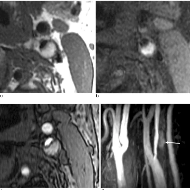
Fig. 2. Hemorrhagic plaque in a 64-year-old male with vascular dementia. The plaque signal intensity is bright on a T1-weighted image (a), T1-weighted image with fat-saturation (b), and source image of time-of-flight MR angiography (c). On a maximum intensity projection image (d), there is a halo sign (arrow) due to intraplaque hemorrhage.
were independently associated with a worse (intact thin or ruptured) stage of fibrous cap status.
Patients with carotid artery lesions and intraplaque hemorrhage tend to be at higher risk of a subsequent ipsilateral ischemic event. Risk evaluation of carotid artery disease should include plaque characteristics (17). Intraplaque hemorrhage evaluated by MRI identified neurologically unstable patients with increased levels of high-sensitivity C-reactive protein regardless of the degree of carotid stenosis (18).
Virani et al. (19) assessed the relationship between regulated on activation, normal T-cell expressed and secreted (RANTES) and carotid atherosclerotic plaque burden and plaque characteristics. Among 1769 inclusive participants, multivariable regression analysis revealed that total wall volume [beta-coefficient ( ) = 0.09, p = 0.008], maximum wall thickness ( = 0.08, p
= 0.01), vessel wall area ( = 0.07, p = 0.02), mean minimum fibrous cap thickness ( = 0.11, p = 0.03), and high-sensitivity C-reactive protein ( = 0.09, p =
a b
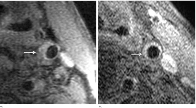
Fig. 3. Necrotic plaque in a 73-year-old male with stroke.
a.A source image of time-of-flight MR angiography shows severe luminal narrowing (arrow) of the left internal carotid artery.
b.contrast-enhanced T1-weighted MR image shows no significant enhancement in the plaque except the outer layers.
a b
Fig. 4. Plaque regression after statin treatment in a 73-year-old male.
a.T1-weighted image with fat-saturation at 1.5T shows an eccentric plaque in the left internal carotid artery.
b.T1-weighted image with fat-saturation at 3T shows significant improvement in plaque thickness after 3-year low dose statin treatment.
0.01) were positively associated with RANTES.
Therapy monitoring
Carotid MRI helps improve risk stratification for stroke in patients and monitor the response to medical therapies; assessing efficacy of medical treatment at individual and population levels. Using ultrasound, computed tomography or MRI, plaques may be measured to monitor regression and progression of plaque volumes or plaque components (Fig. 4).
Contrast-enhanced ultrasound, dynamic contrast- enhanced MRI, and FDG-PET are being explored to target plaque inflammation and neovascularization.
Noninvasive imaging and other advances in risk strati- fication aim to improve and individualize the manage- ment of patients with carotid atherosclerosis (20).
Saam et al. (21) found that increased normalized wall index on MRI and the use of statin therapy are associ- ated with reduced rates of plaque progression amongst individuals with advanced, asymptomatic carotid atherosclerosis. According to Lee et al., nicotinic acid (high dose, 2 g) significantly reduced carotid wall area on MRI compared with placebo (adjusted treatment difference: -1.64 mm2[95% confidence interval: -3.12 to -0.16]; p = 0.03) (22).
In conclusion, high-resolution MRI enables visualiza- tion of carotid atherosclerotic plaque characteristics prone to transient ischemic attack and stroke. Carotid MRI allows quantification of plaque volumes and plaque components and thereby monitoring of response to therapy.
References
1. Underhill HR, Yuan C. Carotid MRI: a tool for monitoring individual response to cardiovascular therapy? Expert Rev Cardiovasc Ther 2011;9:63-80
2. Hatsukami TS, Yuan C. MRI in the early identification and classification of high-risk atherosclerotic carotid plaques.
Imaging Med 2010;2:63-75
3. Yim YJ, Choe YH, Ko Y, et al. High signal intensity halo around the carotid artery on maximum intensity projection images of time-of-flight MR angiography: a new sign for intraplaque hemorrhage. J Magn Reson Imaging 2008;27:1341-1346 4. Young VE, Patterson AJ, Tunnicliffe EM, et al. Signal-to-noise
ratio increase in carotid atheroma MRI: a comparison of 1.5 and 3 T. Br J Radiol 2012;85:937-944
5. Mihai G, Winner MW, Raman SV, Rajagopalan S, Simonetti OP, Chung YC. Assessment of carotid stenosis using three- dimensional T2-weighted dark blood imaging: initial experience.
J Magn Reson Imaging 2012;35:449-455
6. Costanzo P, Cleland JG, Atkin SL, Vassallo E, Perrone-Filardi
P. Use of carotid intima-media thickness regression to guide therapy and management of cardiac risks. Curr Treat Options Cardiovasc Med 2012;14:50-56
7. Dong L, Wang J, Yarnykh VL, et al. Efficient flow suppressed MRI improves interscan reproducibility of carotid atherosclero- sis plaque burden measurements. J Magn Reson Imaging 2010;32:452-458
8. Yang EY, Polsani VR, Washburn MJ, et al. Real-time co-registra- tion using novel ultrasound technology: ex vivo validation and in vivo applications. J Am Soc Echocardiogr 2011;24:720-728 9. Duivenvoorden R, de Groot E, Elsen BM, et al. In vivo quantifi-
cation of carotid artery wall dimensions: 3.0-Tesla MRI versus B-mode ultrasound imaging. Circ Cardiovasc Imaging 2009;2:235-242
10. Horie N, Morikawa M, Ishizaka S, et al. Assessment of carotid plaque stability based on the dynamic enhancement pattern in plaque components with multidetector CT angiography. Stroke 2012;43:393-398
11. Tartari S, Rizzati R, Righi R, et al. High-resolution MRI of carotid plaque with a neurovascular coil and contrast-enhanced MR angiography: one-stop shopping for the comprehensive assessment of carotid atherosclerosis. AJR Am J Roentgenol 2011;196:1164-1171
12. Kwee RM, van Oostenbrugge RJ, Hofstra L, et al. Identifying vulnerable carotid plaques by noninvasive imaging. Neurology 2008;70:2401-2409
13. Patterson AJ, Tang TY, Graves MJ, Muller KH, Gillard JH. In vivo carotid plaque MRI using quantitative T2* measurements with ultrasmall superparamagnetic iron oxide particles: a dose- response study to statin therapy. NMR Biomed 2011;24:89-95 14. Virani SS, Catellier DJ, Pompeii LA, et al. Relation of choles-
terol and lipoprotein parameters with carotid artery plaque characteristics: the Atherosclerosis Risk in Communities (ARIC) carotid MRI study. Atherosclerosis 2011;219:596-602
15. Muller M, van der Graaf Y, Algra A, Hendrikse J, Mali WP, Geerlings MI. Carotid atherosclerosis and progression of brain atrophy: the SMART-MR study. Ann Neurol 2011;70:237-244 16. Ota H, Yu W, Underhill HR, et al. Hemorrhage and large lipid-
rich necrotic cores are independently associated with thin or ruptured fibrous caps: an in vivo 3T MRI study. Arterioscler Thromb Vasc Biol 2009;29:1696-1701
17. Kurosaki Y, Yoshida K, Endo H, Chin M, Yamagata S.
Association between carotid atherosclerosis plaque with high signal intensity on T1-weighted imaging and subsequent ipsilat- eral ischemic events. Neurosurgery 2011;68:62-67; discussion 67
18. Albuquerque LC, Narvaes LB, Maciel AA, et al. Intraplaque hemorrhage assessed by high-resolution magnetic resonance imaging and C-reactive protein in carotid atherosclerosis. J Vasc Surg 2007;46:1130-1137
19. Virani SS, Nambi V, Hoogeveen R, et al. Relationship between circulating levels of RANTES (regulated on activation, normal T-cell expressed, and secreted) and carotid plaque characteris- tics: the Atherosclerosis Risk in Communities (ARIC) Carotid MRI Study. Eur Heart J 2011;32:459-468
20. Degnan AJ, Young VE, Gillard JH. Advances in noninvasive imaging for evaluating clinical risk and guiding therapy in carotid atherosclerosis. Expert Rev Cardiovasc Ther 2012;10:37-53
21. Saam T, Yuan C, Chu B, et al. Predictors of carotid atheroscle- rotic plaque progression as measured by noninvasive magnetic
resonance imaging. Atherosclerosis 2007;194:e34-42
22. Lee JM, Robson MD, Yu LM, et al. Effects of high-dose modified-release nicotinic acid on atherosclerosis and vascular
function: a randomized, placebo-controlled, magnetic resonance imaging study. J Am Coll Cardiol 2009;54:1787-1794
통신저자 : 최연현, (135-710) 서울시 강남구 일원동 50, 삼성서울병원 영상의학과
Tel. (02) 3410-2509 Fax. (02) 3410-2559 E-mail: [email protected]
경동맥 경화판의 고해상도 자기공명영상
성균관대학교 의과대학 삼성서울병원 영상의학과 신 원 선∙김 성 목∙최 연 현
고해상도 경동맥 자기공명영상(MRI)을 이용하여 경화판 특성을 파악할 수 있다. MRI는 경화판의 활동성 염증이나 경화판내 출혈을 비침습적으로 진단할 수 있는 능력이 있다. 3T MRI는 1.5T MRI에 비해 신호 대 잡음비와 대조도 대 잡음비가 높다. 동맥벽의 면적이나 표준화된 동맥벽 면적을 MRI로 측정하면 약물 치료 후 반응을 평가할 수 있다.
결론적으로 고행상도 MRI는 일과성 허혈이나 뇌졸중을 발생하기 쉬운 경화판의 진단과 치료 후 평가에 유용하다.
대한자기공명의과학회지 16:97-102(2012)