Introduction
Extraction of the lower third molars(LM3s) is one of the most frequent procedures in oral surgery.1 This surgical pro- cedure may lead to damage to the inferior alveolar nerve (IAN) in 0.4%-8% of cases, resulting in complications such as hypoesthesia and dysesthesia.1,2 Horizontal angulation, deep impaction, less experience of the operator, and close proximity of the inferior alveolar canal(IAC) to the LM3 have been suggested as risk factors for IAN injury.3
Complications can be predicted before surgery by careful
preoperative radiographic analysis.2 Although panoram- ic radiographs(PRs) are the most widely used method by oral surgeons to determine the risk of IAN injury,1 cone- beam computed tomography(CBCT) provides excellent localization of the IAC and LM3 in 3 dimensions without overlapping, distortion, and magnification,2,4 with lower radiation exposure than medical CT.5 However, CBCT has higher costs, less availability, and a higher radiation dose than PR.6
CBCT was found to be a reliable imaging modality for determining the anatomical relationships between the IAC and LM3.5 Three-dimensional imaging studies revealed the following 3 reliable radiological predictors of IAN injury:
the shape of the IAC, the position of IAC, and the absence of cortication between the IAC and LM3.7 Moreover, it was reported that the combined use of these factors could
Diversion of the mandibular canal: Is it the best predictor of inferior alveolar nerve damage during mandibular third molar surgery on panoramic radiographs?
Melek Tassoker 1,*
1Department of Oral and Maxillofacial Radiology, Faculty of Dentistry, Necmettin Erbakan University, Konya, Turkey
ABSTRACT
Purpose: The aim of this study was to evaluate the relationship between the mandibular canal and impacted mandibular third molars using cone-beam computed tomography(CBCT) and to compare the CBCT findings with signs on panoramic radiographs(PRs).
Materials and Methods: This retrospective study consisted of 200 mandibular third molars from 200 patients who showed a close relationship between the mandibular canal and impacted third molars on PRs and were referred for a CBCT examination of the position of the mandibular canal. The sample consisted of 124 females and 76 males, with ages ranging from 18 to 47years(mean, 25.75±6.15years). PRs were evaluated for interruption of the mandibular canal wall, darkening of the roots, diversion of the mandibular canal, and narrowing of the mandibular canal.
Correlations between the PR and CBCT findings were statistically analyzed.
Results: In total, 146 cases(73%) showed an absence of canal cortication between the mandibular canal and impacted third molar on CBCT images. A statistically significant relationship was found between CBCT and PR findings (P<0.05). The absence of canal cortication on CBCT images was most frequently accompanied by the PR sign of diversion of the mandibular canal(96%) and least frequently by interruption of the mandibular canal wall(65%).
Conclusion: CBCT examinations are highly recommended when diversion of the mandibular canal is observed on PR images to reduce the risk of mandibular nerve injury, and this sign appears to be more relevant than other PR signs.
(Imaging Sci Dent 2019; 49: 213-8)
KEY WORDS: Mandibular Nerve; Tooth, Impacted; Cone-Beam Computed Tomography; Radiography, Panoramic
Copyright ⓒ 2019 by Korean Academy of Oral and Maxillofacial Radiology
This is an Open Access article distributed under the terms of the Creative Commons Attribution Non-Commercial License(http://creativecommons.org/licenses/by-nc/3.0) which permits unrestricted non-commercial use, distribution, and reproduction in any medium, provided the original work is properly cited.
Imaging Science in Dentistry·pISSN 2233-7822 eISSN 2233-7830 Received April 29, 2019; Revised July 8, 2019; Accepted July 17, 2019
*Correspondence to : Prof. Melek Tassoker
Department of Oral and Maxillofacial Radiology, Faculty of Dentistry, Necmettin Erbakan University, Ankara Street, 42050 Konya, Turkey
Tel) 90-332-2200025, E-mail) [email protected]
Melek TASSOKER: https://orcid.org/0000-0003-2062-5713
increase the accuracy of predicting IAN injury.7
Determining risk factors before surgery is important for surgeons and patients. Some specific signs that may be obser ved on PRs may suggest a close relationship between the IAC and LM3.8 These radiographic signs include interrup- tion of the mandibular canal wall, darkening of the roots, di- version of the mandibular canal, and narrowing of the man- dibular canal.8 The present study aimed to evaluate the rela- tionship between the IAC and impacted LM3 using CBCT and to compare the CBCT findings with signs on PR.
Materials and Methods
Study design and sample
This retrospective study was approved by the Necmet- tin Erbakan University Research Ethics Committee and complied with the guidelines laid out in the Declaration of Helsinki(decision no: 2019/03). A total of 200 patients
were selected randomly, with a mean age of 25.75±6.15 years(range, 18-47years). Of the 200 individuals, 76(38%) were male(mean age, 26.76±6.13years) and 124(62%) were female(mean age, 25.12±6.11years). Patients were referred to our radiology department to obtain PRs required for dental examinations. CBCT analysis was performed as part of their oral examination for third molar surgery when PR signs suggested a close relationship of the LM3 with the IAC.
Image assessment
200 impacted LM3s were evaluated from 200 patients presenting a close relationship between the LM3 and IAC on PR. Patients with a pathology such as tumor or cyst around the LM3 or incomplete root formation of the LM3 were excluded.
Initially, the most common signs related to higher risk of IAN injury were recorded, including interruption of the
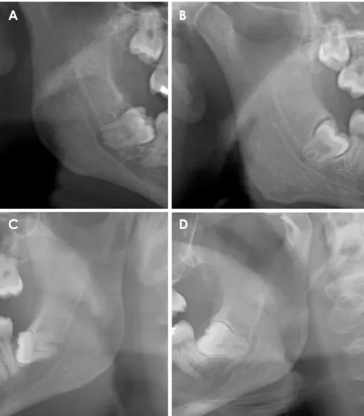
Fig. 1. Cropped panoramic images show darkening of the roots(A), diversion of mandibular canal(B), interruption of the mandibular ca- nal wall(C), and narrowing of the mandibular canal(D).
A B
C D
mandibular canal wall, darkening of the roots, diversion of the mandibular canal, and narrowing of the mandibular canal(Fig. 1).9,10 They were defined as follows: 1) interrup- tion of the mandibular canal wall: loss of the cortical mar- gin of the IAC where it crossed the LM3; 2) darkening of the roots: increased radiolucency of the root of LM3 where the IAC crossed it; 3) diversion of the mandibular canal:
change in the direction of the IAC due to the root of LM3; 4) narrowing of the mandibular canal: a decrease in the width of the IAC while it crosses the LM3.
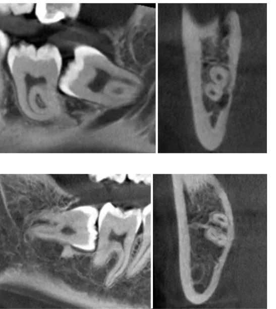
After that, the presence or absence of cortication between the LM3 and IAC was evaluated on cross-sectional CBCT slices(Figs. 2 and 3). Cross-sectional CBCT slices were preferred because these slices visualize the relationship be- tween LM3 and adjacent anatomy most clearly.11 Loss of bone tissue between LM3 root and IAC was defined as the absence of cortication.2,7,9,10,12
All observations were performed by the same maxillofa- cial radiologist, with at least 5 years of experience. The age and sex of the patients, region of the impacted LM3(right- left), angulation of LM3(mesioangular, distoangular, hor- izontal, or vertical), and course of the IAC relative to the roots of LM3(buccal, lingual, inferior, or interradicular) were also recorded.
Scanning and screening procedures
All PRs were taken with a dental X-ray machine(Morita Veraviewepocs 3D R100-P, J Morita MFG Corp., Kyoto, Japan) at 70kVp, 10mA, and 10s according to the manu- facturer’s recommended protocol.
CBCT images were acquired in a sitting position using a Morita 3D Accuitomo 170 device(J Morita MFG Corp., Kyoto, Japan), which was operated at 90kVp and 5mA, with 17.5seconds of rotation time, a voxel size of 0.25mm,
Fig. 2. Lingually positioned inferior alveolar canal without cortication.
Fig. 3. Inferiorly located inferior alveolar canal with cortication.
and a field-of-view of 100mm, according to the manufac- turer’s recommended protocol.
The PR images and CBCT slices were evaluated by the same investigator in a darkened room using a 27-inch flat panel color display with resolution of 2560×1600 pixels (U2711HTM; Dell, Round Rock, TX, USA). All CBCT im- ages were evaluated using i-Dixel software(J Morita MFG Corp., Kyoto, Japan) in all 3 planes(sagittal, axial, and cor- onal). Cross-sectional reconstructions were also used.
Statistical analyses
The PR and CBCT results were evaluated using the Pearson chi-square test. Logistic regression analysis was applied and odds ratios(ORs) were calculated for the PR signs. The kappa test was done to test intraobserver consis- tency. Statistical analysis was performed with SPSS soft- ware version 21.0(IBM Corp, Armonk, NY, USA) with a significance level of P<0.05.
Results
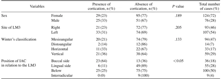
The kappa values were excellent(between 0.82 and 0.94) for all observations. Of the 200 teeth examined on cross- sectional CBCT sections, 54(27%) of the LM3s showed cortication and 146(73%) of them did not. Sex, site of the LM3, and Winter’s classification were not found to be asso- ciated with cortication status. The distribution of the study parameters is given in Table 1. The majority of the cases had an IAC course inferior to the LM3(n=100; 50%).
There was a statistically significant relationship between IAC course and cortication status. The interradicular IAC position showed the highest percentage of absence of corti- cation(100%) and the buccal IAC position showed the low-
est(36%).
The most frequently observed PR sign was interruption of the mandibular canal wall. There was a statistically significant relationship between PR signs and cortication status(P<0.05)(Table 2). The PR sign of diversion of the mandibular canal was the only risk factor for the absence of cortication(OR=12.41; 95% CI, 1.60 to 96.27; P=0.016;
P<0.05)(Table 2).
Discussion
The absence of cortical bone in the IAC may not be clearly evident on PR. Moreover, it is impossible to deter- mine whether its course is buccal or lingual to the roots or between the roots.13 At our dental faculty, the standard preoperative examination relies on PR. If the radiological signs on PR are suggestive of an close relationship between the LM3 and IAC, additional imaging is recommended for a further examination.8 However, some surgeons routinely obtain preoperative CBCT before LM3 surgery to avoid le- gal issues. Although PR does not provide any information regarding the buccolingual dimension,8 it can sometimes be used as the sole preoperative examination for LM3 sur- gery due to the lower availability of CBCT, especially in developing countries considering their socioeconomic con- ditions.8 Turkey is a developing country and CBCT is not easy to access in every region. For this reason, the present study aimed to evaluate the relationship between the IAC and impacted LM3 using CBCT and to compare CBCT findings with signs of PR.
Sedaghatfar et al.14 showed that the following 4 PR fea- tures were significantly associated with mandibular nerve exposure following third molar extraction: darkening of the
Table 1. Distribution of the study parameters
Variables Presence of
cortication, n(%) Absence of
cortication, n(%) P value Total number of cases(%)
Sex Female 29(23) 95(77) .189 124(72)
Male 25(33) 51(67) 76(28)
Site of LM3 Right 21(23) 72(77) .205 93(46)
Left 33(31) 74(69) 107(54)
Winter’s classification Mesioangular 20(21) 74(79) .133 94(47)
Distoangular 2(14) 12(86) 14(7)
Horizontal 11(33) 22(67) 33(17)
Vertical 21(36) 38(64) 59(29)
Position of IAC
in relation to the LM3 Buccal side 23(64) 13(36) <0.05 36(18)
Lingual side 6(11) 49(89) 55(28)
Below 25(25) 75(75) 100(50)
Interradicular 0(0) 9(100) 9(4)
IAC: inferior alveolar canal, LM3: lower mandibular third molar
root, interruption of the white line of the mandibular canal wall, diversion of the mandibular canal, and narrowing of the mandibular canal. These PR signs were analyzed in this study, and the most frequent PR sign was interruption of the mandibular canal wall, with a percentage of 44%.
In a recent study,12 the cortication status between the LM3 and IAC was found to be a reliable predictor of IAN injury. In the present study, CBCT analyses were per- formed when PR signs suggested a close relationship of the LM3 to the IAC. In total, 54(27%) of the LM3s showed cortication and 146(73%) of them did not. There was a sta- tistically significant relationship between PR signs and cor- tication status(P<0.05)(Table 2). The PR sign of diver- sion of the mandibular canal was found to be the only risk factor for the absence of cortication(OR=12.41; 95% CI, 1.60-96.27; P=0.016; P<0.05), with a roughly 12 times higher risk than other PR signs for the absence of cortica- tion around IAN. In a clinical study after LM3 extraction, it was shown that diversion of the mandibular canal had a similar OR(10.41) to that reported in the current study.
Excessive hemorrhage, a procedure-related parameter, was found to be the riskiest condition for IAN injury in that study, with a OR of 99.04. In another study,15 it was also found that diversion of the mandibular canal was the best diagnostic marker, followed by darkening of the root and interruption of the mandibular canal wall. Diversion of the mandibular canal is indicative of a nerve running between the roots, or a nerve sandwiched between the root and the mandibular cortical bone.2
The best diagnostic PR sign is quite controversial in the literature. Hasani et al.2 was indicated that interruption of the mandibular canal wall was the best indicator of risk for IAN injury. The variable results across previous studies might be attributed to sample variations, differences in ob- server experience, the use of different devices, and method- ological diversity.2
IAN injury has been reported to be associated with pro- cedure-related and radiographic factors, rather than demo-
graphic factors such as age and sex. Similarly, we found that sex was not related; however, the course of IAC was related to cortication status, which is an important predictor of IAN injury. This study revealed that the riskiest course of IAC in relation to the LM3 was interradicular. Nine IACs had a course that was identified as interradicular on cross-sectional CBCT examinations, and none of those IACs showed cortication. The majority of the IACs had an inferior course, corresponding to half of the sample(100 cases). The literature shows variable results in this regard.1 In a recently reported conflicting study, lingually-posi- tioned and dumbbell-shaped IACs were reported to pose a high risk for IAN damage. Our results showed that the site (right-left) and angulation of LM3 were also not associated with cortication status.
Panoramic imaging has inherent limitations such as dis- tortion, magnification, and overlapping. However, it is widely accessible and has low cost and low radiation ex- posure compared to CBCT.16 It was found that CBCT was superior to panoramic imaging in predicting neurovascular bundle exposure.10 However, in a recent meta-analysis,6 it was concluded that 3-dimensional imaging neither reduces patients’ risk of experiencing IAN injuries nor affects their prognosis. It might only be useful for the diagnosis and surgical plan of LM3s.
As a limitation, this was a cross-sectional radiological study. Although diversion of the mandibular canal and an interradicular position of the IAC were found to be related with absence of cortication, there was no clinical correla- tion after LM3 surgery.
In conclusion, considering the frequent absence of cor- tication(73%) detected on cross-sectional CBCT slices, surgeons should pay attention during LM3 surgery regard- less of whether a CBCT scan is obtained. The PR sign of diversion of the mandibular canal, which is related to a 12- fold higher risk of absence of cortication should be con- sidered as a potential predictive parameter of IAN injury.
When this specific PR sign is observed, 3-dimensional im-
Table 2. Relationships between signs on panoramic images and cortication status Signs on panoramic images Presence of
cortication, n(%) Absence of
cortication, n(%) OR(95% CI) P value Total, n(%) Interruption of the mandibular canal wall 30(35) 58(65) 1(reference) .079 88(44.0)
Darkening of the root 13(30) 31(70) 1.23(0.56-2.70) .600 44(22.0)
Diversion of the mandibular canal 1(4) 24(96) 12.41(1.60-96.27) <0.05 25(12.5)
Narrowing of the mandibular canal 10(24) 33(76) 1.70(0.74-3.92) .209 43(21.5)
Total 54 146 200(100.0)
Chi-square test; P=0.024, P<0.05
aging is highly recommended. If this radiological finding is recognized on PR and there is no possibility of a CBCT examination, the surgeon may be advised to plan alterna- tive low-risk procedures such as coronectomy. Due to the fact that an interradicular course of the IAC was found to be related with the absence of cortication, surgeons should be cautious regarding possible IAN damage when this IAC position is observed.
References
1. Hasegawa T, Ri S, Shigeta T, Akashi M, Imai Y, Kakei Y, et al.
Risk factors associated with inferior alveolar nerve injury after extraction of the mandibular third molar-a comparative study of preoperative images by panoramic radiography and comput- ed tomography. Int J Oral Maxillofac Surg 2013; 42: 843-51.
2. Hasani A, Ahmadi Moshtaghin F, Roohi P, Rakhshan V. Diag- nostic value of cone beam computed tomography and panoram- ic radiography in predicting mandibular nerve exposure during third molar surgery. Int J Oral Maxillofac Surg 2017; 46: 230-5.
3. Szalma J, Lempel E, Jeges S, Szabó G, Olasz L. The prognostic value of panoramic radiography of inferior alveolar nerve dam- age after mandibular third molar removal: retrospective study of 400 cases. Oral Surg Oral Med Oral Pathol Oral Radiol Endod 2010; 109: 294-302.
4. Ghai S, Choudhury S. Role of panoramic imaging and cone beam CT for assessment of inferior alveolar nerve exposure and subsequent paresthesia following removal of impacted mandib- ular third molar. J Maxillofac Oral Surg 2018; 17: 242-7.
5. Korkmaz YT, Kayıpmaz S, Senel FC, Atasoy KT, Gumrukcu Z.
Does additional cone beam computed tomography decrease the risk of inferior alveolar nerve injury in high-risk cases undergo- ing third molar surgery? Does CBCT decrease the risk of IAN injury? Int J Oral Maxillofac Surg 2017; 46: 628-35.
6. Clé-Ovejero A, Sánchez-Torres A, Camps-Font O, Gay-Escoda C, Figueiredo R, Valmaseda-Castellón E. Does 3-dimensional imaging of the third molar reduce the risk of experiencing infe- rior alveolar nerve injury owing to extraction?: A meta-analysis.
J Am Dent Assoc 2017; 148: 575-83.
7. Tachinami H, Tomihara K, Fujiwara K, Nakamori K, Noguchi M. Combined preoperative measurement of three inferior al- veolar canal factors using computed tomography predicts the
risk of inferior alveolar nerve injury during lower third molar extraction. Int J Oral Maxillofac Surg 2017; 46: 1479-83.
8. Elkhateeb SM, Awad SS. Accuracy of panoramic radiographic predictor signs in the assessment of proximity of impacted third molars with the mandibular canal. J Taibah Univ Med Sci 2018;
13: 254-61.
9. Neves FS, Souza TC, Almeida SM, Haiter-Neto F, Freitas DQ, Bóscolo FN. Correlation of panoramic radiography and cone beam CT findings in the assessment of the relationship between impacted mandibular third molars and the mandibular canal.
Dentomaxillofac Radiol 2012; 41: 553-7.
10. Tantanapornkul W, Okouchi K, Fujiwara Y, Yamashiro M, Maruoka Y, Ohbayashi N, et al. A comparative study of cone- beam computed tomography and conventional panoramic radiography in assessing the topographic relationship between the mandibular canal and impacted third molars. Oral Surg Oral Med Oral Pathol Oral Radiol Endod 2007; 103: 253-9.
11. Şekerci AE, Şişman Y. Comparison between panoramic radi- ography and cone-beam computed tomography findings for as- sessment of the relationship between impacted mandibular third molars and the mandibular canal. Oral Radiol 2014; 30: 170-8.
12. Shiratori K, Nakamori K, Ueda M, Sonoda T, Dehari H. As- sessment of the shape of the inferior alveolar canal as a marker for increased risk of injury to the inferior alveolar nerve at third molar surgery: a prospective study. J Oral Maxillofac Surg 2013; 71: 2012-9.
13. Jung YH, Nah KS, Cho BH. Correlation of panoramic radio- graphs and cone beam computed tomography in the assessment of a superimposed relationship between the mandibular canal and impacted third molars. Imaging Sci Dent 2012; 42: 121-7.
14. Sedaghatfar M, August MA, Dodson TB. Panoramic radio- graphic findings as predictors of inferior alveolar nerve expo- sure following third molar extraction. J Oral Maxillofac Surg 2005; 63: 3-7.
15. Ghaeminia H, Meijer GJ, Soehardi A, Borstlap WA, Mulder J, Bergé SJ. Position of the impacted third molar in relation to the mandibular canal. Diagnostic accuracy of cone beam computed tomography compared with panoramic radiography. Int J Oral Maxillofac Surg 2009; 38: 964-71.
16. Xu GZ, Yang C, Fan XD, Yu CQ, Cai XY, Wang Y, et al. An- atomic relationship between impacted third mandibular molar and the mandibular canal as the risk factor of inferior alveolar nerve injury. Br J Oral Maxillofac Surg 2013; 51: e215-9.