Inhibitory Effect of Snake Venom Toxin on Colorectal Cancer HCT116 Cells Growth through Induction of Intrinsic
or Extrinsic Apoptosis ※
Kyung Tae Kim and Ho Sueb Song*
Department of Acupuncture & Moxibustion Medicine, College of Oriental Medicine, Gachon University
[Abstract]
I investigated whether snake venom toxin(SVT) from Vipera lebetina turanica enhances the apoptosis ability of tumor necrosis factor(TNF)-related apoptosis-inducing ligand(TRAIL) in cancer cells. TRAIL inhibited HCT116 cell growth in a dose-dependent manner. Consistent with cell growth inhibition, the expression of TRAIL receptors; DR4 and DR5 was significantly increased as well as apoptosis related proteins such as cleaved caspase-3, 8, 9 and Bax.
However, the expression of survival proteins(eg, cFLIP, survivin, XIAP and Bcl2) was suppressed by the combination treatment of SVT and TRAIL. Pretreatment with the reactive oxygen species(ROS) scavenger N-acetylcysteine reduced the SVT and TRAIL-induced upregulation of DR4 and DR5 expression and expression of the apoptosis related protein such as caspase-3 and-9 as well as cell growth inhibitory effects. The collective results suggest that SVT facilitates TRAIL-induced apoptosis in human colorectal cancer HCT116 cells through up-regulation of the TRAIL receptors; DR4 and DR5 via ROS pathway signals.
Key words : SVT;
TRAIL;
DR4;
DR5;
ROS
Received : 2013. 1. 17.
Revised : 2013. 1. 24.
Accepted : 2013. 1. 25.
On-line : 2013. 2. 20.
※This research was supported by the Gachon University Research Fund in 2012(GCU-2012-R270)
* Corresponding author : Department. of Acupuncture and Moxibustion Medicine, Gil Oriental Medicine Hospital of Gachon Univercity, 1200-1, Guwol-dong, Namdong-gu, Incheon, 405-760, Republic of Korea
Tel : +82-70-7120-5012 E-mail : [email protected]
This is an Open-Access article distributed under the terms of the Creative Commons Attribution Non-Commercial License (http://creativecommons.org/licenses/by-nc/3.0) which permits unrestricted non-commercial use, distribution, and reproduction in any medium, provided the original work is properly cited.
Copyright © 2013 KAMMS. Korean Acupuncture & Moxibustion Medicine Society. All rights reserved.
Ⅰ. Introduction
Colorectal cancer is one of the most prevailed human malignancies which affects both sex equally and accounts for approximately 9.4 % in a worldwide cumulative incidence rate as a second leading cause of cancer deaths
1,2). It usually develops in the cecum, colon and rectum and mostly recovered by enucleation of tumoral tissue in the local lesions confined within the intestinal wall
3). However, To cope with the advanced metastasized stage, effective chemotherapy should be essentially accompanied by surgical options
3,4). Although pathogenesis and chemoprevention is under intense investigation, current development of standard anti-cancer drug for colorectal cancer such as 5-fluorouracil, leucovorin, irinotecan, and oxaliplatin has the limitation due to the existence of relatively rare, highly chemo-resistance and its quiescent or slow proliferating cancer stem cells
4-6). Thus, a novel therapeutic strategy for this cancer is urgently needed to resolve the tough challenge and to discover a new agent on the basis of better under- standing of the molecular mechanisms of apoptosis.
Selective and specific induction of death receptor signaling apoptosis involving intrinsic or extrinsic apoptotic pathway has been more and more recognized as a promising therapeutic approach for many cancers, including colorectal cancer
7). Tumor necrosis factor(TNF)-α-related apoptosis-inducing ligand(TRAIL), a member of the TNF ligand super family, can representatively induce selective and specific apoptosis in vitro and in vivo with no harm on normal cells
8). This is why TRAIL has increasingly attracted attention and it can be a potential target in cancer therapy and the discovery of agents
7,8). In spite of its usefulness, another challenge remains to be overcomed, which is the resistance of death receptor 4(DR4) and death receptor 5(DR5) to TRAIL because of decreased or mutated DR4 and DR5 or damaged distal signaling cascades
9,10). For these reasons, it is recommended not to use TRAIL alone but to use it together with
other chemotherapeutic agents that can upregulate TRAIL receptors. That is, Sensitizing cancer cells by subtoxic concentrations of chemotherapeutic drugs is helpful for TRAIL to restore its sensitivity through increase of DR4 and DR5 expessions
11,12). Several recent studies
13-18)have reported that the DR4 and DR5 TRAIL receptors are up-regulated by different mechanisms, such as ROS generation by agents such as ursolic acid, Gossypol, curcumin, baicalein and 15-deoxy-delta-prostaglandin J
2in cancer cells.
In the extrinsic apoptotic pathway, DR4 and DR5
exists on the cell surface of Tumors and they
becomes acivated or oligomerized upon binding to
its ligand TRAIL or overexpression as soon as
receiving ROS stimuli and subsequently signals
apoptosis through caspase-8-mediated rapid activation
of caspase cascades
19,20). They also initiated a
mitochondrial regulated apoptotic pathway simul-
taneously by translocation of a truncated form of
Bid(tBid) into mitochondria, inducing enhanced
mitochondrial outer membrane permeability following
increased Bax/Bcl-2, cytochrome C release, consec-
utively activated caspase cascade system
21-25). Many
previous studies demonstrated that natural toxins
sensitize cancer cells to TRAIL-mediated apoptotic
cell death
26,27). many researchers believe that natural
snake venom toxin(SVT) is helpful as a biological
resource due to several pharmacologically active factors
with potential therapeutic value regardless of appre-
hension about its safety
28-30). Recently there have
been a lot of studies substantiating the above such as
antihypertensive drug, anticoagulant drug, and drugs
of heparin-induced thrombocytopenia and stroke
31,32).
In particular, SVT from Vipera lebetina turanica was
previously demonstrated as a possible chemother-
apeutic agent against the growth of human prostate
cancer cell and neuroblastoma cell through induc-
tion of apoptotic cell death that is mediated by the
modulated expression of apoptosis regulatory pro-
teins
28,33). Previously, we found that SVT increases
DRs expression in colon cancer cells
34). Therefor, In
this study, I confirmed cancer cell response toward
SVT from Vipera lebetina turanica combined with
TRAIL in TRAIL sensitive colorectal cancer HCT
116 cells, and I also investigated mechanisms of enhancing sensitivity of them by SVT.
Ⅱ. Materials and method
A. Materials
SVT from Vipera lebetina turanica, N-acetyl- cysteine(NAC) and SP600125 were purchased from Sigma Chemical Co(Saint Louis, USA). Soluble Recombinant human Apo2L/TRAIL was purchased from Peprotech (RockyHill, NJ). all of the secondary antibodies such as Bax, Bcl-2, survivin, c-FLIP, XIAP caspase-3, -9, -8, cleaved caspase-3, -9, -8, DR4, DR5 used in Western blot analysis were purchased from Santa Cruz Biotechnology(Santa Cruz, CA). T4 polynucleotide kinase was obtained from Promega(Madison, WI). Poly(dI-dC), horseradish peroxidase-labeled donkey anti-rabbit secondary antibody, and ECL detection reagent were obtained from Amersham Pharmacia Biotech(Piscataway, NJ). All other reagents were purchased from Sigma unless otherwise stated.
B. Cell culture
The Human colorectal cancer HCT116 cell lines were purchased from the American Type Culture Collection(Manassas, VA). They were cultured in RPMI-1640 medium supplemented with 10 % fetal bovine serum(FBS) and penicillin/streptomycin (100 U/ml). Cell cultures were then maintained at 37 ℃ in a humidified atmosphere with 5 % CO
2.
C. Cell viability assay
To determine the cell number, Human colorectal cancer HCT116 cells were plated in 24-well plates(5×10
4cells/well)with or without TRAIL, and subconfluent cells were subsequently treated with SVT(0.1, 0.5, 1, 2 and 5 ㎍/ml) for 24 hr. After treatment, cells were trypsinized and pelleted by
centrifugation for 5 min at 1,500 rpm, resuspended in 5 ml of phosphate-buffered saline(PBS), and 0.1 ml of 0.2 % trypan blue was added to the cancer cell suspension in each of the solutions (0.9 ml each). Subsequently, a drop of suspension was placed into a Neubauer chamber and the living cancer cells were counted. Cells that showed signs of staining were considered to be dead, whereas those that excluded trypan blue were considered viable. Each assay was carried out in triplicate.
D. Measurement of ROS
Generation of ROS was assessed by 2, 7-dich- lorofluorescein diacetate(DCFH-DA, Sigma Aldrich, St Louis, MO, USA), anoxidation-ensitive fluorescent probe. Intracellular H2O2 or low-molecular-weight peroxides can oxidize 2,7-dichlorofluorescein diacetate to the highly fluorescent compound dichlorofluores- cein(DCF). Briefly, cells were plated in 6 well plates(5×10
4cells/well), and subconfluent cells were subsequently treated with SVT and TRAIL for 30 min. After the cells were trypsinized, the 1x10
4cells were plated in black 96 well plate and incubated with 10 μMDCFH-DA at 37 ℃ for 4 h.
The fluorescence intensity of DCF was measured in a microplate-reader at anex citation wave length of 485 nm and an emission wave length of 538 nm.
E. Western blot analysis
Western blot analysis was performed as described previously
28). The membranes were immunoblotted with the following primary antibodies: mouse monoclonal antibodies directed against cleaved caspase-8(1 : 1,000 dilutions; Cell Signaling Technology, Beverly, MA), against Bax(1 : 500 dilutions; Santa Cruz Biotechnology), and against XIAP, survivin, bcl2, cleaved caspase-3, -9 and c-FLIP(1 : 1,000 dilutions; Cell Signaling Technology, Beverly, MA).
The blot was then incubated with the corresponding
anti-rabbit/goat immunoglobulin G-horseradish peroxidase-
conjugated secondary antibody(Santa Cruz Biotech-
nology Inc.). Immunoreactive proteins were detected
with the Enhanced Chemiluminescence Western blotting detection system (Amersham Pharmacia Biotech, Inc. Buckingham shire, United Kingdom).
F. Apoptosis evaluation
Human colorectal HCT116 cells(2.5×10
5cells/well) were cultured on 8-chamber slides. the cells were treated with SVT(0.5 ㎍/ml). The cells were washed twice with PBS and fixed by incubation in 4 % paraformaldehyde in PBS for 1 hr at room temperature. Membrane was permeabilized by exposure to 0.1 % Triton X-100 in phosphate- buffered saline for 5 min at room temperature.
TdT-mediated dUTP nick and labeling(TUNEL) assays were performed by using the in situ Cell Death Detection Kit(Roche Diagnostics GmbH, Mannheim, Germany) according to the manufacturer’s instructions. For 4'-6-Diamidino-2-phenyl indole(DAPI) staining, slides were incubated for 15 min at room temperature in the dark with mounting medium for fluorescence containing DAPI(Vector Laboratories, Inc, Burlingame, CA). The cells were then observed through a fluorescence microscope(Leica Microsystems AG, Wetzlar, Germany).
G. Data analysis
The data were analyzed using the GraphPad Prism 4 ver. 4.03 software(GraphPad Software, La Jolla, CA).
Data are presented as mean±SD. The differences in all data were assessed by one-way analysis of vari- ance(ANOVA). When the P value in the ANOVA test indicated statistical significance, the differences were assessed by the Dunnett’s test. A value of
p<0.05 was considered to be statistically significant.Ⅲ. Results
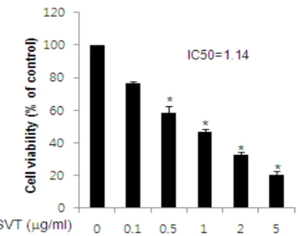
A. Effect of SVT on cell growth in colorectal cancer cells
To assess the inhibitory effect of snake venom
Fig. 1. Effect of SVT on cell viability in colorectal cancer cells
Concentration-dependent effect of SVT was shown on the cell viability assay in colorectal cancer HCT116 cells.
After treatment of SVT(0.1, 0.5, 1, 2 and 5 ㎍/ml) for 24 hr, the cells were harvested by trypsinization and stained with 0.2 % trypan blue. Relative cell survival rate was determined by counting live and dead cells. The results were expressed as a percentage of viable cells. Columns, means of three experiments, with triplicates of each experiment; bars, SD. *, p<0.05, significantly different from untreated control cells.
toxin(SVT) on cell growth of colorectal cancer HCT116 cells, we analyzed cell viability by direct cell counting. The cells were treated with several concentrations of SVT(0.1, 0.5, 1, 2, and 5 ㎍/ml) for 24 hr. As shown in Fig. 1, SVT significantly inhibited cell proliferation of colorectal cancer cells 0.5, 1, 2, and 5 ㎍/ml of it in a concentration- dependent manner with IC50 value of 1.14 ㎍/ml (Fig. 1).
B. Effect of TRAIL on cell growth in colorectal cancer cells
To assess the inhibitory effect of TRAIL on cell
growth of colorectal cancer HCT116 cells, we
analyzed cell viability by direct cell counting. The
cells were treated with several concentrations of
TRAIL(0.1, 1.25, 2.5 and 5 ng/ml) for 24 hr. As
shown in Fig. 2, TRAIL inhibited cell proliferation
of colorectal cancer cells in a concentration-
dependent manner, and 5 ng/ml of TRAIL
significantly hindered the cell growth with IC50
value of 1.14 ㎍/ml(Fig. 2).
Fig. 2. Effect of TRAIL on cell viability in colorectal cancer cells
Concentration-dependent effect of TRAIL was shown on the cell viability assay in colorectal cancer HCT116 cells.
After treatment of TRAIL(0.1, 1.25, 2.5 and 5 ng/ml) for 24 hr, the cells were harvested by trypsinization and stained with 0.2 % trypan blue. Relative cell survival rate was determined by counting live and dead cells.
The results were expressed as a percentage of viable cells. Columns, means of three experiments, with triplicates of each experiment; bars, SD. *, p<0.05, significantly different from untreated control cells.
C. Synergic effect of SVT and TRAIL on cll growth in colorectal cancer cells
To assess the inhibitory effect of SVT and TRAIL on cell growth of colorectal cancer HCT116
Fig. 3. SVT enhanced TRAIL-induced cytotoxicity
Synergic effect of SVT-combined TRAIL was shown on the cell viability assay in colorectal cancer HCT116 cells.After pre-treatment of SVT(0.5 ㎍/ml) for 24 hr, and then TRAIL(1.25 ng/ml) was treated following the medium was removed. the cells were harvested by trypsinization and stained with 0.2 % trypan blue. Relative cell survival rate was determined by counting live and dead cells. The results were expressed as a percentage of viable cells.
Columns, means of three experiments, with triplicates of each experiment; bars, SD. *, p<0.05, significantly different from untreated control cells.
cells, we analyzed cell viability by direct cell counting. The cells were treated with TRAIL(1.25 ng/ml) or SVT(0.5 ㎍/ml) or TRAIL(1.25 ng/ml) plus SVT(0.5 ㎍/ml) for 24 hr. As shown in Figure 3, SVT(0.5 ㎍/ml) significantly inhibited cell proliferation of colorectal cancer cells, compared to control and TRAIL(1.25 ng/ml) plus SVT(0.5 ㎍/ml) also significantly hindered cell proliferation of colorectal cancer cells with IC50 value of 1.14 ㎍/ml, compared to TRAIL(1.25 ng/ml) alone(Fig. 3).
D. Abolishment of synergized effect of SVT-combined TRAIL on cell growth in colorectal cancer cells
To assess whether the synergized inhibitory effect of SVT and TRAIL on cell growth of colorectal cancer HCT116 cells was reversed by strong anti-oxidative agent, NAC(1 or 10 mM), we analyzed cell viability by direct cell counting. The cells were treated with TRAIL(1.25 ng/ml) plus
Fig. 4. SVT combined TRAIL-induced cytotoxicity was reversed by anti-oxidative NAC
Synergic effect of SVT-combined TRAIL was shown on the cell viability assay in colorectal cancer HCT116 cells.
After pre-treatment of SVT(0.5 ㎍/ml) for 24 hr, and subsequently TRAIL(1.25 ng/ml) was treated following the medium was removed, and then NAC(1 or 10 mM) was added. the cells were harvested by trypsinization and stained with 0.2 % trypan blue. Relative cell survival rate was determined by counting live and dead cells. The results were expressed as a percentage of viable cells.
Columns, means of three experiments, with triplicates of each experiment; bars, SD. *, p<0.05, significantly different from untreated control cells, #. p<0.05, significantly different from TRAIL(1.25 ng/ml) plus SVT(0.5 ㎍/ml).
SVT(0.5 ㎍/ml) for 24 hr with(1 and 10 mM) or without NAC. As shown in Fig. 4, TRAIL(1.25 ng/ml) plus SVT(0.5 ㎍/ml) significantly inhibited cell proliferation of colorectal cancer cells, compared to control. However, they did not inhibit the cell proliferation representing its increase by NAC reversely and concentration-dependently. Moreover, NAC(10 mM) significantly the synergized inhibitory effect of SVT and TRAIL with IC50 value of 1.14
㎍/ml, compared to TRAIL(1.25 ng/ml) plus SVT(0.5 ㎍/ml) (Fig. 4).
E. Abolishment of synergized effect of SVT-combined TRAIL on ROS generation in colorectal cancer cells
To assess whether the synergized inhibitory effect of SVT and TRAIL on ROS generation was reversed by strong anti-oxidative agent, NAC(10
Fig. 5. ROS dependent SVT combined TRAIL- induced apoptosis was reversed by anti-oxidative NAC
Synergic effect of SVT-combined TRAIL on the ROS generation was shown on the ROS measurement in colorectal cancer HCT116 cells. After treatment of SVT and TRAIL for 30 min, the HCT116 cells were incubated with 10 μM DCF-DA at 37 ℃ for 4 h, and then washed twice with PBS. In case of adding anti-oxidative NAC, HCT116 cells were pretreated with NAC(10 mM) for 1 h, and then cells were treated with SVT for 24 h and then treated with TRAIL. The fluorescence intensity of DCF was measured in a microplate reader at an excitation wavelength of 485 nm and an emission wavelength of 538 nm. Columns, means of three experiments, with triplicates of each experiment; bars, SD. *, p<0.05, significantly different from untreated control cells, #. p<0.05, significantly different from TRAIL(1.25 ng/ml) plus SVT(0.5 ㎍/ml).mM) on cell growth of colorectal cancer HCT116 cells, we analyzed ROS measurement as described in materials and method. The cells were treated with TRAIL(1.25 ng/ml) plus SVT(0.5 ㎍/ml) for 30 min with(10 mM) or without NAC. As shown in Figure 5, TRAIL(1.25 ng/ml) plus SVT (0.5 ㎍/ml) significantly increased ROS generation, compared to control. However, they conversely and significantly decreased it following NAC (10 mM) treatment, compared to TRAIL(1.25 ng/ml) plus SVT(0.5 ㎍/ml) (Fig. 5).
F. Synergic Effect of SVT and TRAIL on apoptosis in colorectal cancer cells
I also determined whether SVT enhances TRAIL-induced apoptotic cell death of TRAIL- sensitive colorectal cancer HCT116 cells. I found that SVT and TRAIL treatments alone induced 32.4 % and 27.5 % apoptosis in HCT116 cells, respectively.
In agreement with cell growth inhibition pattern, combination treatment with SVT and TRAIL enhanced apoptotic cell death to 65.8 % in HCT116 cells(Fig. 6).
Fig. 6. SVT enhanced TRAIL-induced apoptosis in colorectal cancer HCT116 cells
HCT116 cells were pretreated with SVT(0.5 mg/ml) for 24 h, the media were removed, and the cells were exposed to TRAIL(1.25 ng/ml in HCT116) for 24 h.
Apoptosis was analyzed byTUNELassay. The green color in the fixed cells marks TUNEL-labeledcells. Data means± SD expressed as percentage of control value, which is set to 100 %. At least three independent experiments were carried out in triplicate.*, p<0.05, significantly different from control cells.
G. Effect of SVT on the expression of death receptors 4 and 5 in
colorectal cancer HCT116 cells
To ascertain the underlying mechanism that may be responsible for enhancement of TRAIL-induced apoptotic cell death by SVT, I examined the effect of SVT on the expression of death receptors. SVT induced both DR4 and DR5 in a dose dependent manner in TRAIL-sensitive HCT116(Fig. 7). In addition, we found that the expression of DR4 and DR5 was increased in TRAIL-sensitive HCT116 cells by treatment of TRAIL, but not in TRAIL- resistant cell lines(Fig. 7). But, the expression of DR4 and DR5 was further increased in TRAIL- sensitive by the combination treatment of SVT and TRAIL(Fig. 7). These results demonstrated that the up-regulation of DR4 and DR4 by SVT can
Fig. 7. SVT enhanced the expression of DR4 and DR5 in the colorectal cancer HCT116 cells
HCT116 cells were treated with SVT(0.1, 0.5, 1 μg/ml) for 24 h, then equal amounts of total proteins(50 μg/lane) were subjected to 12 % SDS-PAGE. Expression of DR4, DR5 and b-actin was detected by Western blotting using specific antibodies. HCT116 cells were pretreated with SVT(0.5 mg/ml) for 24h, the media were removed, and the cells were exposed to TRAIL(1.25 ng/ml in HCT116) for 24 h, then equal amounts of total proteins (50 μg/lane) were subjected to 12 % SDS-PAGE. Expression of DR4, DR5 and b-actin was detected by Western blotting using specific antibodies.overcome the TRAIL resistance by induction of DR4 and DR5.
H. Effect of SVT on TRAIL-induced expression of apoptosis related proteins
I selected the HCT116 cell line that was TRAIL sensitive. I confirmed the effect of SVT and/or TRAIL on the activation of caspase-8, caspase-3, caspase-9 cleavage and the expression of Bax in the TRAIL sensitive colon cancer(HCT116) cell.
Although SVT and TRAIL alone had little effect on
Fig. 8. SVT enhanced TRAIL-induced apoptotic proteins expressions in colorectal cancer HCT116 cells
HCT116 cells were pretreated with SVT for 24 hand washed out. After that the cells were treated with TRAIL for 24 h, and whole cell extracts were analyzed by western blotting using antibodies against caspase-3, caspase-8, caspase-9, Bax and b-actin. b-actin protein was used an internal control. Each band is representative for three experiments. HCT116 cells were treated with snake venom toxin(0.1, 0.5, 1 μg/ml) for 24 h, Equal amounts of total proteins(50 μg/lane) were subjected to 12 % SDS-PAGE.
Expression of cFLIP, survivin, XIAP, Bcl2, Bax and b-actin was detected by Western blotting using specific antibodies. b-actin protein was used an internal control.
Each band is representative for three experiments.
the activation of caspases cleavage and Bax expression, the combination treatment significantly increased expression of the apoptotic cell death regulatory proteins(Fig. 8). Various antiapoptotic proteins including survivin, Bcl2, XIAP and cFLIP have been shown to induce resistance to TRAIL-induced apoptosis
35,36). I examined whether SVT sensitized cancer cells to TRAIL through down-regulation of the expression of these cell survival proteins. As shown in Fig. 8, SVT decreases expression of XIAP, survivin, bcl-2 and cFLIP. These results indicated that the SVT enhances TRAIL-induced apoptotic cell death through the over-expression of DR4 and DR5, as well as the down- regulation of anti-apoptotic protein expression.
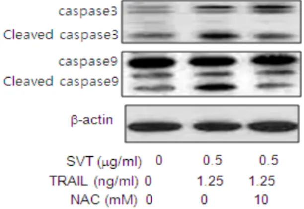
I. Abolishment of synergized effect of SVT-combined TRAIL on
TRAIL-induced expression of apoptosis related proteins
HCT116 cells were pretreated with SVT for 24 hand washed out. After that the cells were treated with TRAIL for 24 h, and whole cell extracts were
Fig. 9. The Expressions of SVT combined TRAIL- induced apoptotic protein was reversed by anti- oxidative NAC
HCT116 and HT-29 cells were pretreated with NAC(10 mM) for 1 h, and then cells were treated with SVT for 24 h and then treated with TRAIL. Whole cell extracts were analyzed by western blotting using the antibodies against caspase-3, caspase-9 and b-actin. b-actin protein was used an internal control. Each band is representative for three experiments.
analyzed by western blotting using antibodies against caspase-3, caspase-9,b-actin. b-actin protein was used an internal control. Each band is representative for three experiments. I found that pretreatment(1 h) of ROS scavenger N-acetylcysteine(NAC, 10 mM) reversed the activation of caspase-3 and -9 cleavage by the combination treatment of SVT and TRAIL(Fig. 9).
J. Abolishment of synergized effect of SVT-combined TRAIL on
RAIL-induced expression of DR4 and DR5
HCT116 cells were pretreated with SVT for 24 hand washed out. After that the cells were treated with TRAIL for 24 h, and whole cell extracts were analyzed by western blotting using antibodies against DR4, DR5 and b-actin. b-actin protein was used an internal control. Each band is representative for three experiments. I found that pretreatment(1 h) of ROS scavenger N-acetyl- cysteine(NAC, 10 mM) reversed the activation of DR4 and DR5 by the combination treatment of SVT and TRAIL (Fig. 10).
Fig. 10. The Activation of DR4 and DR5 was reversed by anti-oxidative NAC
HCT116 and HT-29 cells were pretreated with NAC(10 mM) for 1 h, and then cells were treated with SVT for 24 h and then treated with TRAIL. Whole cell extracts were analyzed by western blotting using the antibodies against DR4, DR5 and b-actin. b-actin protein was used an internal control. Each band is representative for three experiments.