J. Exp. Biomed. Sci. 14 (2008) 179–186
Cytochalasin D-induced Matrix Metalloproteinase-2 Regulates Articular Chondrocytes Dedifferentiation
In Kyu Choi, Seon Mi Yu and Song Ja Kim†
Department of Biological Sciences, College of Natural Sciences, Kongju National University, Korea
Matrix metalloproteinases (MMPs), also designated matrixins, hydrolyze components of the extracellular matrix.
These proteinases play a central role in many biological processes, such as embryogenesis, normal tissue remodeling, wound healing, and angiogenesis, and in diseases such as atheroma, arthritis, cancer, and tissue ulceration. In previous data, disruption of the actin cytoskeleton by cytochalasin D (CD) inhibited NO-induced apoptosis, dedifferentiation, cyclooxygenase (COX)-2 expression, and prostaglandin E2 production in chondrocytes cultured on plastic or during cartilage explants culture. In this study, we investigated the effects of the actin cytoskeleton architecture on MMP-2 expression and dedifferentiation by CD in rabbit articular chondrocytes. Rabbit articular chondrocytes were prepared from cartilage slices of 2-weeks-old New Zealand white rabbits by enzymatic digestion. CD was used as a disruptor of actin cytoskeleton. In this experiments measuring CD dose response, primary chondrocytes were treated with various concentrations of CD for 24 h. The actin disruption was determined by immunostaining. MMP-2 expression levels were determined by immunoblot analysis and Reverse transcriptase-Polymerase chain reaction (RT-PCR) and MMP-2 activity was determined by gelatin zymography. We found that cell morphological change and up-regulation of MMP-2 expression by CD as determined via immunostaining, gelatin zymography and immunoblotting. Moreover, CD induced MMP-2 transcription was detected by RT-PCR. Also, CD-induced type II collagen expression was inhibited by MMP-2 inhibitor I treatment. Our results indicate that CD up-regulated MMP-2 activation causes dedifferentiation of articular chondrocyte.
Key Words: Actin, Cytoskeleton, Cytochalasin D, Matrix Metalloproteinase-2
서 론
연골세포는 배아발생과정 중 중간엽 줄기세포로부터 분화하여 생성된다 (Sandell, 1999; DeLise et al., 2000). 분 화된 연골세포의 표현형은 type II collagen이나 proteo- glycan과 같은 연골세포 특이적 세포외부 기질의 합성, 침적 및 유지로 묘사할 수 있다. 이러한 연골세포의 세 포외부 기질의 합성은 연골조직의 발생, 유지, 재생에 매우 중요한 역할을 하는 것으로 알려져 있다. 이와 같 은 연골세포는 염증성 사이토카인인 IL-1β (Goldring et al., 1994), 산화질소 (Amin and Abramson, 1998), Retinoic Acid
(Cash et al., 1997; Weston et al., 2000), 계대배양 (Lefebvre et al., 1990; Yoon et al., 2002) 등 여러 가지 원인에 의해 외 부 기질 합성의 불균형을 야기시키며, 이는 연골세포의 탈분화 및 세포사멸을 일으킨다. 이러한 세포 수준의 손 상으로 인해 조직이 파괴되며 이는 류마티스 관절염 혹 은 골 관절염의 발병원인이 된다.
Matrix Metalloproteinase (MMP)는 세포외부 기질을 분 해하는 분해효소로써 세포외부로 분비되며 외부 기질의 중요한 분해 단백질로 알려져 있다. 이러한 MMP는 효 소의 특이성에 따라 collagenase, gelatinase, stromelysin, matrilysin, membrane-type MMP 등의 종류로 나눠진다 (Visse and Nagase, 2003). MMP-2는 젤라틴 분해효소 A 혹 은 72 kDa Type IV collagen 분해효소로 잘 알려져 있으며 발생, 분화, 염증반응, 조직재생, 암 전이 및 침투, 등에 중요한 역할을 하는 것으로 알려져 있다. 이러한 MMP-2 의 촉매 도메인은 MMP-9의 촉매 도메인과 매우 유사한 것으로 알려져 있으며 이러한 유사 도메인은 hemopexin
*논 문 접 수: 2008년 8월 13일 수정재접수: 2008년 9월 8일
†교신저자: 김송자, (우) 314-701 충남 공주시 신관동 182번지, 국립공주대학교 자연과학대학 생명과학과
Tel: 041-850-8507, Fax: 041-850-0927 e-mail: Ksj85@kongju.ac.kr
유사 도메인을 포함하여 다른 MMP들의 도메인에서는 드물게 보이는 fibronectin type II module (FII) 등이 있다.
비활성 상태의 MMP-2는 활성부위를 감싸고 있는 pro- domain의 절단에 의해 활성부위가 노출되며, 활성부위에 아연 이온이 결합하면서 단백질 활성화가 이루어진다.
이러한 구조는 최근 X-ray 결정화 모델과 NMR에 의해 밝혀졌다 (Briknarova et al., 2001). 이전 연구에서 MMP들 과 같은 분비 단백질의 분비는 세포골격을 따라 움직이 는 분비 단백질 운반 소포체들에 의해 분비되며, 따라 서 MMP의 분비는 세포골격의 구조와 많은 연관이 있 다는 것으로 생각된다 (Schnaeker et al., 2004). 암세포에서 tubulin 세포골격의 중합을 억제하는 paclitaxel은 MMP-2 와 MMP-9의 분비를 억제하며 암 전이 및 침투 또한 억 제하는 것으로 밝혀졌다 (Benya et al., 1988).
Actin 세포골격의 구조는 연골세포의 표현형을 결정하 는 중요한 조절인자로 생각된다. Retinoic acid나 세포 계 대배양에 의해 탈분화된 연골세포에서는 actin 세포골격 의 변화를 관찰할 수 있다. CD는 actin의 붕괴를 통하여 중합을 억제한다고 알려져 있다 (Bray, 1992). 그러므로, CD는 actin의 기능적인 역할을 연구하는데 있어 많이 사용되고 있다. Dihydrocytochalasin B에 의한 actin 세포골 격의 붕괴는 탈분화된 연골세포의 재분화를 일으킨다 (Brown and Benya, 1988). 뿐만 아니라, 이러한 actin 세포 골격은 관절 연골세포에서 골 형성 단백질이나 산화질 소에 의한 표현형의 변화를 야기시킨다 (Vinall et al., 2002). 또한 CD에 의한 actin 세포골격의 붕괴는 기존 연 구에서 발생과정 중 날개부분의 중간엽 줄기세포로부터 연골 형성화를 촉진시키며, 분화된 연골세포의 표현형을 유지시킨다 (Clancy, 1999). CD에 의한 actin 세포골격의 붕괴는 PI3-Kinase, AKT, MAP Kinase, PKC-α와 PKC-ξ와 같은 세포 신호 전달과정을 조절하여 산화질소에 의한 연골세포의 탈분화와 세포자가사멸을 억제하며 염증반응 에 중요한 역할을 하는 COX-2의 발현을 저해하는 것으 로 밝혀졌다 (Loty et al., 1995; Lim et al., 2000, 2003). 하지 만 JAS에 의한 actin 세포골격의 재구성은 오히려 연골 세포의 탈분화와 염증반응 단백질인 COX-2의 발현을 유도하는 것으로 밝혀졌다 (Kim et al., 2003).
본 연구에서는 토끼 무릎관절 연골세포에서 CD에 의 한 actin 세포골격의 붕괴에 의한 actin 세포골격의 재구 성이 연골세포외부 기질의 중요한 조절인자인 MMP-2의 발현과 활성화에 어떠한 영향을 주며, 이러한 MMP-2의 활성이 연골세포의 탈분화를 조절하는지에 대해 알아보
고자 하였다. 본 연구결과에 따르면 CD에 의한 actin 세포골격의 붕괴는 MMP-2의 단백질 발현과 활성을 유 도하였으며, CD에 의해 유도된 연골세포분화 지표인 sulfated glycosaminoglycan의 축적과 type II collagen의 발 현 증가는 MMP-2 저해제에 의하여 억제되어짐을 확인 할 수 있었다. 따라서 본 연구결과를 통하여, 연골세포 에서의 actin 세포골격단백질의 붕괴는 세포외 기질의 중 요한 조절인자인 MMP-2의 발현과 활성화 유도에 중요 하며, 이러한 MMP-2의 활성은 연골세포의 탈분화를 조 절하는 것으로 생각된다.
재료 및 방법
1. 토끼관절 연골세포의 단층배양 및 분화측정
토끼관절 연골세포의 수집은 2주된 New Zealand White 종의 토끼를 사용하였다. 수집방법은 치사량의 diethyl ether로 토끼를 치사 시킨 후 뒷다리 관절부분을 포함한 요골과 장골을 적출한다. 적출한 관절로부터 연골을 절 편하여 0.2% collagen 분해효소 (381 U/mg, Sigma)가 함유 된 PBS 완충용액에 넣고 37℃에서 6시간 동안 반응시 켜 세포를 조직에서 분리시킨다. 세포는 원심분리를 이 용하여 수집한다. 수집된 세포는 10%의 FBS, 50 μg/ml의 Streptomycin, 50 units/ml의 Penicillin이 함유된 Dulbecco's modified Eagle's medium (DMEM, Gibco-BRL, Gaithersburg, MD)에서 4×10⁵의 밀도로 배양한다. 배양액은 이틀에 한번씩 교체하여 주었으며, 배양접시의 세포밀도가 거 의 80%가 되었을 때 시약을 처리하였다. 연골세포의 분 화 정도는 alcian blue 염색액을 이용하여 sulfated glyco- saminoglycan의 축적량으로 결정하였다 (Oh et al., 1999;
Yoon et al., 2000).
2. Immunoblot Analysis
단백질의 정량분석을 위하여 세포를 50 mM Tris-HCl, pH 7.4, 150 mM NaCl, 1% Nonidet P-40, 0.1% Sodium dodecylsulfate가 함유된 세포 용해용액에 단백질 분해효 소 억제제 [10 μg/ml leupeptin, 10 μg/ml pepstatin A, 10 μg/
ml aprotinin, 1 mM 4-(2-aminoethyl) benzensulfonyl fluoride]
와 인산 분해효소 저해제 [1 mM NaF, 1 mM Na3VO4]를 첨가하여 단백질을 추출하여 사용하였다. 분리된 단백질 은 SDS-polyacrylamide gel에 전기영동하여 Nitrocellulose 막으로 이동시켰다. MMP-2와 type II collagen 항체는 Santa Cruz Biotechnology사에서 구입하여 사용하였다
(Santa Cruz, CA).
3. RT-PCR
모든 RNA는 Trizol (Invitrogen, Gronongen, Netherland)을 이용하여 추출하였다. 추출한 RNA는 M-MLV (Promega, Madison, WI) 역전사 효소를 사용하여 중합하였다. 사용 된 MMP-2 primer는 다음과 같다: 5'-AGC CTT CTC ACC CCC ACC TG-3' (sense), 5'-GCC CTT ATC CCA CTG CCC C-3' (antisense).
4. Immunofluorescence
연골세포에서 CD에 의한 actin 세포골격의 형태를 관 찰하기 위하여 Immunofluorescence를 수행하였다. 배양된 연골세포는 3.5% paraformaldehyde로 10분 동안 상온에서 고정시켰으며, 0.1% triton X-100을 이용하여 세포막 투 과성을 높여주었다. 고정된 세포는 rhodamine과 결합된 phalloidin을 사용하여 염색하였으며, 형광현미경으로 관 찰하였다.
5. Zymography
배양된 연골세포에서 CD에 의한 MMP-2의 분비와 활 성변화를 측정하기 위하여 gelatin zymography를 수행하 였다. 세포는 시약 처리 전 2% 우 태아혈청 DMEM으로 배지를 교체해주었으며, 시약 처리 24시간 후 세포 배양 액을 모았다. 모은 배양액은 최종농도 5%로 Tricholoro- acetic Acid (TCA, Sigma)를 처리하여 단백질을 침전시켰 다. 원심분리로 침전된 단백질을 모은 후 상등액을 제거 한다. 수집한 단백질은 4℃의 acetone으로 2번 수세하여 TCA를 제거한다. 수세한 단백질은 상온에서 1시간 동안 방치하여 acetone을 증발시켜 주었으며, 건조된 단백질은 1 X Zymography sample buffer [125 mM Tris-HCl, pH 6.8, 5% Glycerol, 4% SDS, 0.005% Bromophenol Blue]에 용해시 켜 10배 농축하였다. 준비된 단백질은 0.1% gelatin SDS- polyacrylamide gel에서 전기영동 하였다. 전기영동이 끝 난 gel은 2.5% Triton X-100으로 상온에서 30분씩 3번 수 세 후 증류수로 1시간 동안 수세하였다. 수세가 끝난 gel 은 incubation buffer [50 mM Tris base, 0.2 M NaCl, 5 mM CaCl2, 0.02% Brij 35]에 37℃에서 14시간 동안 반응시킨 다. 반응이 끝난 gel은 Coomassie Blue로 염색하여 결과를 확인하였다.
6. 결과 분석 및 평균화
본 실험의 결과는 여러 차례의 실험 중 평균적인 결과 를 사용하였다.
결 과
1. Cytochalasin D와 Jasplakinolide에 의한 Actin 세포 골격 단백질의 형태변화
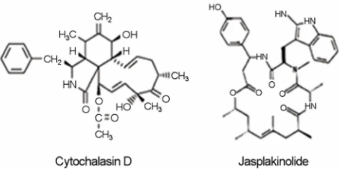
본 실험에서 사용된 CD는 균계 생물체에서 대사산물 로 만들어지는 물질로서 actin에 매우 높은 친화도를 가 지고 있어 actin 세포골격의 중합과 탈중합을 억제하며 세포투과성 성질을 가지고 있다. Jasplakinolide (JAS)는 해양생물인 Jaspis johnstoni의 대사산물로부터 추출된 cyclodepsipeptide 단백질로서 phalloidin과 경쟁적으로 actin 에 결합하여 중합상태를 안정화시켜 주어 중합을 촉진 하는 것으로 밝혀져 있다. Fig. 1은 본 실험에 사용되어진 CD와 JAS의 화학적 구조를 나타낸 것으로서 이의 용매 제로는 유기용제인 Dimethyl sulfoxide (DMSO)가 사용되 어진다. 토끼관절 연골세포를 일차배양 후 CD와 JAS 처 리시 actin 세포골격 단백질의 형태변화를 형광현미경으 로 관찰하였다. 초기 배양한 연골세포의 actin 세포골격 구조는 stress-fiber 형태로 길게 뻗어 있으며, 이러한 구 조는 세포 전체적인 부분에서 나타난다. 반면, 초기 배 양세포에 CD 0.1 μM를 24시간 동안 처리한 세포에서의 actin 세포골격은 위와 같은 stress-fiber와 같은 형태가 사라지며, actin이 파괴되어 뭉쳐져 있는 형태로 세포에 서 불균등하게 분포되어 있음을 관찰할 수 있었다. CD 처리에 의한 세포의 사멸 등과 같은 세포독성 효과는 없 었다 (data not shown). 세포에 JAS 100 μM를 24시간 동안 처리하였을 때는 세포 전반적인 부분에서 actin이 염색되 는 것을 관찰하였다 (Fig. 2).
Fig. 1. Structure of cytochalasin D (M.W., 507.6; M.F., C30H37NO6) and Jasplakinolide (M.W., 709.6; M.F., C36H45BrN4O6).
2. Cytochalasin D와 Jasplakinolide에 의한 Actin 세포 골격 단백질의 형태변화에 의한 MMP-2의 발현 증가
이전 연구결과에서 CD와 JAS는 actin 세포골격 단백질 의 붕괴와 재구성을 통하여 연골세포의 탈분화, 염증, 세포사멸에 영향을 주는 것으로 보고하였다 (Kim et al.,
2003). 이에, 본 실험에서는 이전의 실험결과를 토대로 연골세포의 탈분화에 직간접적으로 역할을 할 것으로 기 대되는 MMP에 대해 조사하기로 하였다. 먼저 연골조직 에 풍부하다고 알려진 MMP-1, 2, 9, 13에 대한 발현 정도 를 토끼관절 연골세포에서 Western blot으로 확인한 결과, MMP-2, 9은 높은 발현량을 보였으나, MMP-1, 13은 낮은 발현량을 보였다. 이에 연골세포에 CD를 처리하여 발현 량을 확인한 결과, MMP-2만이 높은 발현량 증가를 보였 고, MMP-1, 9, 13의 발현량에는 변화가 없는 것을 확인하 였다 (data not shown). 이에 본 실험에서는 MMP-2가 세 포골격 단백질의 형태변화에 의한 연골세포의 탈분화와 관련이 있을 것으로 생각되어 이후 MMP-2를 중심으로 실험을 수행하였다. 초기 배양한 연골세포에서 CD를 농 도 별로 처리한 후 24시간 동안 배양한 후 MMP-2 단백 질의 발현량을 조사해본 결과, CD의 처리농도가 높아질 수록 MMP-2 단백질의 발현이 농도의존적으로 증가하는 것을 관찰할 수 있었다 (Fig. 3A). MMP-2의 발현량을 전 사수준에서 확인하고자 RT-PCR을 행한 결과 전사수준에 서도 MMP-2의 발현량이 증가함을 확인할 수 있었으며, 이는 MMP-2의 발현이 전사수준에서 조절되어짐을 말해 주는 것이라 하겠다 (Fig. 3B). 연골기질 단백질인 collagen 의 또 다른 주요 분해효소로 알려진 MMP-9의 단백질 발현 및 활성화를 조사해 본 결과 발현이나 활성화에 변 화가 없음을 확인하였다 (data not shown). 그러나, JAS에 의한 actin 세포골격의 변화는 MMP-2의 발현에 아무런 영향을 주지 않음을 확인하였다 (Fig. 3C). 이는 actin 세 포골격 단백질의 중합저해가 MMP-2의 발현변화에 관 여하고 있음을 나타내는 것이라 하겠다.
Fig. 2. Phenotype of actin cytoskeleton. Articular chondrocytes were untreated (Control) or treated with 1 μM CD or with 50 nM JAS for 24 h. Cells were stained for F-actin with rhodamine-conjugated phalloidin. Data are presented as results of a typical experiment.
B
C A
Fig. 3. CD upregulated MMP-2 expression in articular chondro- cytes. (A). Chondrocytes were treated with the indicated concen- trations of CD for 24 h. The expression of MMP-2 and actin was determined by Immunoblot analysis. (B). Chondrocytes were treated with 1 μM CD for 24 h. The expression of MMP-2 and actin was determined by RT-PCR. (C). Chondrocytes were treated with the indicated concentrations of JAS for 24 h. The expression of MMP-2 and actin was determined by immunoblot analysis.
The data represent the results of a typical experiment conducted at least three times with similar results.
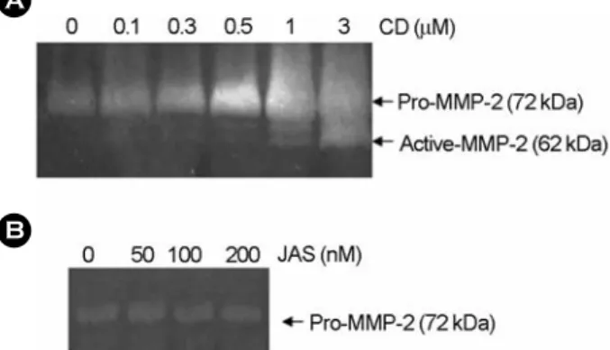
3. Cytochalasin D와 Jasplakinolide에 의한 Actin 세포 골격 단백질의 형태변화에 의한 MMP-2의 활성 증가
위의 실험을 토대로 CD와 JAS에 의한 actin 세포골격 의 변화가 MMP-2의 분비와 활성화에도 어떠한 영향을 주는지 gelatin zymography를 통하여 알아보았다. 먼저, 초기 배양한 연골세포에 CD를 농도 별로 24시간 처리 하여 분비된 MMP-2의 활성과 분비량을 측정해본 결과 CD의 처리 농도의존적으로 MMP-2의 활성과 분비가 증 가하는 것을 관찰할 수 있었다 (Fig. 4A). 그러나, JAS에 의한 MMP-2의 활성변화는 관찰할 수 없었다 (Fig. 4B).
이는 actin 세포골격 단백질의 붕괴가 MMP-2의 활성과 분비에도 관여하고 있음을 나타내는 것이라 하겠다.
4. Cytochalasin D에 의한 MMP-2의 활성이 연골세포 의 탈분화 조절
본 실험에서는 CD에 의한 MMP-2의 활성 증가가 연 골세포의 탈분화에 관여하는지를 조사하기로 하였다. 먼 저, 연골세포에 CD를 농도 별로 처리하여 연골세포의 분화정도를 sulfated glycosaminoglycan 축적과 type II collagen 발현으로 확인하였다. 기대한 것처럼, CD 처리 농도의존적으로 glycosaminoglycan의 축적과 type II colla- gen의 발현량이 증가하는 것을 확인할 수 있었다 (Fig.
5A). 즉, 연골세포의 탈분화가 저해되어짐을 확인하였다.
이에, 연골세포 탈분화와 MMP-2 활성과의 상관관계를 조사하기 위하여, MMP-2의 활성을 선택적으로 억제하는 것으로 알려진 MMP-2 inhibitor I (Calbiochem, San Diego, CA; Polette et al., 1990; Suzuki et al., 1997)를 30분 연골세포 에 전 처리하고, CD를 24시간 처리 배양하였다. CD 처 리에 의한 sulfated glycosaminoglycan의 축적과 type II collagen의 발현 증가는 MMP-2 저해제에 의하여 감소하 는 것을 확인할 수 있었다 (Fig. 5B). 즉, CD에 의해 유 도되어진 연골세포의 탈분화는 MMP-2의 활성 저해제에 의하여 회복되어짐을 확인할 수 있었다. 위의 결과를 종합해 볼 때, CD에 의해 유도된 MMP-2의 활성은 연골 세포의 탈분화를 조절하는 것으로 생각된다.
고 찰
Actin 세포골격 단백질의 중합저해는 세포의 여러 가 Fig. 4. CD induced MMP-2 activity in articular chondrocytes.
Chondrocytes were treated with the indicated condentrations of CD (A) or JAS (B) for 24 h. The activity of MMP-2 was determined by gelatin-zymography. The data represent the results of a typical experiment conducted at least three times with similar results.
A B
Fig. 5. CD-induced dedifferention effect is linked to active MMP-2 in articular chondrocytes. (A). Chondrocytes were treated with the indicated concentrations of CD for 24 h. (B). Chondrocytes were treated with 1 μM CD for 24 h in the absence or presence of 20 μM MMP-2 inhibitor I. Accumulation of sulfated glycosaminoglycan was quantified by Alcian blue staining. The expression of type II collagen was determined by immunoblot analysis. The data represent the average values with standard deviation (n=4).
B A
지 기능을 조절하는 것으로 알려지고 있다. CD 처리에 의한 actin의 중합저해는 MMP-2의 발현을 유도하는 것 으로 여러 세포에서 알려지고 있으며 (Ailenberg et al., 1994; Ailenberg and Silverman, 1996, 2003; Tomasek and Halliday, 1997), 이렇게 유도된 MMP-2는 세포외 기질의 분해를 통하여 세포 증식, 사멸, 분화에도 관여하는 것 으로 보고되고 있다 (Cowan et al., 1999; Jones et al., 1997, 2000). 지난 연구에서 토끼관절 연골세포에 CD를 처리하 여 actin 세포골격 단백질의 형태변화 유도 시 연골세포 의 탈분화, 염증반응, 세포사멸을 조절한다는 결과를 보 고한 바 있다 (Kim et al., 2003). 이에 본 연구에서는, 토끼 관절 연골세포에서 actin 세포골격 단백질의 형태변화에 의해 야기되는 탈분화에 많은 영향을 미칠 것으로 기대 되는 MMP-2의 단백질 발현 및 활성화를 조사하고자 하 였으며, 이러한 MMP-2의 활성이 연골세포의 탈분화 조 절에 관여하는지에 대해 알아 보고자 하였다. CD와 JAS 에 의한 actin 세포골격의 중합억제와 중합상태를 유지시 켰을 때 MMP-2의 발현변화를 살펴본 결과, CD 처리에 의한 actin 세포골격의 중합억제는 MMP-2의 전사와 단 백질 발현량을 증가시켰으며, 단백질 분비 및 활성화 또 한 증가시키는 것으로 나타났다. 반면 JAS에 의한 actin 세포골격의 중합 유지는 MMP-2의 발현에 아무런 영향 을 주지 않는 것으로 나타났다. 이와 같은 결과는, MMP- 2의 발현이 단순한 actin 세포골격의 구조적 변화에 의 하여 조절되는 것이 아닌 세포골격의 중합억제에 의한 세포 신호전달 체계에 의하여 조절되는 것으로 생각된 다. 또한, CD에 의해 유도된 연골세포의 탈분화 저해는 MMP-2의 활성을 저해하였을 경우 회복되는 것을 확인 할 수 있었다. 즉, MMP-2는 연골세포의 탈분화를 조절 하는 것으로 생각된다. 지난 연구에서 (Kim et al., 2003) CD 처리에 의한 연골세포 사멸이 ERK-1/-2 kinase의 활 성과는 무관하게 p38 kinase의 활성에 의존적으로 저해 되어지는 것을 확인하였다. 이는 연골세포의 탈분화가 MMP의 활성 및 세포사멸 등에 기인하여 이루어짐을 고려해 볼 때, CD에 의한 MMP-2의 활성 및 발현 증가 에 의한 연골세포의 탈분화는 주요 MAP kinase 중 하나 인 p38 MAP kinase에 의해 조절될 것으로 생각된다. 따 라서, 앞으로의 연구에서는 CD에 의한 MMP-2의 활성 및 발현 유도와 이렇게 유도된 MMP-2에 의해 조절되는 연골세포의 탈분화를 두 개의 주요 MAP kinase를 중심으 로 어떠한 신호전달계가 관련이 있는지를 밝혀야 할 것 으로 생각된다. 또한, 연골세포에서 발현이 확인된 MMP
family (MMP-1, 2, 9, 13) 중 MMP-2만이 세포골격 붕괴에 의한 탈분화 유도에 관여하는지는 차후 더 연구되어야 할 것으로 사료된다.
감사의 글
This work was supported by a Korea Research foundation Grant funded by the Korean Government (MOEHRD, Basic Research Promotion Fund) (KRF-2005-202-100654), and the SRC/ERC program of Most/KOSEF (R11-2002-098-05001-0).
REFERENCES
Ailenberg M, Silverman M. Cellular activation of mesangial gelatinase A by cytochalasin D is accompanied by enhanced mRNA expression by both gelatinase A and its membrane associated gelatinase A activator (MT-MMP). Biochem J.
1996. 313: 879-884.
Ailenberg M, Silverman M. Cytochalasin D disruption of actin filaments in 3T3 cells produces an anti-apoptotic response by activating gelatinase A extracellularly and initiating intra- cellular survival signals. Biochimica Biophysica Acta. 2003.
1593: 249-258.
Ailenberg M, Weinstein T, Li L, Silverman M. Activation of pro-collagenase IV by cytochalasin D and concanavalin A in cultured rat mesangial cells: linkage to cytoskeletal re- organization. J Am Soc Nephrol. 1994. 4: 1760-1770.
Amin AR, Abramson SB. The role of nitric oxide in articular cartilage breakdown in osteoarthritis. Curr Opin Rheumatol.
1998. 10: 263-268.
Benya PD, Brown PD, Padilla SR. Microfilament modification by dihydrocytochalasin B causes retinoic acid-modulated chon- drocytes to reexpress the differentiated collagen phenotype without a change in shape. J Cell Biol. 1988. 106: 161-170.
Bray D. Actin filaments: structure and assembly, Cell movement.
1992. pp. 75-92. Garland Publishing. NY. USA
Briknarova K, Gehrmann M, Banyai L, Tordai H, Patthy L, Llinas M. Gelatin-binding region of human matrix metalloproteinase 2: solution structure, dynamics, and function of the COL-23 two-domain construct. J Biol Chem. 2001. 276: 27613-27621.
Brown PD, Benya PD. Alterations in chondrocyte cytoskeletal architecture during phenotypic modulation by retinoic acid and dihydrocytochalasin B-induced reexpression. J Cell Biol.
1988. 106: 171-179.
Cash DE, Bock CB, Schughart K, Linney E, Underhill TM.
Retinoic acid receptor alpha function in vertebrate limb skeletogenesis: a modulator of chondrogenesis. J Cell Biol.
1997. 136: 445-457.
Clancy R. Nitric oxide alters chondrocyte function by disrupting cytoskeletal signaling complexes. Osteoarthritis Cartilage.
1999. 7: 399-400.
Cowan KN, Jones PL, Rabinovitch M. Regression of hyper- trophied rat pulmonary arteries in organ culture is associated with suppression of proteolytic activity, inhibition of tenascin-C, and smooth muscle cell apoptosis. Circ Res.
1999. 84: 1223-1233.
DeLise AM, Fischer L, Tuan RS. Cellular interactions and signaling in cartilage development. Osteoarthritis Cartilage. 2000. 8:
309-334.
Goldring MB, Birkhead JR, Suen LF, Yamin R, Mizuno S, Glowacki J, Arbiser JL, Apperley JF. Interleukin-1 beta- modulated gene expression in immortalized human chon- drocytes. J Clin Invest. 1994. 94: 2307-2316.
Jones PL, Crack J, Rabinovitch M. Elastase and matrix metallo- proteinase inhibitors induce regression, and tenascin-C antisense prevents progression, of vascular disesase. J Clin Invest. 2000. 105: 21-34.
Jones PL, Crack J, Rabinovitch M. Regulation of tenascin-C, a vascular smooth muscle cell survival factor that interact with avb3 integrin to promote epidermal growth factor receptor phosphorylation and growth. J Biol Chem. 1997. 139: 279 -293.
Kim SJ, Hwang SG, Kim IC, Chun JS. Actin cytoskeletal archite- cture regulates nitric oxide-induced apoptosis, dedifferentiation, and cyclooxygenase-2 expression in articular chondrocytes via mitogen-activated protein kinase and protein kinase C pathways. J Biol Chem. 2003. 278: 42448-42456.
Lefebvre V, Peeters-Joris C, Vaes G. Production of collagens, collagenase and collagenase inhibitor during the dedifferen- tiation of articular chondrocytes by serial subcultures.
Biochim Biophys Acta. 1990. 1051: 266-275.
Lim YB, Kang SS, An WG, Lee YS, Chun JS, Sonn JK.
Chondrogenesis induced by actin cytoskeleton disruption is regulated via protein kinase C-dependent p38 mitogen- activated protein kinase signaling. J Cell Biochem. 2003. 88:
713-718.
Lim YB, Kang SS, Park TK, Lee YS, Chun JS, Sonn JK.
Disruption of actin cytoskeleton induces chondrogenesis of mesenchymal cells by activating protein kinase C-alpha signaling. Biochem Biophys Res Commun. 2000. 273: 609
-613.
Loty S, Forest N, Boulekbache H, Sautier JM. Cytochalasin D induces changes in cell shape and promotes in vitro chondrogenesis: a morphological study. J Biol Chem. 1995.
83: 149-161.
Oh CD, Chang SH, Yun YM, Lee SJ, Lee YS, Kang SS, Chun JS.
Opposing role of mitogen-activated protein kinase subtypes, erk-1/2 and p38, in the regulation of chondrogenesis of mesenchymes. J Biol Chem. 1999. 275: 5613-5619.
Polette M, Huet E, Birembaut P, Maquart FX, Homebeck W, Emonard E. Influence of oleic acid on the expression, activation and activity of gelatinase A produced by oncogene- transformed human bronchial epithelial cells. Int J Cancer.
1990. 80: 751-755.
Sandell LJ, Adler P. Developmental patterns of cartilage. Front Biosci. 1999. 4: D731-742.
Schnaeker EM, Ossig R, Ludwig T, Dreier R, Oberleithner H, Wilhelmi M, Schneider SW. Microtubule dependent matrix metalloproteinase-2/matrix metalloproteinase-9 exocytosis:
prerequisite in human melanoma cell invasion. Cancer Res.
2004. 64: 8924-8931.
Suzuki I, Ligo M, Ishikawa C, Kuhara T, Asamoto M, Kunimoto T, Moore MA, Yazawa K, Araki E, Tsuda H. Inhibigtory effects of oleic and decosahexaenoic acids on lung metastasis by colon-carcinoma-26 cells are associated with reduced matrix metalloproteinase-2 and 9 activities. Int J Cancer.
1997. 73: 607-612.
Tomasek J, Halliday N. Gelatinase A activation is regulated by the organization of the polymerized actin cytoskeleton. J Biol Chem. 1997. 272: 7482-7487.
Vinall RL, Lo SH, Reddi AH. Regulation of articular chondrocyte phenotype by bone morphogenetic protein 7, interleukin 1, and cellular context is dependent on the cytoskeleton. Exp Cell Res. 2002. 272: 32-44.
Visse R, Nagase H. Matrix metalloproteinases and tissue inhibitors of metalloproteinases: structure, function, and biochemistry.
Circ Res. 2003. 92: 827-839.
Weston AD, Rosen V, Chandraratna RA, Underhill TM. Regulation of skeletal progenitor differentiation by the BMP and retinoid signaling pathways. J Cell Biol. 2000.148: 679-690.
Yoon YM, Oh CD, Kim DY, Lee YS, Park JW, Huh TL, Kang SS, Chun JS. Epidermal growth factor negatively regulates chondrogenesis of mesenchymal cells by modulating the protein kinase C-alpha, Erk-1, and p38 MAPK signaling pathways. J Biol Chem. 2000. 275: 12353-12359.
Yoon YM, Kim SJ, Oh CD, Ju JW, Song WK, Yoo YJ, Huh TL, Chun JS. Maintenance of differentiated phenotype of articular chondrocytes by protein kinase C and extracellular signal-
regulated protein kinase. J Biol Chem. 2002. 277: 8412 -8420.