J. Exp. Biomed. Sci. 12 (2006) 1–7
Effect of Mineral-induced Alkaline Reduced Water on Sprague-Dawley Rats Fed on High-fat Diet
Dan Jin1,5, Seung Kyu Park2, Young Mi Lee4, Yang Suk Yoon4, Dong Heui Kim3, Young Kun Deung3 and Kyu Jae Lee2†
1Department of Microbiology, 2Department of Parasitology and Institutes for Basic Medical Science, and
3Department of Basic Science, Wonju College of Medicine, Yonsei University, Wonju, Gangwon 220-701, Korea.
4Biotech Research Institute, HDr Co., Ltd., 1214-6, Heung Yang-Ri, Socho-myun, Wonju, Gangwon 220-836, Korea.
5Department of Microbiology and Immunology, Yanbian University College of Medicine, Yanji 133000, China
Mineral-induced alkaline-reduced water (MRW) is generated by the chemical reaction of water with alkaline earth metals and characterized by high pH and low oxidation-reduction potential. As ROS are believed to have a role in the pathogenesis of obesity, we attempted to determine the effect of MRW on obesity in Sprague-Dawley (SD) rats fed on a high-fat diet. The body weight of the MRW group was significantly lower than that of the control group in most periods of the examination (P<0.05). Serum level of triglycerides (P<0.05) and fat deposition in the livers of the MRW group were found to have been significantly reduced. This suggests that MRW down-regulates lipid metabolism, thereby suppressing obesity. Possible mechanisms of MRW related to reactive oxygen species were also discussed. Our results suggest that MRW is effective in the alleviation of obesity in SD rats fed on high-fat diet.
Key Words: Mineral-induced alkaline-reduced water (MRW), High-fat diet, Obesity
INTRODUCTION
Alkaline-reduced water (ARW) is produced at the cathode during electrolysis of water. The ARW is characterized by high pH, low oxidation-reduction potential (ORP), high dissolved hydrogen and low dissolved oxygen (Shirahata et al., 1997). The ARW includes supersaturated colloidal hy- drogen (Kikuchi et al., 2001), which is believed to be related to low ORP as well as the scavenging of reactive oxygen species (ROS). In previous reports, the ARW is effective on the promotion of growth in fetal and neonatal SD rats (Watanabe, 1995), suppression of metastasis of B16 melanoma cells in C57BL/6 mice (Lee et al., 2003), and reduction of oxidative stress in renal disease patients during hemodialysis (Huang et al., 2003).
The ARW is also generated by chemical reactions of alkaline earth metal such as magnesium. Exposure to gran- ulated magnesium metal gives the water characteristics of high pH and low ORP (Park et al., 2004). In order to di- stinguish the ARW according to its generating method, we define that "electrolyzed reduced water (ERW)" is the water generated at cathode by electrolysis of water, and that
"mineral-induced alkaline-reduced water (MRW)" is the water generated by reaction of alkaline earth metal such as magnesium.
We have previously reported that the MRW is effective on the reduction of body fat rates in human (Lee et al., 2004). We have also found that oral administration of the MRW significantly reduces body fat rate in broiler chickens and downregulates lipid parameters in blood in OLETF rats (unpublished data). Although the MRW is very alkaline in the pH, pH-retaining potential of the MRW is so low that titration of the MRW readily neutralize the pH, which im- pose little or no affect on the acidic gastric environment (Park et al., 2005).
In this study, we attempted to determine the effects of the MRW on the obesity induced by a high-fat diet. Growing
*Received: December 18, 2005
Accepted after revision: February 17, 2006
†Corresponding author: Kyu Jae Lee, Department of Parasitology and Institutes for Basic Medical Science, Wonju College of Medicine, Yonsei University, Wonju, Gangwon, 220-701, Korea.
Tel: +82-33-741-0331, Fax: +82-33-731-6953 e-mail: [email protected]
Sprague-Dawley (SD) rats were permitted to the high-fat diet and the MRW, and the changes in the body weight of the rats, changes in the weight of inguinal white adipose tissue (IWAT), epididymal adipose tissue (EDAT) and blood parameters representing obesity were also measured. Lipid depositions in the liver were examined by histostaining.
MATERIALS AND METHODS 1. Animals
Three week-old male SD rats were purchased from Folas International (Korea), and kept at Animal Facility at Wonju College of Medicine, Yonsei University according to the provided Animal Care Regulations. The rats were then ran- domly divided to control (n=10) or MRW (n=10) groups.
Twelve hour light and dark cycle was maintained throughout this study. Temperature was controlled between 22~24℃.
Humidity was controlled at 40%.
2. MRW
MRW was generated by dipping Alkalogen® Stick (HDr, Korea) in 240 ml water bottle containing 2 mM NaCl (con- trol water) for more than 30 min. Alkalogen® Stick is cylind- rical polyprostyrene housing (Φ17 mm in inside) containing magnesium granules (1.5 mm in mean diameter). While the control water was supplied to the rats in the control group, the MRW was supplied to SD rats in the MRW group after confirmation that the pH is higher than 9.5 and the ORP lower than -100 mV. The water bottles containing the MRW were changed every 24 hrs, and ICP-MS analysis of retrie- ved MRW revealed that the concentration of magnesium was lower than 0.3 ppm.
3. Administration of high-fat diet
SD rats in the control and MRW groups were ad libitum permitted to control or high-fat diet, respectively (Table 1).
Body weights were measured every week. On day 83, the rats were sacrificed, and the bloods were collected from their hearts for serological examination. The weights of in- guinal white adipose tissue (IWAT) and epididymal adipose tissue (EDAT) were also measured.
4. Serological examination
Sera from the SD rats of both groups were assayed for GOT, GPT, glucose, triglyceride (TG), total cholesterol,
HDL and LDL using Hitachi 7150 analyzer (Japan) as in- structed in user's manual.
5. Histostaining
Sections of frozen liver from the control and MRW groups were cut to a thickness of 3 m and stained with oil red O and hematoxylin to assess for the presence of lipid droplets.
6. Statistics
Differences between the two groups were assessed by Student's t-tests, using Prism version 3.0 (GraphPad Soft- ware, USA). All data is expressed as the mean ± SD.
RESULTS
1. Effects of MRW on body weight and adipose tissues Until day 35, the body weights of the control and MRW groups increased steeply, then slowed down until the end of the study, on day 83 (Fig. 2). From day 14 to day 63, the body weights of the rats in the MRW group were signifi- cantly lower than those in the control group (P<0.05), except for on day 28. However, in the last two examinations on days 77 and 83, no significant differences were found. The weights of the IWAT of the control and MRW groups were 6.50±0.14 g and 6.79±1.05 g, respectively. The weights of the EDAT of the control and MRW groups were 5.77±
0.11 g and 5.36±0.69 g, respectively. There were no signi- ficant differences in the weights of IWAT and EDAT bet- ween the two groups (Fig. 3).
Table 1. Composition of normal and high-fat diet Control diet1) High-fat diet2) Composition Weight
(%) Calorie
(%) Weight
(%) Calorie (%) Total protein 20 38.6% 19.0% 26.2%
Total fat 4.5 8.7% 34.9% 48.1%
Total fiber 6 3.3%
Carbohydrate 61.5 52.7% 42.0% 25.7%
Total 93% 100.0% 100.1% 100.0%
1) Control diet (Samyang Oil & Feed Co., Ltd, Inchon, Korea) was composed of 7.0% animal products, 86% cereals, 1.0% vitamins and minerals and 6.0% other nutrients.
2) High-fat diet was mixture of the normal diet and butter (Seoul Milk Co., Seoul, Korea) that is composed of 1.0% carbohydrate, 1.0% protein, 83% fat and 0.5% sodium
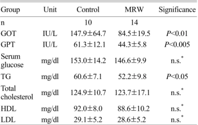
2. Serological examination
The GOT and GPT levels in the MRW group were 84.5
±19.5 IU/l and 44.3±5.8 IU/l, respectively, significantly lower than those of the control group, which were 147.9±
64.7 IU/l (P<0.01) and 62.3±12.1 IU/l (P<0.001), respec- tively. Glucose levels of the control and MRW groups were 153.0±14.2 mg/dl and 146.6±9.9 mg/dl, respectively, and this was not statistically significant. With regard to lipid parameters, the level of TG in the MRW group was 52.2±
9.8 mg/dl, which was significantly lower than that seen in the control group, 60.6±7.1 mg/dl (P<0.05; Table 2). Levels of other parameters, including total cholesterol, HDL and LDL, were not significantly different, as shown in Table 2.
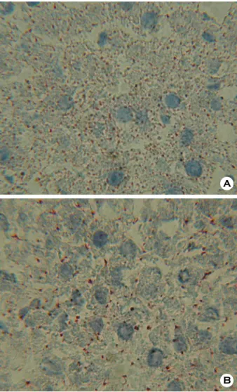
3. Histological changes in liver tissue
Lipid droplets in the liver parenchyma were stained by oil red O as seen in Fig. 4. In the control groups, the liver showed microvesicular steatosis of densely scattered dro- plets (Fig. 4A), In the MRW group, the liver showed macro- vesicular steatosis of relatively rare droplets compared with that of the control group (Fig. 4B).
DISCUSSION
In present study, we demonstrated for the first time that the MRW suppresses increases in body weight and liver fat deposition in SD rats fed on high-fat diet. At the beginning of present study, the body weights of the three-week-old SD rats in the control and MRW groups increased rapidly until day 35, because the rats were in their growing stage.
With the exception of day 28, the body weights of the rats Table 2. Change of serological parameters
Group Unit Control MRW Significance
n 10 14
GOT IU/L 147.9±64.7 84.5±19.5 P<0.01 GPT IU/L 61.3±12.1 44.3±5.8 P<0.005 Serum
glucose mg/dl 153.0±14.2 146.6±9.9 n.s.* TG mg/dl 60.6±7.1 52.2±9.8 P<0.05 Total
cholesterol mg/dl 124.9±10.7 123.7±17.1 n.s.*
HDL mg/dl 92.0±8.0 88.6±10.2 n.s.*
LDL mg/dl 29.1±5.2 28.6±5.2 n.s.*
* n.s.: not significant
Fig. 2. Change in body weight. Three week-old SD rats were fed on the control water (n=10) or MRW (n=10) for 83 days. Data was expressed as the means ± SEM. (*P<0.05)
Fig. 3. Changes in the weight of adipose tissues. On day 83, the weight of the IWAT and EDAT of SD rats were measured. Data are expressed as means ± SEM.
Fig. 1. ROS scavenging effect of MRW. 0 or 50% MRW was assayed in the HX/XOD system. The experiment was performed in triplicate, and the data was expressed as mean ± SEM.
in the MRW group between day 14 and day 63 were found to be significantly lower than those of the control group (Fig. 2). This difference in body weight was so marginal that, in the last two measurements, on days 77 and 83, the differences were not statistically significant. However, this result strongly suggests that the MRW can exert in vivo biological effects on the reduction of body weight of SD rats fed on the high-fat diet.
Microvesicular steatosis in the control group and macro- vesicular steatosis in the MRW group shows the effect of the MRW on suppression of obese progression. Physiopa- thology of microvesicular and macrovesicular steatosis can be summarized as followed (Fromenty et al., 1997; Fro- menty and Pessayre, 1995). Severe impairment of mito- chondrial fatty acid β-oxidation accumulates poorly oxidi-
zed nonesterified fatty acid (NEFA). Amphiphilic NEFA is supposed to form microvesicules by capsuling hepatic TG, resulting in microvesicular steatosis. Meanwhile, NEFA and its metabolites exert detrimental effect on mitochondrial function, resulting in reduction of local energy output and reduction of ketone body and glucose delivery to pheripheral tissues. As the result, progress of microvesicular steatosis in diverse organs including liver causes hepatic failure, coma and death. In the other hand, macrovesicular steatosis is mainly occurred in alcohol abuse, obesity, diabetes and some dyslipidemia, and is supposed to be resulted by in- creased mobilization of fat from adipose tissue, increased hepatic synthesis of fatty acid, increased esterification of fatty acid into TG and/or decreased egress of TG from the liver (Fromenty and Pessayre, 1995). Macrovesicular stea- tosis without other liver lesion is classified as benign con- dition.
However, continuous ingestion of high-fat diet increases incomplete β-oxidation in the mitochondria (Koves et al., 2005) and increased amount NEFA in the cells, resulting in microvesicular steatosis (Fig. 4). In the present study, the control group shows microvesicular hepatic steatosis with many small lipid droplets, which is compared with that of the MRW group showing macrovesicular hepatic steatosis with little large lipid droplets. This difference is supposed by that activated lipid metabolism in hepatic mitochondria by MRW administration is sufficient for esterification of the over-ingested lipid. However, it is to be elucidated how the MRW activates the lipid metabolism in the rats.
It is suggested as a clue that obesity is intensified by oxi- dative stress, which can be neutralized by administration of the MRW. Oxidative stress is increased in accumulated fat in obese subjects, which is one of the important cause of systemic oxidative stress (Furukawa et al., 2004). Adipose tissue under oxidative stress downregulates the expression of peroxisome proliferators-activated receptor (PPAR)-γ and resulting downregulation of adiponectin expression (Furu- kawa et al., 2004; Saltiel, 2001). Adiponectin is an adipo- cytokine expressed only in adipose tissue, and expected to suppress insulin resistance and dyslipidemia (Saltiel, 2001).
Authors had shown that the MRW scavenges ROS gene- rated by in vitro HX/XOD system, and reduces lung meta- stasis of B1BL6 melanoma cells in mouse model (Lee et al., 2003; Park et al., 2004). Huang et al. (2003) have repor- ted that electrolyzed reduced water that has similar charac- A
B
Fig. 4. Lipid deposition in liver. Five µm liver sections from the control (A) and MRW (B) groups were stained with hematoxylin and oil red O.
teristics in pH and ORP with MRW significantly reduced hemodialysis-induced oxidative stress of end-stage renal disease patients. These facts support that the reduction of oxidative stress in SD rats fed on high-fat diet by admini- stration of the MRW increases the expression of adiponectin that is effective on suppression of obesity.
The MRW is basic water with the pH of over 9.0. Ho- wever, its pH-retaining potential is so weak that it is readily neutralized by the addition of acidic agents. This ensures that administered MRW is quickly neutralized in the acidic environment of stomach. This explanation is supported by the fact that nine volumes of the MRW did not result in more than 0.1 unit increase in the pH of RPMI 1640 me- dium (pH 7.4) (unpublished data). Also, the administration of the MRW resulted in no increases in the serum levels of GOT and GPT, both of which are parameters for liver damage (Table 2). Although the levels of GOT and GPT were found to significantly decreased, we give less impor- tance on the results because the lowered values were within normal range. It is rather important that the liver was not found to have been significantly damaged after administra- tion of the MRW.
Among the serum lipid parameters examined in this study, the TG levels of the MRW group were significantly reduced.
TG is known to be related to obesity, steatosis and insulin resistance, and TG accumulation in the liver have been shown in many cases (Browning and Horton, 2004; Ma- rceau et al., 1999; Wanless and Lentz, 1990). In fatty indivi- duals, the robust activity of lipogenic enzymes is expected.
This tends to increase the biosynthesis of fatty acids in the liver, thereby stimulating the biosynthesis of TG, and in- creasing the concentration of TG in the blood (Coppack et al., 1994). Therefore, decrease in TG in this result implies that oral administration of the MRW downregulates lipid metabolism in the liver, resulting in the decrease of TG levels in the blood. This assumption is proven by the oil red O staining of the liver section. Red spots, oil red O-stained lipid droplets, in the control group were found to be very densely scattered around the hepatocytes, whereas those of the MRW group were fewer in the number (Fig. 4).
We had previously investigated on the effects of the ERW on the reduction of the body weights in OLETF rats, and found that the mean TG and total cholesterol levels in the sera were reduced in the ERW-fed rats (Kim et al., 2003). However, it is still remained to be elucidated what
MRW mechanism biologically affects on the suppression of steatosis. The chemical and electrochemical properties of the MRW are similar to those of the ERW (Fig. 1), thus the effect of the MRW can also be explained by the effects of the ERW. Actually, almost all of the investigations regar- ding the ERW have been conducted at the viewpoint of chemistry or electrochemistry. Only one clinical study has focused on reduction of oxidative stress of an end stage renal disease patients during hemodialysis (Huang et al., 2003). Hemodialysis by-products such as hydrogen pero- xide, hypochlorite (HOCl) and the resulting oxidized pro- ducts of lipids and proteins were significantly reduced by using ERW, resulting in less oxidative stress on the patients.
The authors have explained the result by noting that ERW exhibits activities of ROS-neutralizing enzyme such as superoxide dismutase and catalase against superoxide and hydrogen peroxide, which originate from the 'hydrogen atoms' generated by electrolysis. Although the 'hydrogen atom theory' has a matter of controversy, the ROS scaven- ging effects of the ERW are evident in the results of other investigations (Hanaoka, 2001; Shirahata et al., 1997).
Relatively little is known regarding the role of ROS in obesity. Fenster et al. (Fenster et al., 2002) have proposed a hypothesis that explains the relation of ROS and obesity as follows. Oxidative stress in obesity may result, in part, from the accumulation of intracellular TG, which has been pro- posed to elevate superoxide production in the electron tran- sport chain via the inhibition of the adenosine nucleotide transporter (Bakker et al., 2000). Inhibition of the adeno- sine nucleotide transporter results in the reduction of the levels of mitochondrial ADP, which is the proton acceptor in the ATP synthesis reaction (Fenster et al., 2002). As the result, electrons transported from the electron transport chain react with oxygen to form superoxides (Fenster et al., 2002).
In this study, rats were fed on high-fat diet for 83 days.
Excess calorie supplementation may serve to promote in- creased TG synthesis that is the causative agent of insulin resistance (Chen and Farese, 2004). However, the MRW administration resulted in a significant reduction in serum TG levels (Table 2), which were positively proportional to liver TG levels (Coppack et al., 1994). As the result, the reduction of TG in the sera may represent the reduction not only of insulin resistance and obesity but also of superoxide generation in the mitochondria. Reduction of body weight of the SD rats in the MRW group in this study was con-
sistent with the results of our previous study, in which signi- ficant reductions in body weights were observed in volun- teers to whom one thirty volume of ERW against their body weights was administered everyday (Lee et al., 2004). We have also reported that mean ROS levels in the livers of B16BL6 melanoma-metastased C57BL6 mice fed on the MRW were decreased compared with control mice (Lee, et al., 2003). In fatty individuals, the activity of superoxide dismutase decreased significantly, although the body weight of the rats did not change significantly (Beltowski et al., 2000).
In this study, we evaluated, for the first time, the effects of the MRW on obesity in SD rats fed on a high-fat diet.
The body weights of the rats decreased significantly, alth- ough not until the end of present study. Reduced levels of serum TG suggest further investigations on the effect of the MRW on fatty acid metabolism and obesity. Steatosis was greatly reduced in the rats of the MRW group. We expect that these results are attributable to uncover the ROS sca- venging properties of the MRW.
Acknowledgements
This work was supported in part by Yonsei University Research Fund of 2004.
REFERENCES
Bakker SJ, RG IJ, Teerlink T, Westerhoff HV, Gans RO, Heine RJ.
Cytosolic triglycerides and oxidative stress in central obesity:
the missing link between excessive atherosclerosis, endothe- lial dysfunction, and beta-cell failure? Atherosclerosis 2000.
148: 17-21.
Beltowski J, Wojcicka G, Gorny D, Marciniak A. The effect of dietary-induced obesity on lipid peroxidation, antioxidant enzymes and total plasma antioxidant capacity. J Physiol Pharmacol. 2000. 51: 883-896.
Browning JD, Horton JD. Molecular mediators of hepatic steatosis and liver injury. J Clin Invest. 2004. 114: 147-152.
Casteilla L, Rigoulet M, Penicaud L. Mitochondrial ROS metabo- lism: modulation by uncoupling proteins. IUBMB Life 2001.
52: 181-188.
Chen HC, Farese RV, Jr. Inhibition of Triglyceride Synthesis as a Treatment Strategy for Obesity. Lessons From DGAT1- Deficient Mice. Arterioscler Thromb Vasc Biol. 2004.
Coppack SW, Jensen MD, Miles JM. In vivo regulation of lipo-
lysis in humans. J Lipid Res. 1994. 35: 177-193.
Fenster CP, Weinsier RL, Darley-Usmar VM, Patel RP. Obesity, aerobic exercise, and vascular disease: the role of oxidant stress. Obes Res. 2002. 10: 964-968.
Fromenty B, Berson A, Pessayre D. Microvesicular steatosis and steatohepatitis: role of mitochondrial dysfunction and lipid peroxidation. J Hepatol. 1997. 26 Suppl 1: 13-22.
Fromenty B, Pessayre D. Inhibition of mitochondrial beta-oxidation as a mechanism of hepatotoxicity. Pharmacol Ther. 1995. 67:
101-154.
Furukawa S, Fujita T, Shimabukuro M, Iwaki M, Yamada Y, Nakajima Y, Nakayama O, Makishima M, Matsuda M, Shi- momura I. Increased oxidative stress in obesity and its impact on metabolic syndrome. J Clin Invest. 2004. 114: 1752-1761.
Hanaoka K. Antioxidant effects of reduced water produced by electrolysis of sodium chloride solution. J Appl Electrochem.
2001. 31: 1307-1313.
Huang KC, Yang CC, Lee KT, Chien CT. Reduced hemodialysis- induced oxidative stress in end-stage renal disease patients by electrolyzed reduced water. Kidney Int. 2003. 64: 704 -714.
Kikuchi K, Takeda H, Rabolt B, Okaya T, Ogumi Z, Saihara Y, Noguchi H. Hydrogen particles and supersaturation in alka- line water from an Alkali-Ion-Water electrolyzer. J Elec- troanal Chem. 2001. 506: 22-27.
Kim JW, Moon WW, Park SK, Kim DH, Deung YK, Kim SK, Kim HW, H. CC, Lee KJ, The reducing effect of alkaline reduced water on lipid components and glucose level in blood of OLETF rats. Autumn Meeting of the Korean Society of Med Biochem Mol Biol (Seoul) (2003).
Koves TR, Li P, An J, Akimoto T, Slentz D, Ilkayeva O, Dohm GL, Yan Z, Newgard CB, Muoio DM. PPARgamma coactivator- 1alpha -mediated metabolic remodeling of skeletal myocytes mimics exercise training and reverses lipid-induced mito- chondrial inefficiency. J Biol Chem. 2005.
Lee KJ, Kim JW, Kim GY, Ryang YS, Kim GH, Park SK, Cho HC, Jin D, Kim SK. Oral fed ARW (Alkaline Reduced Water) inhibits B16 melanoma metastasis and tumor growth in C57BL/6 mice. Kor J Physiol Pharmacol. 2003. 7: 76.
Lee KJ, Park SK, Sung JS, Kim DH, Deung YK, Kim MC, Yang EJ, Lim SJ, Ryang YS, Kim HW. Effect of electrolyzed- reduced water: in vivo and in vitro examination and clinical trials. The 3rd Asia Pacific Conference on Evidence-Based Medicine (Hong Kong) #93 (2004).
Marceau P, Biron S, Hould FS, Marceau S, Simard S, Thung SN,
Kral JG. Liver pathology and the metabolic syndrome X in severe obesity. J Clin Endocrinol Metab. 1999. 84: 1513-1517.
Park SK, Kim DH, Deung YK, Jin D, Yang EJ, Lim SJ, Ryang YS.
Studies on the safety of oral administration of alkaline-reduced water. The 13th Federation Meeting of Korean Basic Medical Scientists (Seoul) P118 (2005).
Park SK, Kim DH, Deung YK, Yang EJ, Ryang YS, Kim HW, Lee KJ. The change of pH and oxidation-reduction potential (ORP) of alkaline reduced water. The Newest Med J. 2004.
47: 24-30.
Park SK, Lee KJ, Yang EJ, Lim SJ, Ryang YS, Jin D, Kim HW, Deung YK, Kim DH. Effect of mineral-induced alkaline- reduced water: scavenging of reactive oxygen species, sup- pression of B16BL6 metastasis and decrease of blood glocose
concentration. International Symposium on Current Topics in Biomedicine (Seoul) M-15 (2004).
Saltiel AR. You are what you secrete. Nat Med. 2001. 7: 887-888.
Shirahata S, Kabayama S, Nakano M, Miura T, Kusumoto K, Gotoh M, Hayashi H, Otsubo K, Morisawa S, Katakura Y.
Electrolyzed-reduced water scavenges active oxygen species and protects DNA from oxidative damage. Biochem Biophys Res Commun. 1997. 234: 269-274.
Wanless IR, Lentz JS. Fatty liver hepatitis (steatohepatitis) and obesity: an autopsy study with analysis of risk factors. Hepa- tology 1990. 12: 1106-1110.
Watanabe T. Effect of alkaline ionized water on reproduction in gestational and lactational rats. J Toxicol Sci. 1995. 20: 135 -142.