Relationship of Foot Type to Callus Location in Healthy Subjects
Do-young Jung, M.Sc., P.T.
Dept. of Prosthetics and Orthotics, Suncheon First College Moon-hwan Kim, B.H.Sc., P.T.
Dept. of Rehabilitation Medicine, Wonju Christian Hospital, Wonju College of Medicine, Yonsei University In-su Chang, M.Sc., P.T., C.P.O.
Dept. of Prosthetics and Orthotics, Suncheon First College
Abstract
1)The purpose of this study was to determine whether a relationship existed between foot type and the location of plantar callus in healthy subjects. Twenty-five healthy subjects with plantar callus were re- cruited for this study. Foot deformities were classified according to the operational definitions as 1) a compensated forefoot varus, 2) an uncompensated forefoot varus or forefoot valgus, or 3) a compensated rearfoot varus. The location of plantar callus was divided into two regions. Fourteen of the 19 feet with compensated forefoot varus and six of the 9 feet showed plantar callus at the second, third or fourth metatarsal head. Five of the 6 feet with uncompensated forefoot varus and twenty of the 16 feet with forefoot valgus showed plantar callus at the first or fifth metatarsal head. A significant relationship was found between foot type and location of callus (p<.01). The results support the hypothesis that certain foot types are associated with characteristic patterns of pressure distribution and callus formation. We believe diabetic patients with insensitive feet and with the types of foot deformity should be fit with foot orthoses and footwears that accommodate their respective deformity in a position as near to the subtalar joint as possible with the goal of preventing plantar ulceration.
Key Words: Diabetic foot ulceration; Foot deformitiy; Foot orthosis; Plantar callus; Subtalar joint neutral position.
Introduction
Foot problems are a major cause of morbidity in people with diabetics and may result in lengthy peri- ods of hospitalisation and amputation (Bild et al, 1989). The factors that predispose to the develoment of diabetic foot ulcers include sensory, motor and autonomic nerve dysfunction and small and large vessel vascular disease (Reiber et al, 1999).
Biomechanical dysfunction resulting from the changes in foot posture and leading to abnormal weight bear- ing is also important especially in the presence of sensory neuropathy (Reiber et al, 1999). Plantar cal- lus is frequently observed in people with diabetes. It
is a result of hypertrophy of the startum corneum with excess keratinisation and indicates abnormal foot pressure (Pataky et al, 2002). Plantar callus is an early sign of potential future problems in people with diabetes who often have other factors which place the foot at an increased risk. The high dynam- ic vertical pressure and shear stress at the site of callus formation may ultimately result in ulcer for- mation (van Schie, 2005).
Root et al (1977) and Tiberio (1998) have de- scribed clinically observed patterns of plantar callus formations, which they attribute to increased plantar stresses characteristic of certain foot abnormalities.
These authors theorize that different types of foot
Corresponding author: Do-young Jung [email protected]
abnormalities cause specific patterns of weight bearing and subsequent stresses on the plantar surface of the foot, resulting in identifiable patterns of callus formation.
Forefoot varus deformity has been defined as a midtarsal joint abnormality in which the forefoot is in an inverted position in relation to the bisector of the posterior aspect of the calcaneus when the sub- talar joint (STJ) is in a neutral position (Root et al, 1977; Thomas, 1997; Tiberio, 1998) These authors indicate that, for the medial aspect of the forefoot to make contact with the ground during stance, com- pensation must occur at the triplanar STJ axis.
Subtalar joint pronation permits the medial forefoot to bear weight. This condition has been referred to as compensated forefoot varus (Root et al, 1977;
Thomas, 1997; Tiberio, 1998). The characteristics of this type of foot abnormality are a flattened arch, a prominent navicular tuberosity, and an everted calca- neus; this condition is commonly called "flatfoot." In the presence of a compensated forefoot varus, some authors speculate that the peroneus longus muscle is unable to stabilize the first ray (first metatarsal and medial cuneiform) and the first ray becomes hypermobile. During gait, the compensatory pronation at the STJ causes increased medial foot weight bearing, A transfer of increased medial weight bear- ing to the second or third metatarsal heads is be- lieved to occur because the first metatarsal is hypermobile. Thomas (1997) and Tiberio (1998) in- dicate that keratomas will develop under the central metatarsal heads as a result of increased loading and abnormal shear over an excessive period of time. A different weight-bearing pattern will occur if the STJ has inadequate range of motion and is unable to compensate for the forefoot varus deformity.
Uncompensated forefoot varus occurs if the STJ is unable to pronate enough to bring the medial forefoot into full contact with the ground. In an un- compensated forefoot varus, primary weight-bearing forces remain lateral, placing excessive stresses on the lateral side of the foot. It has been suggested that limited STJ eversion associated with callus un-
der the fifth metatarsal head is a good clinical in- dicator of this uncompensated state (Root et al, 1977;
Thomas, 1997; Tiberio ,1998).
Forefoot valgus deformity has been defined as a midtarsal joint abnormality in which the forefoot is in an everted position in relation to the bisector of the posterior calcaneus when the STJ is in a neutral position. With this foot abnormality, the medial as- pect of the forefoot (particularly the first metatarsal head) will load before the lateral forefoot, resulting in supinatory compensation at the STJ and a lateral shift of weight-bearing forces to the fifth metatarsal head (Root et al, 1977; Thomas, 1997; Tiberio, 1998).
This condition is in contrast to forefoot varus in which the lateral aspect of the forefoot (particularly the fifth metatarsal head) will load before the medial forefoot, resulting in a pronatory compensation as the STJ and a medial shift of weight-bearing forces.
The weight-bearing patterns of a forefoot valgus will be somewhat different, depending on whether the forefoot valgus is rigid, flexible, or accompanying a plantar-flexed first ray. If the forefoot valgus is rigid or occurs in conjunction with a plantar-flexed first ray, callus formation would be expected under the first of fifth metatarsal head. The pattern of cal- lus formation observed with a flexible forefoot valgus is variable, dependent on the amount of pronatory compensation available at the STJ (Root et al, 1977;
Thomas, 1997; Tiberio, 1998).
These abnormalities may allow increased, repetitive pressure in the previously described patterns. If these patterns exist, a higher incidence of ulceration would be expected in the areas where pressure is theoret- ically increased. The theory of callus formation spe- cific to certain foot types is based on the clinical observations of several authors. Therefore, the pur- pose of this study was to determine whether there was a relationship between the foot type and the lo- cation of callus in healthy subjects.
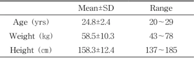
Mean±SD Range
Age (yrs) 24.8±2.4 20~29
Weight (㎏) 58.5±10.3 43~78
Height (㎝) 158.3±12.4 137~185
Table 1. General characteristics of the subjects (N=25)
Methods
Subjects
Twenty five healthy subjects with plantar callus were recruited for the study. None of the subjects suffered from any complaints of the musculoskeletal system and none had a history of foot trauma or previous foot surgery. The age, height, and weight of the subjects are summarized in Table 1.
Procedures
All measurements of STJ ROM, forefoot-to-rearfoot relationship were taken by the same two physical therapists. In order to assess the interrater reliability, the subjects were first randomly tested by one of the two testers and then by the other tester. To assess the intrarater reliability, each subject was measured three times in a random order by one of the testers.
Each evaluation took about 15 minutes to complete.
Testers were placed in separate rooms when the measurements were taken. Both examiner underwent a lengthy training period in order to conduct the evaluation. Training sessions consisted of the evalua- tion of patients by each therapist independent of the other. Each patient's measurements were discussed and remeasured jointly if discrepancies existed.
A standard biomechanical examination procedure described by Elveru et al (1988) was used.
Goniometric measurements were taken with gravity goniometer and plastic goniometer with 6-inch arms and the scale marked in 2 degree increments. Briefly, patients were positioned prone with the leg not being examined flexed at the knee, laterally rotated, and abducted at the hip. The lower third of the tested leg and the calcaneus were bisected. For passive calcaneal inversion and eversion, the calcaneus was
secured with one hand, maximally invered or everted, and measured from anatomical zero. The goniometer arms were aligned with the bisection of the lower third of the leg. The palpated subtalar joint neutral (STJN) is the position of inversion or eversion of which the calcaneus assumes, in relationship to the tibia, when the talus position is congruent between palpating fingers. The foot is passively pronated and supinated until the medial and lateral sides of the talar head are neither protruding nor depressed. The fourth and fifth metatarsal heads are then passively dorsiflexed until soft end-feel was encountered, lock- ing the midtarsal joints, and the calcaneal position is then measured as described previously. To determine the FF/RF relationship, first the STJN position is located. The goniometer is then placed with one arm parallel to the plane of the metatarsal heads and the other arm aligned perpendicular to the bisection of the calcaneus. The subject was then asked to stand, and calcaneal stance and tibial varum were measured by using gravity goniometer aligning one arm of the goniometer with the calcaneal bisect and the other arm parallel with the floor. All marks on the leg were then washed off, and the subject was measured by the next examiner.
Foot deformities were classified according to the operational definitions as 1) a compensated forefoot varus, 2) an uncompensated forefoot varus or forefoot valgus, or 3) a compensated rearfoot varus. The de- formity was classified as a compensated forefoot varus if the foot demonstrated a forefoot varus de- formity of at least 2 degrees and a greater amount of STJ eversion than the forefoot varus deformity. The deformity was classified as an uncompensated fore- foot varus or forefoot valgus if the foot demonstrated less STJ eversion than the amount of forefoot varus or at least 2 degrees of a rigid forefoot valgus.
Data Analysis
The intrarater and interrater reliability were as- sessed using intrarater and interrater intraclass cor- relation coefficients (ICCs) for all goniometric
Measurement Interrater Intrarater
Left Right Left Right
Calcaneal inversion .907 .916 .867 .897
Calcaneal eversion .930 .945 .751 .841
Tibial varum .973 .955 .832 .776
Calcaneal stance .948 .909 .634 .636
Table 2. Interrater and intrarater intraclass correlation coefficents (ICCs) in four goniometric measurements (N=25)
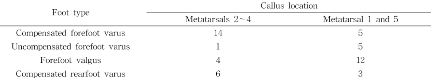
Foot type Callus location
Metatarsals 2~4 Metatarsal 1 and 5
Compensated forefoot varus 14 5
Uncompensated forefoot varus 1 5
Forefoot valgus 4 12
Compensated rearfoot varus 6 3
aSignificant relationship between foot type and callus location by chi-square test of association (χ2=11.809, df=2, p<.01).
Table 3. Relationship of foot type to callusa (N=25) measurements. The Pearson chi-square test of asso-
ciation was used to determine whether a relationship existed between foot deformity and the location of callus. The alpha level of significance for all com- parisons was set at .05.
Results
The results of the interrater and intrarater ICCs for goniometric measurements are summarized in Table 2.
Intrarater reliability coefficients ranged from .63 to .89, and interrater reliability coefficients ranged from .91 to .97. Fourteen of the 19 feet with compensated forefoot varus and six of the 9 feet showed plantar callus at the second, third or fourth metatarsal head (Figure 1).
Five of the 6 feet with uncompensated forefoot varus and twenty of the 16 feet with forefoot valgus showed plantar callus at the first or fifth metatarsal head (Figure 2). These results indicated a significant relationship between the foot type and the location of callus (χ2=11.809, df=2, p<.01).
Discussion
Measurements of the passive range of motion of the subtalar joint, relaxed calcaneal stance position, and the tibial varum are often part of a physical therapy evaluation. These measurements may be used in treatment planning, such as in the pre- scription of specialized shoes or foot orthoses.
Therefore, the reliability of these measurements, as they are obtained clinically, must be determined (Somers et al, 1997), Various authors have published report about reliability of the biomechanical foot evaluation (Lohmann et al, 1987; Mcpoil et al, 1988;
Picciano et al, 1993). Picciano et al (1993) reported that when performed by inexperienced testers, no re- liability of open and closed chain subtalar joint neu- tral positions measurement was established.
Therefore, clinicians should practice these measure- ment techniques. Mcpoil et al (1988) found high in- trarater reliability (ICC=.90~.96) for three techniques used to measure a condition commonly referred to as
"tibia varum" which the authors state is more accu- rately termed "tibiofibular varum". No interrater reli- ability was reported. Lohmann et al (1987) analyzed
Figure 1. Location of callus at second and third metatarsal head in subject with compensated forefoot varus.
Figure 2. Location of callus at first and fifth metatarsal head in subject with forefoot valgus.
tibial varum measurements obtained by two raters while healthy subjects assumed three different positions. Intrarater reliability coefficients by position were .46 and .83 for each of the raters, and inter- rater reliability coefficients by position were .41, .49, and .58. Our results also showed that intrarater reli- ability coefficients range from .63 to .89, and inter- rater reliability coefficients ranged from .91 to .97.
We attempted to minimize our measurement varia- bility by undergoing extensive training in the meas- ures being used.
In addition, our research results indicated that a significant statistical relationship existed between foot classifications and location of the plantar callus. All of the subjects classified with forefoot and rearfoot deformity demonstrated callus under the metatarsal heads. Location of the plantar callus was divided into two regions. Fourteen of 19 feet with a compensated forefoot varus and six of 9 feet showed plantar cal- lus at the second, third or fourth metatarsal head (Figure 1). Five of 6 feet with a uncompensated forefoot varus and twenty of 16 feet with a forefoot valgus showed plantar callus at the first or fifth metatarsal head (Figure 2). The callus that were lo- cated inconsistent with the hypotheses in this study seemed to occur in feet with abnormal other struc- ture such as tibia, knee, and hip which can influence weight-bearing forces on the plantar aspect of the foot. As noted previously, the number of foot classi-
fications evaluated in this study was limited. The classifications chosen were those most definable and most representative of the crux of the hypotheses.
Further research is needed to identify other relation- ships between type of deformity and location of in- creased plantar pressure points. This study did not actually measure increased pressure. Increased pres- sure was inferred by the location of the callus.
Although studies have been conducted to assess pressure at various subjects foot, further research is needed to quantify the pressure distribution as it re- lated to foot type or available foot and ankle range of motion during gait. In addition, deformities were not quantified beyond classification as one of four foot deformity types. The severity of each deformity was not considered in establishing a relationship.
Additional research is required to quantify the degree of deformity and the relationship to callus formation.
These results have several implications for the treatment of the diabetic foot. In general, foot abnor- malities and deformities should be accommodated with the foot positioned as close to the STJ neutral positioned as possible. One reason total-contact cast- ing may be successful in healing plantar ulcers is that loads are spread over an increased area and the foot is immobilized in a position as close to STJ neutral as possible. Proper footwear for an in- sensitive foot often requires an insole using a com- bination of several materials. Firmer material can be
used to improve the alignment of a forefoot varus or valgus, but the firm material must be covered with softer materials, as described by Bowker and Pfeifer (2001) and Lockard (1987). Footwear and orthotic rec- ommendations according to foot deformity, sensitivity, and history of ulceration have been proposed to assist the clinician in prescription of appropriate footwear.
Additional research is required to determine the ef- fects of exercise and various types of footwear on plantar pressures so that optimal treatment strategies can be devised to treat and prevent foot problem.
Conclusion
A significant relationship between foot classification and the location of callus was found in twenty five healthy subjects. The results of this study support the hypothesis that certain foot types are associated with characteristic patterns of pressure distribution and callus formation. We believe that the diabetic pa- tients with insensitive feet and with type of foot de- formity should be fit with foot orthoses and foot- wears that accommodate their respective deformity in a positoin as near to the STJ neutral as possible with the goal of preventing plantar ulceration.
References
Bild DE, Selby JV, Sinnock P, et al. Lower-ex- tremity amputation in people with diabetes.
Epidemiology and prevention. Diabetes Care.
1989;12(1):24-31.
Boulton AJ, Hardist0y CA, Betts RP, et al. Dynamic foot pressure and other studies as diagnostic and management aids in diabetic neuropathy.
Diabetes Care. 1983;6(1):26-33.
Bowker JH, Pfeifer MA. Levin and O'Neal's The Diabetic Foot. 6th ed. Mosby, 2001.
Elveru RA, Rothstein JM, Lamb RL, et al. Methods for taking subtalar joint measurements. A clin-
ical report. Phys Ther. 1988;68(5):678-682.
Lockard MA. Foot orthoses. Phys Ther.
1987;68(12):1866-1873.
Lohmann KN, Rayhel HE, Schneiderwind WP, et al.
Static measurement of tibia vara. Reliability and effect of lower extremity position. Phys Ther.
1987;67(2):196-202.
McPoil TG, Schuit DKnecht HG. A comparison of three positions used to evaluate tibial varum. J Am Podiatr Med Assoc. 1988;78(1):22-28.
Mueller MJ, Diamond JE, Delitto A, et al.
Insensitivity, limited joint mobility, and plantar ulcers in patients with diabetes mellitus. Phys Ther. 1989;69(6):459-462.
Pataky Z, Golay A, Faravel L, et al. The impact of callosities on the magnitude and duration of plantar pressure in patients with diabetes mellitus. A callus may cause 18,600 kilograms of excess plantar pressure per day. Diabetes Metab. 2002;28(5):356-361.
Picciano AM, Rowlands MS, Worrell T. Reliability of open and closed kinetic chain subtalar joint neutral positions and navicular drop test. J Orthop Sports Phys Ther. 1993;18(4):553-558.
Reiber GE, Vileikyte L, Boyko EJ, et al. Causal pathways for incident lower-extremity ulcers in patients with diabetes from two settings.
Diabetes Care. 1999;22(1):157-162.
Root ML, Orien WP, Weed JH. Clinical Biomechanics: Normal and abnormal function of the foot. LA, Clinical Biomechanics Co., 1977.
Sinacore DR, Mueller MJ, Diamond JE, et al.
Diabetic plantar ulcers treated by total contact casting. A clinical report. Phys Ther.
1987;67(10):1543-1549.
Somers DL, Hanson JA, Kedzierski CM, et al. The influence of experience on the reliability of go- niometric and visual measurement of forefoot position. J Orthop Sports Phys Ther.
1997;25(3):192-202.
Thomas C. Foot Orthoses and Other Forms of Conservative Foot Care. Lippincott, Williams &
This article was received October 4, 2006, and was accepted October 30, 2006.
Wilkins, 1997.
Tiberio D. Pathomechanics of structural foot deformities. Phys Ther. 1988;68(12):1840-1849.
van Schie CH. A review of the biomechanics of the diabetic foot. Int J Low Extrem Wounds.
2005;4(3):160-170.