Inter-Rater Reliability of Abdominal Muscles Thickness Using Ultrasonography for Different Probe Locations
and Thickness Measurement Techniques
One-bin Lim, B.H.Sc., P.T.
Ji-a Hong, B.H.Sc., P.T.
Dept. of Physical Therapy, The Graduate School, Yonsei University
Chung-hwi Yi, Ph.D., P.T.
Heon-seock Cynn, Ph.D., P.T.
Dept. of Physical Therapy, College of Health Science, Yonsei University
Dept. of Ergonomic Therapy, The Graduate School of Health and Environment, Yonsei University
Doh-heon Jung, B.H.Sc., P.T.
Il-woo Park, B.H.Sc., P.T.
Dept. of Physical Therapy, The Graduate School, Yonsei University
Abstract 22)
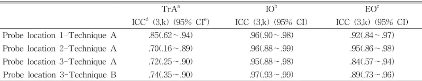
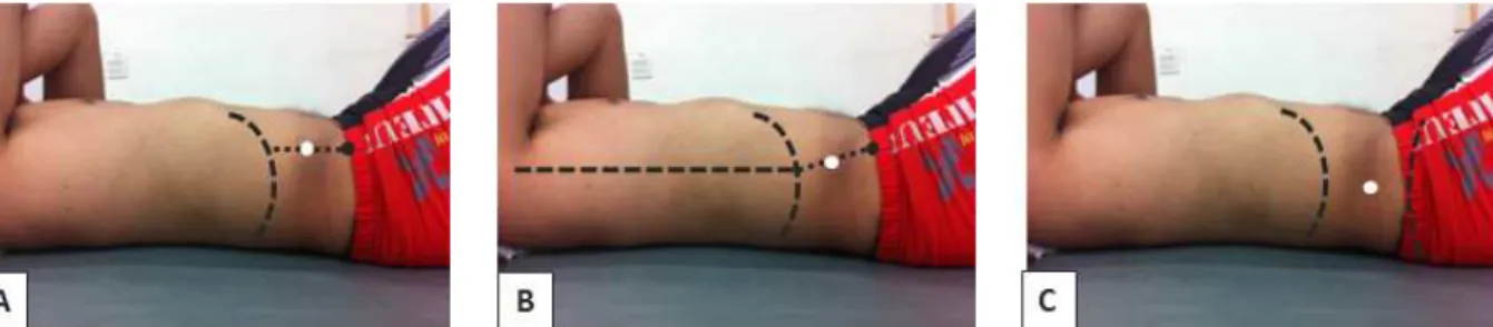
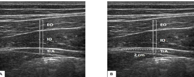
Ultrasonography (US) is a recent technique that has proven to be useful for assessing muscle thickness and guiding the rehabilitation decision-making of clinicians and researchers. The purpose of this study was to determine the inter-rater reliability of the US measurement of transversus abdominis (TrA), internal oblique (IO), and external oblique (EO) thicknesses for different probe locations and measurement techniques. Twenty healthy volunteers were recruited in this study. Muscle thicknesses of the transversus TrA, IO, and EO were measured three times in the hook-lying position. The three different probe locations were as follows: 1) Probe location 1 (PL1) was below the rib cage in direct vertical alignment with the anterior superior iliac spine (ASIS). 2) Probe location 2 (PL2) was halfway between the ASIS and the ribcage along the mid-axillary line. 3) Probe location 3 (PL3) was halfway between the iliac crest and the inferior angle of the rib cage, with adjustment to ensure the medial edge of the TrA. The two different techniques of thickness measurement from the captured images were as follows: 1) Muscle thickness was measured in the middle of the muscle belly, which was centered within the captured image (technique A; TA). 2) Muscle thickness was measured along a horizontal reference line located 2 ㎝ apart from the medial edge of the TrA in the captured image (technique B; TB). The intraclass correlation coefficient (ICC [3,k]) was used to calculate the inter-rater reliability of the thickness measurement of TrA, IO and EO using the values from both the first and second examiner. In all three muscles, moderate to excellent reliability was found for all conditions (probe locations and measurement techniques) (ICC=.70~.97). In the PL1-TA condition, inter-rater reliability in the three muscle thicknesses was good to excellent (ICC=.85~.96). The reliability of all measurement conditions was excellent in IO (ICC=.95~.97).
Therefore, the findings of this study suggest that TA can be applied to PL1 by clinicians and researchers in order to measure the thickness of abdominal muscles.
Key Words: External oblique; Internal oblique; Inter-rater reliability; Muscle thickness;
Transversus abdominis; Ultrasonography.
Introduction
Many clinicians and researchers have used ultra-
sonography (US) for therapeutic intervention aimed at
improving neuromuscular function, and clinical re-
habilitative research to inform clinical practice (Costa et
Corresponding author: Chung-hwi Yi [email protected]
al, 2009; Teyhen, 2006; Whittaker et al, 2007). Recently, researchers have increasingly studied associations be- tween underlying neuromuscular control deficits and neuromusculoskeletal disorders such as low back pain (LBP) (Ferreira et al, 2007; Hodges and Moseley, 2003;
Teyhen, 2006; Teyhen, 2007; Whittaker et al, 2007).
However, valid and reliable non-invasive measurement methods to assess neuromuscular control deficits that could be employed in a clinical setting have been in- sufficient (Teyhen, 2011). Evidence supporting the use of US as a method to assist patients with neuro- musculoskeletal disorders is growing (Teyhen, 2011).
Although US is less sophisticated in terms of res- olution than computed tomography (CT) and mag- netic resonance imaging (MRI), it has advantages as a non-invasive, inexpensive, portable, safe, and clin- ically accessible method for gathering data about the static characteristics of muscle and muscle behavior during dynamic activities (Hides et al, 2001; Kiesel et al, 2007; Koppenhaver et al, 2009; Teyhen, 2011;
Whittaker et al, 2007). As such, it shows promise as a measurement in musculoskeletal examination, eval- uation, and intervention. Furthermore, in contrast with CT, US does not expose the patient to ionizing radiation and is well tolerated by patients (Whittaker et al, 2007). A characteristic unique to US is its dy- namic capability to scan in real time, which makes it superior to CT and MRI for evaluating movable structures such as muscles, nerves, and tendons, and it may become an important tool for directing suit- able clinical decision-making. Still, US is not without weaknesses, and is very examiner dependent.
Possibly the most promising characteristics of US is its feasibility and accessibility, as it is easy for clinicians to acquire the skills needed to incorporate its use into clinical practice. Nevertheless, before widespread routine clinical use can be promoted, the evidence of validity and reliability for the use of US in different applications within rehabilitation is need- ed (Whittaker et al, 2007).
US is useful for evaluating the function of deep abdominal muscles (Critchley and Coutts, 2002; Henry
and Westervelt, 2005; Rankin et al, 2006). The trans- versus abdominis (TrA), internal oblique (IO), and external oblique (EO) are deep segmental muscles re- sponsible for lumbar stability (Rankin et al, 2006). It is not possible to take direct force measurements to compare the strength of the abdominal muscles, but their thicknesses may provide an indirect measure- ment of force-generating capacity (Rankin et al, 2006). US has been used to measure abdominal mus- cle thickness in respiratory and biomechanical re- search (Krag et al, 1987; McGill et al, 1996).
Studies on the measurement of abdominal muscle thickness have used different probe locations and measurement techniques (Ferreira et al, 2004;
Koppenhaver et al, 2009; Rankin et al, 2006;
Springer et al, 2006; Teyhen et al, 2011). Rankin et al (2006) introduced two different probe locations:
One probe was located immediately below the rib cage in direct vertical alignment with the anterior superior iliac spine (ASIS), whereas the other probe was located halfway between the ASIS and the rib- cage along the mid-axillary line. Ferreira et al (2004) located a probe halfway between the iliac crest and the inferior angle of the rib cage, and then adjusted it to ensure the medial edge of the TrA. Springer et al (2006) and Teyhen et al (2011) measured the thickness of the abdominal muscles at the middle of the muscle belly, centered within the captured image. Ferreira et al (2004) determined the thickness of the abdominal muscles along a hori- zontal reference line located 2 ㎝ apart from the medial edge of the TrA.
Although studies have been conducted on ab-
dominal muscle thickness measurement, however,
the inter-rater reliability of US measurements of
TrA, IO, and EO thicknesses using different probe
locations and measurement techniques have not
been reported in the literature. Therefore, the pur-
pose of this study was to compare the inter-rater
reliability of the US measurement of TrA, IO, and
EO thicknesses for different probe locations and
measurement techniques.
Characteristics Mean±SD
Age (yrs) 21.2±2.4
Height (㎝) 167.2±8.0
Weight (㎏) 58.0±9.4
BMI
a(㎏/㎡) 20.6±2.1
a
Body Mass Index.
Table 1. General characteristics of subjects (N=20)
TrA
aIO
bEO
cICC
d(3,k) (95% CI
e) ICC (3,k) (95% CI) ICC (3,k) (95% CI) Probe location 1-Technique A .85(.62~.94) .96(.90~.98) .92(.84~.97) Probe location 2-Technique A .70(.16~.89) .96(.88~.99) .95(.86~.98) Probe location 3-Technique A .72(.25~.90) .95(.88~.98) .84(.57~.94) Probe location 3-Technique B .74(.35~.90) .97(.93~.99) .89(.73~.96)
a