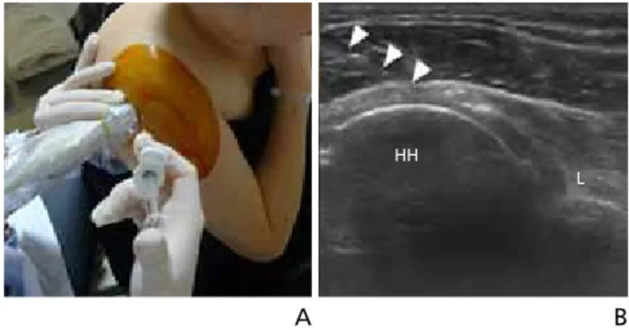
견관절의 초음파검사 방법
전체 글
수치
관련 문서
The steps in diffusion bonding: (a) Initially the contact area is small; (b) application of pressure deforms the surface, increasing the bonded area; (c) grain boundary
Three small identical spheres A, B, and C, which can slide on a horizontal, frictionless surface, are attached to three 200-mm-long strings, which are tied to a ring G. Which
Two matrices A=[a jk ] and B=[b jk ] are equal, written A=B, if and only if they have the same size and the corresponding entries are equal, that is, a 11 =b 11 , a 12 =b 12 ,
(A) Before repair of RCT shows fatty degeneration of supraspinatus (stage 2) and infraspinatus (stage 3) muscle and atrophy (Grade 2) (B) After 18months later
special space lattice- rotation axis and mirror plane - restriction on the cell parameters. ex) 4 z -fold rotation axis- a=b, γ=90 o - simplifications in
(b) Assuming that the allowable load, found in part a, is applied as shown at a point 0.75 in. from the geometric axis of he column, determine the horizontal deflection of the
Figure 12.3.1a shows a simply-supported, linearly-elastic, simple beam with a transverse load Q applied at its midspan and a compressive load P directed along its centroidal
Results: The results demonstrated a significant positive correlation between the muscle atrophy and the tendon tear.(p<0.05) And there was statistically
