I. 서론
치주치료의 궁극적 목적은 치주질환에 의해 상실 된 치주조직의 구조적 ,기능적 재생이라고 할 수 있 다. 이를 위해 골재생 혹은 새로운 부착재생에 대한 연구가 사람과 동물에서 이루어졌으며, 여러 재료와 수술방법들이 이용되어져 왔는데1)2)3)4)5), 차단막의 사용6)7), 골 이식술8), 치근처치9)등이 이용되고 있으 며 그 외에 실험적으로 성장인자10)11)12)를 적용시키 는 방법이 연구되어져 오고 있다.
골 이식술에는 자가골 이식, 동종골 이식, 이종골 이식, 합성골 이식이 사용되고 있다. 이러한 이식재 의 이상적인 조건은 골 형성 및 백악질 형성의 유도 능력이 있어야 하고 염증 반응이 없으며 빠른 혈관 형성이 이루어지고 숙주조직에 대한 친화성이 있어 야 하며 사용과 구입이 용이하면서도 경제적이어야 한다13). 이 중 자가골 이식은 수 년간 치조골 결손 부 위 사용시 임상적으로 성공적인 결과를 나타내었으 나, 이차적인 수술 부위의 필요성, 대량 공급의 어려 움, 골 유착 유발, 치근 흡수등의 제한점이 있다14). 따라서 이를 대신할 동종골 이식에 대한 연구가 이 루어졌으나 흡수시기, 재료 자체의 염증반응, 숙주의
거부 반응, 질병 전이 등의 가능성이 제시되었다14). 또한 치유속도가 일정하지 않으며 제조공정상의 문 제로 결과에 있어서 다양한 변이가 보고되고 있다는 단점이 있다.
한편 이종골 이식재는 양에 제한은 없으나 면역반 응과 조직거부의 위험이 여전히 있으며 골형성 유도 능력이 아주 낮은 단점이 있어서 많이 사용되지 않 는다.
따라서, 이런 여러 이식재들의 단점을 보완하기 위 해 합성골에 대한 관심이 증가되었는데15)16)17)18), Plaster of Paris, Polymers, Calcium carbonate, Ceramics 등이 있다. 이들 중 Ceramics는 골과 동일 한 탄성계수를 가지고 있으며, 골과 결합조직에 결합 하는 능력 때문에 치주치료와 치조골 증대술에서의 사용이 증가되어 왔다19)20)21)22). 가장 연구가 많이 진 행된 ceramics로는 Hydroxyapatite(HA)와 tricalcium phosphate23)가 있다.
최근에는, 정형외과, 성형외과 등에서 사용되어 온 bioactive glass가 치과 분야에서의 응용을 위해 연구 되어지고 있다. Bioactive glass는 입자크기는 nar- row size range(300-355μm)이며 SiO2, CaO, Na2O, P2O5로 이루어진 bioactive ceramics로 치주 골내 결
Bioactive glass의 장기 임상적 평가
이항빈·백정원·김창성·최성호·조규성·김종관·채중규 연세대학교 치과대학 치주과학교실, 치주조직재생연구소
대한치주과학회지 : Vol. 32, No. 1, 2002
*이 논문은 1998년도 연세대학교 학술연구비로 이루어졌음
교신 저자: 채중규, 서울특별시 서대문구 신촌동 134 연세대학교 치과대학 치주과학교실, 우편번호: 120-752
손을 치유하는 골대체물로 가능성을 보이고 있다.
Schepers 등24)25)26)27)28)에 의한 임상적, 조직학적, 방 사선학적 분석에 의하면 bioactive glass는 보호낭을 형성하여 이미 존재하는 골과 상관없이 중앙부에 새 로운 골소주 성장을 나타내는 것으로 보고되었다.
이러한 효과를 골조직 형성 자극능이라고 하는데 Bioactive glass 입자들 사이의 공극으로 미분화 간엽 세포의 조골세포로의 분화가 유도되어 골치유를 위 한 핵 부위로 작용하는 것으로, 이는 narrow size가 갖는 새로운 골조직 형성 자극능력에 의한 것이다.
또한 이 입자 크기는 새로운 혈액 공급을 위한 최상 의 공극을 형성해 준다. 그리고 임상적으로 bioactive glass 입자는 다루기 쉬우며 접촉시 응집력있는 종괴 를 이루어 결손부 내에 충전이 용이하고 출혈시 부 동되지 않으며 출혈을 감소시키는 지혈능도 가지고 있는 것으로 보인다. 또한 Wilson 등29)30)31)32)의 여러 동물 및 임상 실험 연구에 의하면 세포가 bioactive glass 표면에서 어떠한 미만성 염증없이 살 수 있으 며 배양내 세포 분열과 성장을 방해하지 않는 생체 친화적인 재료이다.
현재까지 Bioactive glass를 골내낭에 사용한 임상 증례 보고와 효과에 대한 조절된 연구는 있었지만, 장 기간에 걸친 영향을 평가한 연구는 보고된 바 없다.
이 연구의 목적은 치주 골 결손부위에서 bioactive glass(Biogran®)를 이식재로 재생술식을 시행한 후 1) 치주낭 깊이, 임상부착수준, 골탐침깊이의 변화를 측정하여 bioactive glass (Biogran®)의 장기간(36개 월 이상)의 치료 반응을 임상적으로 평가하고 2) 3개
월마다 치태조절한 군과 치태조절하지 않은 군으로 나누어서 치태조절이 장기간의 치료 반응에 미치는 영향을 알아보는 것이다.
II. 연구 대상 및 연구 방법
1. 연구 대상연세대학교 치과병원 치주과에 내원하여 성인성 치주염으로 진단받은 환자중 11명 환자의 17개의 골 내낭을 이용하며 74개의 부위를 얻었다. 연령분포는 34세에서 68세까지였고 평균나이는 51.6세였다. 남 자가 6명, 여자가 5명이었다. 연구 대상자들은 특이 한 전신적 질환이 없었고 최근 12개월내에 약물투여 를 받고 있지 않았다. 대상치아는 소구치와 대구치 로 골결손부위는 골이식 재생 술식이 적당하다고 판 단한 2벽 혹은 3벽 혹은 환상모양이었다. 구강위생 교육과 치석제거술 후 치주낭 탐침 깊이가 5mm이상 이고 수술시 골내 치주낭 깊이가 3mm이상인 치조골 결손부를 가진 치아를 선택하였다.
2. 연구방법
1) 실험군 설정
술 후 36개월 동안 3개월 간격으로 주기적 내원하 여 치태조절한 환자를 유지관리군(Follow-up group - F/U)으로 주기적 내원하지 않은 환자를 비유지관 리군(Non Follow-up group - Non F/U)으로 하였다.
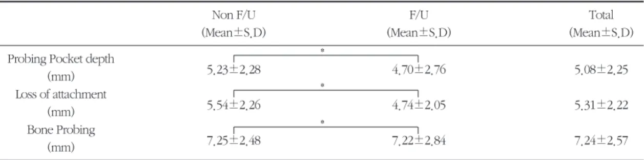
Table 1. Pre-treatment clinical parameters
Non F/U F/U Total
(Mean±S.D) (Mean±S.D) (Mean±S.D)
Probing Pocket depth
5.23±2.28 4.70±2.76 5.08±2.25
(mm) Loss of attachment
5.54±2.26 4.74±2.05 5.31±2.22
(mm) Bone Probing
7.25±2.48 7.22±2.84 7.24±2.57
(mm) S.D: standard deviation
* : no significant difference between F/U and Non F/U groups : P<0.05
*
*
*
2) 임상검사
모든 대상 환자에 대해 치석제거술과 구강 위생 교 육 후 치주낭 깊이, 임상부착수준, 골탐침 깊이를 측 정하였다(Table 1).
치주낭 깊이 : 치은연에서 치주낭저까지의 탐침깊이 임상부착수준 : 법랑-백악경계에서 탐침되는 치주 낭저까지의 거리
골탐침깊이 : CEJ에서 탐침으로 인지할 수 있는 치 조골까지의 거리
골결손의 골충전을 평가하기 위해 골탐침을 시행 하였다37).
수술 전에 측정한 치주낭 깊이, 임상부착수준, 골 탐침깊이는 두 군 간 통계학적으로 유의한 차이 없 이 비슷한 양상을 보였다 (Table 1).
치석 제거술과 구강위생 교육을 하고 재평가 후에 골 이식 수술을 하였다. 치은열구 절개 후 전층 판막 을 거상하여 염증 조직을 제거하고 철저한 치근 활 택술을 시행한 후 골 결손 부위를 Biogran®으로 이식 하였고 판막은 재위치시키거나 약간 치관측으로 위 치시켰다.
수술 후 유지관리군은 3개월 마다 주기적으로 내 원하여 치태조절을 시행하였으며 주기적으로 내원 하지 않은 환자들은 비유지관리군으로 분류하여 술
후 36개월 후에 다시 내원시켰다.
수술 후 36개월 째에 다시 임상검사를 시행하였으 며 치주낭 깊이, 임상부착수준, 골탐침 깊이를 측정 하였다.
3) 통계처리
유지관리군과 비유지관리군에서 술 전과 술 후 36 개월 째의 임상지수를 비교하기 위해 paired t-test를 시행하였고 유지관리군과 비유지관리군 간 비교를 위해 unpaired t-test를 시행하였다.
III. 연구성적
1. 육안적 소견유지관리군과 비유지관리군 모두 술 후 1주에 봉 합사를 제거하였으며 모두 염증 소견없이 정상적인 치유상태를 보였다. 치유 기간 내내 뚜렷한 치은 염 증 소견은 보이지 않았다(Fig. 1).
2. 임상지수 평가
11명 환자의 17개의 결손에서 74개의 측정 부위를 제공 하였다.
a
d
b
e
c
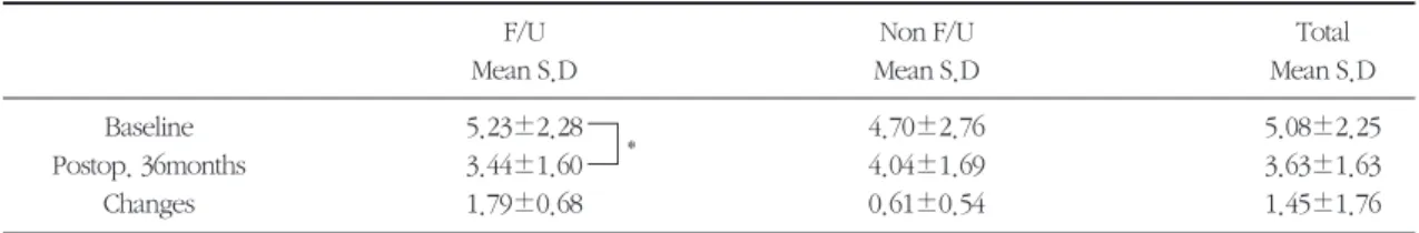
1)치주낭 탐침깊이의 변화
유지관리군에서는 술 전 치주낭 탐침깊이는 5.23
±2.28mm에서 술 후 36개월에는 3.44±1.60mm를 보였고, 비유지관리군에서는 4.70±2.76mm에서 술 후 36개월에는 4.04±1.69mm를 보였으며 두 군의 평균은 5.08±2.25mm에서 3.63±1.63mm를 보였
다.(Table 2, Fig. 2) 유지관리군에서는 술 전과 술 후 에 유의성있는 감소를 보였으나(P<0.05) 비유지관리 군에서는 유의성 있는 차이를 보이지 않았다.
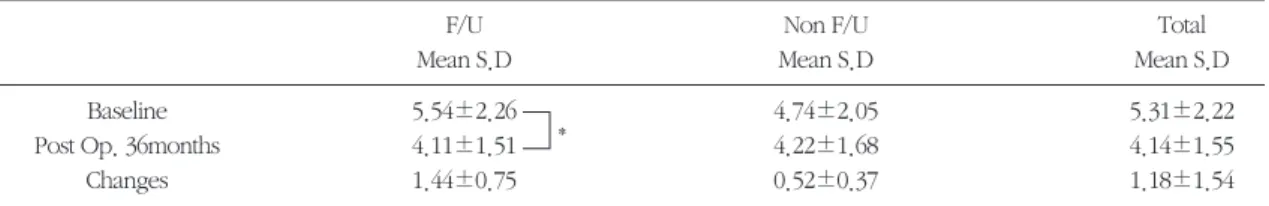
2) 부착획득
유지관리군에서 술 전 부착상실은 5.54±2.26mm
Table 2. Probing pocket depth(㎜)
F/U Non F/U Total
Mean S.D Mean S.D Mean S.D
Baseline 5.23±2.28 4.70±2.76 5.08±2.25
Postop. 36months 3.44±1.60 4.04±1.69 3.63±1.63
Changes 1.79±0.68 0.61±0.54 1.45±1.76
* : significant difference from baseline : P<0.05 S.D: standard deviation
*
Figure 2. Probing pocket depth(㎜)
* : significant difference from baseline : P<0.05 Baseline
mm
8 6 4 2
0 Post Op
36 months
Changes
F/U Non F/U Total
f g
Figure 1. Clinical case of patient 3
a. Clinical periodontal status before surgery, b. Surgical view of 2wall intrabony defect of lower right 1st molar, c. Preoperative radi- ographic view of lower right 1st molar showing significant dental calculus deposition and deep osseous defect, d. Biogran® is placed into the defect, e. Surgical view after suture, f. 36 months postoperative radiographic view demonstrates filling of the mesial osseous defect, g. 36months postoperative clinical periodontal status
Table 3. Loss of attachment(㎜)
F/U Non F/U Total
Mean S.D Mean S.D Mean S.D
Baseline 5.54±2.26 4.74±2.05 5.31±2.22
Post Op. 36months 4.11±1.51 4.22±1.68 4.14±1.55
Changes 1.44±0.75 0.52±0.37 1.18±1.54
* : significant difference from baseline : P<0.05 S.D: standard deviation
*
Figure 3. Loss of attachment(㎜)
* : significant difference from baseline : P<0.05
Table 4. Bone probing depth(㎜)
F/U Non F/U Total
Mean S.D Mean S.D Mean S.D
Baseline 7.25±2.48 7.22±2.84 7.24±2.57
Post Op. 36months 5.89±2.20 6.43±2.29 6.05±2.23
Changes 1.35±0.28 0.78±0.55 1.19±1.18
* : significant difference from base line : P<0.05 S.D: standard deviation
*
Figure 4. Bone probing depth(㎜)
* : significant difference from base line : P<0.05 Baseline
mm
10 8 6 4 2
0 Post Op
36 months
Changes
F/U Non F/U Total
Baseline
mm
12 10 8 6 4 2
0 Post Op
36 months
Changes
F/U Non F/U Total
에서 술 후 36개월에 4.11±1.51mm로 변화하여 1.44±0.75mm의 부착획득을 얻었으며, 비유지관리 군에서는 부착상실이 4.74±2.05mm에서 4.22±
1.68mm로 감소하여 0.52±0.37mm의 부착획득을 얻었다. 두 군의 평균 부착상실은 5.31±2.22mm에 서 4.14±1.55mm로 감소하여 1.18±1.54mm의 부 착획득을 얻었다(Table 3, Fig. 3) 유지관리군에서는 술 전과 술 후 36개월 째에 유의성 있는 감소를 보였 으나(P<0.05) 비유지관리군에서는 유의성 있는 차이 를 보이지 않았다.
3)탐침골 깊이의 변화
유지관리군에서는 치석제거술 후 탐침골 깊이는 7.25±2.48mm에서 술 후 36개월에는 5.89±
2.20mm로, 비유지관리군에서는 7.22±2.84mm에서 6.43±2.29mm로 감소하였으며 두 군의 평균 탐침골 깊이는 7.24±2.57mm에서 6.05±2.23mm로 감소를 보였다.
IV. 총괄 및 고찰
생체 유리는 1970년대에 Hench33)에 의해 처음 개 발된 것으로 종래의 유리와 달리, 40-50%의 낮은 농 도의 SiO2와 높은 Na2O, CaO 그리고 P2O5로 구성되 어진다. 그리고 여러 실험실과 동물 조직배양실험을 통해 생체 유리군 중 무게비 45% SiO2, 24.5% CaO Na2O, 6%P2O5로 조성된 유리가 생체친화성이 있고 골조직 결합능이 가장 뛰어나 이를 45S5 생체유리라 명명하고 임상에서 사용하고 있다34).
Ouhayoun35), 장 등36)은 생체유리가 체액에 노출 시 Na, Ca, P, Si ion 등이 용출 되어 나오면서 국소적 인 pH 변화와 함께 생체유리 표면에 실리카겔 층을 형성하고 그 위에 생체유리에서 나온 Ca, P과 체액에 서 공급되는 Ca, P이 쌓이고, 일정기간이 경과되면 수산화인회석 결정층으로 변화되며 이러한 활성이 골발생을 자극한다고 보고하였다.
Schepers 등37)은 beagle dog에 치밀형 hydroxyap- atite와 porous hydroxyapatite, 그리고 45S5 생체유 리를 이식하여 1,2,3,6,12개월에 조직학적으로 관찰
한 결과 치밀한 hydroxyapatite의 경우 이식재료의 중심부 입자들이 섬유성 조직으로 피막화되며 for- eign body giant cell이 나타났고, porous hydroxyap- atite의 경우는 입자와 성장된 골이 얇은 섬유조직으 로 분리되어 있었으며, 45S5 생체유리의 경우에는 모든 입자들이 골전도성에 의한 골성장에 의해 연결 되고 골조직층 사이에 존재하는 섬유조직에 많은 혈 관이 존재하여 풍부한 혈액 공급이 되고 있음을 알 수 있었다.
박38)등은 인체에서 38개의 치주 골내낭을 대상으 로 Biogran®의 6개월 후의 결과를 비교하여 치주조 직 재생에 미치는 효과를 평가하였다. 6개월 후에 4.1±1.8mm의 치주낭 탐침 깊이변화, 3.0±1.4mm 의 부착상실 변화, 2.8±1.3mm의 탐침골 깊이의 변 화를 보고하였다.
본 연구에서는 45S5 생체유리 가운데 하나인 Biogran®을 치주골 결손에 이식재료로 사용한 후 36개월 후에 결과의 안정성을 평가하며, 수술 후 정 기적 내원을 한 경우와 정기적 내원을 하지 않은 경 우를 비교하여 안정성에 정기적 내원이 미치는 영 향에 대해 평가하였다. 초기 임상 지수는 두 군 간 유의한 차이 없이 비슷한 값으로 나타나 두 군 간 초 기 상태의 차이에 의한 연구 결과의 오차는 없음을 알 수 있다.
치조골 결손부의 골재생의 평가는 방사선학적 평 가, 재수술에 의한 평가, 조직학적 평가 및 탐침골 깊 이를 이용하는 방법이 있는데 본 연구에서는 탐침골 깊이에 의한 평가방법을 이용하였다. 탐침골 깊이는 실제의 치조골의 깊이를 잘 반영해주는 지수이다39).
박 등38)은 bioactive glass(Biogran®)를 이식한 결 과 3.0mm의 부착획득과 2.8mm의 탐침골 깊이 변화 를 보고하였으며 Fox 등40)은 사람의 인접면 골내낭 에 bioactive glass를 이식한 결과 2.6mm의 부착획득 과 1.75mm의 골획득을 보고하였다.
본 연구에서는 유지관리군에서 1.44mm의 부착획 득과 1.35mm의 골획득을 나타냈다. 이 수치는 같은 재료를 사용한 박38)과 Fox의 단기간(6개월)의 연구 와 비교하면 더 적은 효과를 보이지만 망상골을 탈회 냉동건조시켜 단기간(6개월)동안 평가한 Quintero41)
의 연구성적과는 유사한 수치를 나타낸다.
Bioactive glass를 제외한 다른 골이식재료의 장기 임상결과를 보고하는 문헌은 드문 실정이며 그 중에 Biocoral의 5년 후 영향을 평가한 연구42)가 있다. 이 연구에서 술전 임상부착수준이 평균 5.7mm에서 5 년 후에 4.0mm로 1.7mm 감소함을 보였는데 이는 이번실험의 유지관리군의 36개월 동안의 임상부착 수준의 변화량인 1.44mm와 유사하다.
위와 같이 본 연구의 결과는 단기간의 다른 연구 결과들과 비교하였을 때 결과가 약간 퇴보하는 경향 을 보이지만 다른 골이식재료의 장기간 결과와는 유 사함을 보여준다. 단기간의 결과와 비교해서 차이가 나는 이유는, 이개부에서 일반적인 재생술식을 시행 하여도 장기간 후에는 결과가 안정적으로 유지되거 나 향상되기 보다 감소하는 결과를 나타내기 때문이
다43)44). 또한, 환자선택, 결손부의 성질과 범위, 치태
조절, 흡연같은 요인이 작용하였을 것으로 사료된다.
1996년 Tonetti등은45) plaque index와 gingival index가 낮은 환자일수록 재생술식이 더 성공적이 며, 재생술식을 통해 얻어진 부착의 안정성은 구강위 생과 관계 있다고 하였다.
본 연구에서도 이와 유사하게 치주낭 탐침깊이의 변화, 임상부착의 증가가 비유지관리군은 술 전과 36 개월 후에 통계학적으로 유의성이 없는 반면 유지관 리군은 술 전과 36개월 후에 통계학적으로 유의성 있는 차이를 나타내어서 부착의 안정성과 구강위생 과 관계가 있음을 알 수 있었다.
본 연구의 연구 성적을 종합할 때, Biogran®을 이 용한 재생술식 후에 3개월마다 주기적으로 재내원 하여 치태조절이 잘 된다면 수술 후 36개월 후에도 수술 전보다 더 유의성 있는 치주낭 탐침깊이의 감 소, 임상부착수준의 증가, 골탐침깊이의 증가를 가 져옴을 알 수 있다. 그러나 비유지관리군에서는 수 술 후 얻어졌던 양호한 결과들이 관찰기간 후에 다 시 퇴보하여 수술전과 비슷한 수준으로 돌아가서 통계적으로 유의성 있는 차이를 보여주지 못하고 있다.
따라서 적절하고 철저한 수술기법과 적절한 유지 기 치료가 행해진다면, Biogran®이 유용하며 이익이
있는 골대채물질이라는 것과 임상적으로 다른 합성 골대체물과 비슷하거나 더 나은 유용한 골 대체물이 될 수 있음을 보여준다.
만약, 골이식 재료 사이에 큰 차이가 없으며 장기 적으로 안정된다면, Biogran®은 향상된 지혈작용, 훌 륭한 조작성과 조직반응 때문에 좋은 골 이식재의 선택이 될 것으로 사료된다.
그러나, 이 재료의 다른 차단막과 혼합사용과 Biogran®을 사용한 재생술식 후에 얻어진 부착획득 이 정상 치주조직의 부착보다 치주질환의 이환에 더 감수성이 있어서, 같은 환경이라도 치주질환에 더 잘 이환되는지에 대한 연구가 더 필요할 것으로 사 료된다.
V. 결론
치주질환으로 인해 상실된 치주조직의 재생을 위 해 이용되는 골이식재의 하나인 bioactive glass의 효 과를 알아보기 위해 술 후 36개월 후 치료에 대한 임 상적 효과를 11명의17개의 골내낭에서 얻은 74개의 부위를 대상으로 정기적으로 재내원하여 치태조절 이 된 51개의 부위를 유지관리군으로, 정기적으로 재 내원하지 않은 23개의 부위를 비유지관리군으로 하 여 연구하였다.
술전과 술 후 36개월의 치료 효과를 치주낭 탐침 깊이, 부착상실, 탐침골 깊이, 치은 퇴축의 변화로 비 교하여 다음과 같은 결론을 얻었다.
1. 치주낭 탐침깊이의 변화는 유지관리군은 1.79
±0.68mm, 비유지관리군은 0.61±0.54mm, 평 균은 1.45±1.76mm를 보였으며, 유지관리군에 서만 초진에 비해 유의성 있는 감소를 보였 다.(P<0.05)
2. 부착상실의 변화는 유지관리군은 1.44±
0.74mm, 비유지관리군은 0.52±0.37mm, 평균 은 1.18±1.54 mm를 보였으며, 유지관리군에서 만 술 후 36개월에 유의성 있는 감소를 보였 다.(P<0.05)
3. 탐침골 깊이의 변화는 유지관리군은 1.35±
0.28mm, 비유지관리군은 0.78±0.55mm, 평균 은 1.19±1.18mm를 보였으며, 양 군 모두에서 유의성 있는 감소를 보였다(P<0.05).
이상의 결과에서 볼 때 치주 골내낭 처치시 bioac- tive glass를 사용하는 경우 술 후 36개월 후에 부착상 실과 탐침골 깊이가 유의차 있게 감소함을 알 수 있 다. 이는 bioactive glass가 골내낭에서 술 후 주기적 재내원에 의한 치태조절이 잘 유지된다면 일반적으 로 양호한 임상적 결과들을 나타내므로 유용한 골 대채 물질이 될 수 있음을 보여준다.
VI. 참고문헌
1. Bowers G, Chadroff B, Carnevale R, Mellonig J, Corio R, Emerson J, Stevens M, Romberg E.
Histologic evaluation of new attachment appara- tus formation in humans. Part II. J Periodontol 1989;60:675-682.
2. Bowers G, Chadroff B, Carnevale R, Mellonig J, Corio R, Emerson J, Stevens M, Romberg E.
Histologic evaluation of new attachment appara- tus formation in humans. Part III. J Periodontol 1989;60:683-693.
3. Gottlow J, Nyman S, Lindhe F, Karring T, Wennström J. New attachment formation in the human periodontium by guided tissue regenera- tion. Case reports. J Clin Periodont 1986;13:604- 616.
4. Renvert S, Nilveus R, Egelberg J. Healing after treatment of periodontal intraosseous defects. V.
Effect of root planing versus flap surgery, J Clin Periodontol 1985;12:619-629.
5. Schluger S. Osseous resection-A basic principle in periodontal surgery. Oral Surg Oral Med Oral Pathol 1949;2:316-325.
6. Melcher AH. On the repair potential periodontal tissues. J Periodontol 1976;47 :125-131.
7. Yukna RA. Clinical human comparison of e-
PTFE barrier membranes and freeze-dried dura mater allografts for guided tissue regeneration of lost periodontal supports. I. mandibular molar class II furcations. J Periodontol 1992;63:431- 442.
8. Urist MR, States BS. Bone formation by autoin- duction. Science 1965;150:892.
9. Isidor F, Attström R, Karring T. Regeneration of alveolar bone following surgical and non-surgi- cal periodontal treatment. J Clin Periodontol 1985;12:687-696.
10. Becker W, Lynch SE, Lekholm U, Becker B, Caffesse R, Donath C, Sanchez R. A comparison of PTFE membranes alone or in combination with platelet derived growth factor-I, or dem- ineralized freeze-dried bone in promoting bone formation around immediate extraction socket implants : A study in dogs. J Periodontol 1992;63: 929-940.
11. Lynch SE, Williams RC, Polson AM, Howell TH, Reddy MS, Zappa UE and Antoniades HN. A combination of platelet derived and insulin-like growth factors enhances periodontal regenera- tion. J Clin Periodontol 1989;16:545-548.
12. Lynch SE, Williams RC. A possible role for polypeptide growth factors and differentiation factors in periodontal regeneration. Am Acad Periodontol 1990, 1-5.
13. Yukna RA. Synthetic bone grafts in periodontics.
Periodontology 2000 1993;1:92.
14. Schallhorn RG. Postoperative problems associat- ed with iliac transplants. J Periodontolol 1972;43:3-9.
15. 김현영, 채중규. 성견의 3면 골내낭에서 calcium sulfate를 탈회 냉동 건조골과 혼합이식 및 차단 막으로 사용할 때의 치주조직의 치유효과. 연세 치대 논문집, Vol. 10, 1995.
16. Bowen JA. Comparison of decalcified freeze- dried bone allograft and porous particulate
hydroxyapatite in human periodontal osseous defects. J Periodontol 1989;60:647.
17. Carranza FA. Glickman's clinical periodontol- ogy. 7th edition, WB Sander Co. 1990,852-856.
18. Meffert RM, Thomas JR, Hamilton KM, Brownstein CN. Hydroxyapatite as an alloplastic graft in the treatment of human periodontal osseous defects. J Periodontol 1985;56:63-73.
19. Carranza FA, Kenndy EB, Lekovic V, Talamanie E. Histologic study of healing of human peri- odontal defects after placement of porous hydroxyapatite implants. J Periodontol 1987;59:682-688.
20. Kenney EB, Lekovic V, Sa Ferreira JC, Han J, Dimitrijevic B, Carranza FA Jr. Bone formation within porous hydroxyapatite implants in human periodontal defects. J Periodontol 1986;57:76-83.
21. Kenney EB, Lekovic V, Han J, Carranza FA Jr, Dimitrijevic B. The use of porous hydroxyapatite implant in periodontal defects. Clinical result after six months, J Periodontol 1985;56:82.
22. Ouhayoun JP. Influence of biomaterials in the healing pattern of bony defects in miniature pig mandible. J Dent Res 1989;68(special issue):Abstract No. 1944.
23. Yukna RA. Synthetic bone grafts in periodontics.
Periodontology 2000 1993; 1:92-99.
24. Schepers EJG, Ducheyne P. The application of bioactive glass particles of narrow size range as a filler material for bone lesions. a 24 month ani- mal experiment. Bioceramics 1993;6:401.
25. Schepers EJG, Pinruethal P. A comparative study of bioactive glass and porous hydroxylapatite particles in periodontal bone lesions.
Bioceramics 1993;5:113.
26. Schepers EJG. Bioactive glass particles of narrow size range : A new materials for the repair of bone defects. Implant dent 1993;2:151.
27. Schepers EJG. Bioactive glass particulate materi- al as a filler for bone lesions. J Oral Rehabilitation 1991;18:439.
28. Schepers EJG, Ducheyne P, Barbier L. Clinical application of bioactive glass granules of narrow size range on dental osseous lesions.
Bioceramics 1993;6:361.
29. Wilson J, Low SB. Bioactive ceramics for peri- odontal treatment : Comparative studies in the Patus monkey. J Applied Biomat 1992;3:123.
30. Wilson J, Merwin GE. Biomaterials for facial bone augmentation : Comparative studies.
Applied Biomaterials 1988;22:159.
31. Wilson J, Clark AE, Hall M, Hench L. Tissue response to bioglass endosseous ridge mainte- nance implants. J Oral Implantol 1993;19(4):295.
32. Wilson J, Pigott GH, Schoen FJ, Hench LL.
Toxicology and biocompatibility of bioglasses. J Biomed Mater Res 1981;15:805.
33. Hench LL. Bonding mechanisms at the interface of ceramic prosthetic materials. J Biomed Mater Res 1971;Sympo 2 :117.
34. Hench LL. Bioactive ceramics : Theory and clini- cal applications. 7th international symposium on ceramics in medicine. Turku. Finland. July 28, 1994.
35. Ouhayoun JP. Histological evaluation of natural coral skeleton as a grafting material in miniature swine mandible. J Mater Sci in Med. 1992;3:222.
36. 장소희, 고재승, 황성명. 생체유리에서 조섬유세 포의 교원섬유 형성에 관한 전자현미경적 연구.
대한구강해부학회지 1992.
37. Schepers EJG. Bioactive glass particulate material as a filler for bone lesions. J Oral Rehabilitation.
1991;18:439.
38. 박지숙, 최성호, 조규성, 채중규, 김종관. 치주 골 내낭에 bioactive glass 이식효과에 대한 임상연 구. 대한치주과학회지, 1997
39. Kim HY, Yi SW, Choi SH, Kim CK. Bone prob-
ing measurement as a reliable evaluation of the bone level in periodontal defects. J Periodontol 2000;71:729-735.
40. Fox GK, Rosenberg ES. Bioactive glass particles for the repair of human interproximal periodon- tal defects. 1996(impress).
41. Quintero G, Mellonig JT. A six months clinical evaluation of decalcified freeze-dried bone allo- graft in periodontal osseous defect. J Periodontol. 1982;53:726
42. Yukna RA, Yukna CN. A 5-year follow-up of 16 patients treated with coralline calcium carbon- ate(BiocoralTM) bone replacement grafts in infrabony defects. J Clin Periondontol
1998;25(12):1036-40.
43. Gottlow, Nyman S, Karring T. Maintenance of new attachment gained through guided tissue regeneration. J Clin Periodontol 1992:19:315- 317.
44. Metzler D, Seamons B, Mellonig J, Gher M, Gray J. Clinical evaluationof guided tissue regenera- tion in the treatment of maxillary class II molar furcation invasions. J Periodontol 1991;62:353- 360.
45. Cortellini P, Pini-prato G, Tonetti M. Periodontal regeneration of human infrabony defects. J Clin Periodontol 1994;21:606-610.
-Abstract-
Clinical Long-term Assessment of Bioactive Glass Graft
Hang-Bin Lee, Chang-Sung Kim, Seong-Ho Choi, Kyoo-Sung Cho, Chong-Kwan Kim, Choong-Kyoo Chai Department of Periodontology, College of Dentistry, Yonsei University
Research Institute for Periodontal Regeneration
The ultimate goal of periodontal therapy is the regeneration of periodontal tissue which has been lost due to destructive periodontal disease. To achieve periodontal regeneration, various kinds of methods have been investigated and developed, including guided tissue regeneration and bone graft. Bone graft can be catago- rized into autografts, allografts, xenografts, bone substitutes. And materials of all types have different biological activity and the capacity for periodontal regeneration, but ideal graft material has not been developed that fits all the requirement of ideal bone graft material. Recently, bioactive glass that has been utilized in plastic surgery is being investigated for application in dental practice. But, there has not been any long-term assess- ment of bioactive glass when used in periodontal intrabony defects.
The present study evaluates the long-term effects of bioactive glass on the periodontal regeneration in intra- bony defects of human and the effect of plaqu control on long term treatment results after dividing patients into those who underwent 3-month regular check-up and those who didn't under go regular check-up
The clinical effect on 74sites from 17 infrabony pockets of 11 patients were analyzed 36months after treat- ment.
51 sites which underwent regular check up were classified as the Follow-up group(F/U group), and 23 sites which did not undergo regular check up were classified as Non Follow-up group(Non F/U group).
After comparing the probing depth, attachment loss, bone probing depth before and 36months after treat- ment, the following results could be concluded.
1. The changes of probing pocket depth showed a statistically significant decrease between after baseline and 36 months after treatment in F/U group(1.79±0.68mm) and did no show astatistically significant decrease between after baseline and 36months after treatment in Non F/U group(0.61±0.54mm) (P<0.05).
2. The changes of loss of attachment showed a statistically significant decrease between after baseline and 36 months after treatment in F/U group(1.44±0.74mm) and did no show astatistically significant decrease between after baseline and 36months after treatment in Non F/U group(1.18±1.54) (P<0.05).
3. The changes of bone probing depth showed a statistically significant decrease between after baseline and 36 months after treatment in both F/U(1.35±0.28) and Non F/U group(0.78±0.55mm) (P<0.05).
The results suggest that treatment of infrabony defects with bioactive glass resulted in significan reduction of
attachment loss and bone probing depth 36months after the treatment.
The use of bioactive glass in infrabony defects, combined with regular check-up and proper plaque control generally shows favorable clinical results.
This measn that bioactive glass could be a useful bone substitute.
Key words : periodontal tissue regeneration, bone graft material, bioactive glass, intrabony defect, bone prob- ing depth, loss of attachment, long-term assesment