Ⅰ. Introduction
Dental implants are considered a good treatment option as an alternative to conventional reconstruction methods. In recent years, restoration with a dental
implant has become one of the common treatment options
1). Because of the excellent long term results, a topic concerning survival rates of dental implants is not as intriguing as it once used to be. Also, clinicians still focused on the
Peri-implantitis의
regeneration therapy 증례 보고
서울대학교 치의학대학원 치주과학교실
조 영 재Use of Bovine-derived bone mineral (Bio-Oss Collagen ) in surgical treatment of peri- implantitis: A case report
Program in Periodontology, School of dental science, Graduate school, Seoul National University Young Jae Cho DDS, MSD,
The aim of this study was to achieve healing of Peri-implantitis defects and hard tissue augmentation using a bovine-derived bone mineral on the defect site.
Two patients were treated with the surgical approach. With a full muco-periosteal flap elevation, the implant surfaces were exposed and granulation tissue removed around the implant and between the threads. Each surface of the contaminated implant was prepared with the air-abrasive device(PerioFlow ) for decontamination. Bovine-derived bone mineral(Bio-Oss collagen ) was then used to fill the defects and muco-periosteal flaps sutured to achieve transmucosal healing. Radiographs and clinical photographs were taken before and after 6 months of healing and an estimate of bone fill was assessed. Within the limits of the present case report, a surgical approach in treatment of peri-implantitis defects using a collagen form of bovine bone mineral was visited. Although limited, the two cases showed the stability and biocompatibility of a bovine-derived bone mineral and effectiveness of air-abrasive device(PerioFlow ) as a decontamination method.
Key words : Implantitis, dental implant, bone regeneration, bone substitute, bovine-derived xenograft ABSTRACT
Corresponding Author Ki-Tae Koo DDS, MS, PhD
Department of Periodontology, School of Dentistry, Seoul National University
Tel: 82-2-2072-0108; FAX: 82-2-744-0051, E-mail: [email protected]
technical aspects of implant surgery and prosthetic make-up, have somewhat overlooked the importance of maintenance of the implant and the surrounding tissue.
The prevalence of chronic inflammation of soft and hard tissues neighboring implants has been reported at a rate of 8.6 to 9.7% after 5years
2, 3). The pathological conditions which are defined as peri- implant mucositis and peri-implantitis are considered one of the major complications for the dental implant
3).
The clinical features of peri-implantitis are similar to a chronic periodontal inflammation. Gingival bleeding, swelling and supporting bone loss, as well as bacterial etiology are observed similarities which are reported in the literature
4, 5). Thus, similar to periodontitis, peri-implant probing depth(PPD) and radiographic change around the implant fixture are the most common parameters to diagnose peri- implantitis and these parameters are in actual use as an evaluation method of the treatment
6).
Treatment of peri-implantitis has been reported in a variety of ways, but each therapeutic indication or outcome still lacks sufficient evidence. Schwarz defined the classification of bone defects following peri-implantitis and suggested that the treatment method to be chosen should depend on the classification
7). Depending on the configuration, whether it be an intrabony or a supracrestal defect, the approach should be different. In treating
intrabony defects, access surgery may lead to a good response, but with low predicta bility. Therefore, in such instances as the circumferential defects with favorable intrabony components, regeneration the rapy may be indicated to fill the intrabony components and eventually reduce the probing pocket depth around the implants.
In this study, 2 cases of peri-implantitis with a circumferential intrabony component and vertical bone loss have been introduced.
Ⅱ. Case Case 1.
After removal of prosthetics, the healing abutment was connected for surgical convenience(Fig 1, 2). The full-thickness flap was elevated around #37 implant fixture to access the peri-implant defect under anesthesia. The defects were classified as Class Ie and II type bone defects according to the classification by Schwarz
7)(Fig 3).
After the granulation tissue was carefully removed, air-powder abrasives (PerioFlow , EMS) were used for mechanical debridement and decontamination of the contaminated implant surface(Fig 4). The bone defect site was filled with a bovine- derived bone mineral containing collagen(Bio-Oss Collagen Geistlich). This material has shown good maneuverability
Peri-implantitis 의 regeneration therapy 증 례 보 고
and easier adaptability to provide a stable structural integrity that may be advantageous in treating hard to reach intrabony compon ents. The surgical wound was completely covered and sutured to allow transmucosal healing of the
augmented implant sites(Fig 5). The sutures were removed 1 week after the surgery.
During the 4weeks after surgery, no sign of inflammation was observed on the surgical site and the radiographs exhibited
Fig. 1. Pre-operative presentation of the implant with peri-implantitis in the mandibular left second molar position
Fig. 2. Pre-operative periapical radiograph of the affected site. Note the marginal bone loss up to the third or fourth thread.
Fig. 3. The clinical presentation of the affected site after removing the inflammatory tissues following reflection of the flap. Note the circumferential bony defect surrounding the fixture.
Fig. 4. Application of bovine-derived xenograft with collagen in the circumferential bone defect.
Fig. 5. Transmucosal healing following regenerative attempt to augment the defect created by the peri-implantitis lesions.
Fig. 6. Post-operative occlusal view at 4 weeks.
Fig. 7. Post-operative periapical radiograph at 4 weeks
Fig. 8. Post-operative occlusal view at 6 months Fig. 9. Post-operative periapical radiograph at 6 months. Note the bone fill that is maintained prior to re-connection of the prosthesis.
Case 1
an uneventful healing with good space maintenance at the defect site(Fig 6, 7).
Re-osseointegration and bone remodeling, although controversial, is expected around the exposed implant surface.
At 6months following treatment, bone remodeling and maintenance of the grafted bovine bone mineral is observed with stable soft tissue sealing(Fig 8, 9).
Case 2
The patient presented swelling of the peri-implant mucosa and pus discharge
around the healing abutment(Fig 1, 2, 3).
Under local anesthesia, a full thickness muco-periosteal flap was elevated for regeneration surgery. After the flap elevation, the sites showed a large circum ferential-type bone defect with vertical bone loss(Fig 4).
As described above, the same debridement method was applied to remove the granulation tissue and decontamination of the contaminated implant surface(Fig 5).
Bio-Oss Collagen was selected to graft the defect site for the same reasons(Fig 6).
Immediately after surgery, there was
Peri-implantitis 의 regeneration therapy 증 례 보 고
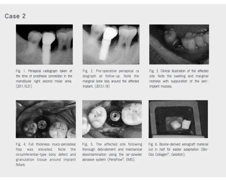
Fig. 1. Periapical radiograph taken at the time of prosthesis connection in the mandibular right second molar area.
(2011.10.21)
Fig. 2. Pre-operative periapical ra diograph at follow-up. Note the marginal bone loss around the affected implant. (2013.1.18)
Fig. 3. Clinical illustration of the affected site. Note the swelling and marginal redness with suppuration of the peri- implant mucosa.
Fig. 4. Full thickness muco-periosteal flap was elevated. Note the circumferential-type bony defect and granulation tissue around implant fixture.
Fig. 5. The affected site following thorough debridement and mechanical decontamination using the air-powder abrasive system (PerioFlow , EMS).
Fig. 6. Bovine-derived xenograft material cut in half for easier adaptation (Bio- Oss Collagen , Geistlich).
Case 2
insufficient amount of soft tissue to achieve primary closure(Fig 7). The sutures were removed 10 days after the surgery, but some exposure of grafting material occurred(Fig 8). Although a dehiscence of the wound occurred with exposure of the grafting material at 4weeks following treatment, there was no infection sign or
‘washout’of the grafting material(Fig 9).
This may have resulted from using the xenograft with collagen with structural integrity. At 8 weeks(Fig 10, 11) and 4(Fig 12, 13), 6 months (Fig 14, 15) after treatment, bone fill is observed on the graft site in radiographs. Re-osseointegration may also be expected to some extent.
Fig. 10. Panoramic radiograph after 8 weeks. Fig. 11. Post-operative periapical radi ograph at 8 weeks. Note the slight decrease in height of the grafted biomaterial that was not protected indirectly suggesting the need for using a resorbable barrier membrane for protection of the wound.
Fig. 12. Soft tissue stability observed at 4 months despite the oral communication at 4 weeks.
Fig. 13. Bone remodeling observed after 4 months of surgery.
Fig. 14. Post-operative periapical radiograph at 6 months.
Fig. 15. Post-operative occlusal view at 6 months.
Case 2
Fig. 7. Suture following augmentation surgery. A transmucosal healing was attempted.
Fig. 8. Post-operative occlusal view at 4 weeks. Note the marginal gap in the mesial side creating a direct communicating passage to the oral cavity increasing susceptibility for bacterial infection.
Fig. 9. Post-operative periapical radio graph at 4 weeks.
Ⅲ. Discussion
Two peri-implantitis cases with associated intrabony components were successfully treated with the guided bone regeneration technique using a bovine- derived bone graft. After 6months, radiographs showed a stable bone fill at the grafting site with the resolution of the inflammation around the implant fixture.
Re-osseointegration on the previously contaminated implant surface that has been exposed to bacterial plaque biofilm is still controversial. Bacterial contamination of the implant surface, which affects the surface energy, as well as the alteration of surface characteristics can have negative effects on tissue regeneration.
To resolve this problem, the efficient decontamination method has been studied in many ways in recent years. Several therapeutic approaches were identified such as mechanical treatment, chemical agent, photodynamic treatment, and laser applications. Mechanical debridement aims to remove the biofilm of surface by means of titanium or plastic curettes, air-powder abrasive system, and ultrasonic devices.
Anti-infective treatments aim to detoxify the implant surface by chemical agents, such as with chlorhexidine, tetracycline, metronidazole, and citric acid. Recently, CO
2or Er:YAG laser has been used to sterilize the surface without causing
damage or alteration
8, 9). Beneficial effects of laser therapy on peri-implantitis have been shown, but this approach needs to be further evaluated.
Air-powder abrasive technique shows improved results and effectiveness com pared to the other mechanical methods
10). In a 5-year long-term study, the air-powder abrasive devices compared to treatment using the laser has been reported to achieve good treatment results
11). In the study of a comparison between the air-powder abrasive technique and Er:YAG laser, the clinical parameter such as bleeding on probing, probing pocket depth around the implant was improved in both groups, but with limited differences. At 6months, the decrease in suppuration was significant in both treatment groups, but with no group difference in the change of suppuration between baseline and at 6 months after treatment
12).
Er:YAG laser shows high-BIC compared to other methods in some studies evaluating re-osseointegration of previously contamin ated surfaces
13). However, the air-powder abrasive device with a high efficiency in cleaning the surface with a minimum damage to the surface is also recommended in the treatment of peri-implantitis.
It has been suggested that the estab lishment of an implant surface conducive to bone formation is a prerequisite for successful regenerative treatment of peri- implantitis
14). In various studies, the
Peri-implantitis 의 regeneration therapy 증 례 보 고
recovery of biocompatibility of the implant surface is required, but there is no definitive gold standard yet. At the 3rd ITI Consensus Conference in 2003, there was no statistically significant difference among the decontamination methods. Thus, recent studies focus on the importance of selecting the right surgical method rather than the methods of decontamination.
As described above, surgical approach for the treatment of peri-implantitis has several advantages compared to the non- surgical treatment. Muco-periosteal flap elevation provides visibility and surgical accessibility to underlying bone and defect site. The stabilization of the defect with a bone substitute may also be advantageous.
For the successful regeneration of defect site, the choice of grafting material should be carefully considered. Autogenous bone material is the most predictable material, but it has some limitations. Harvesting from intraoral or extraoral may be painful and limited with morbidity, and autogenous bone is easy to resorption in the healing process.
Xenograft material has been used quite frequently as bone graft substitutes with good success in recent years. Bovine- derived bone material may serve as an alternative to overcome the disadvantages of autogenous bone. Xenograft is
osteoconductive, readily available and risk free of disease transmission. Most of all, the biomaterial has been shown to maintain the augmented volume for long periods that resist to resorption
15, 16).
Recently, a bovine-derived bone mineral containing collagen has been used in attempts to prevent bone loss and promote bone remodeling in fresh extraction sockets
17). The graft material containing collagen is thought to possess several advantages compared to the particulate bone type biomaterial. The material is easily moldable into the desired form. The augmented material has been shown to maintain the volume for long periods
15, 16). Thus, in the surgical treatment of peri- implantitis defects with favorable intrabony components, the use of such biomaterial with collagen may be one option providing surgical convenience and improving the long term prognosis of the peri-implantitis defects.
Ⅳ. Limitation
This case report focused on the
radiographic bone morphology and limited
clinical parameters were used for
assessment not to mention the small
number of patients invoved. Further
Peri-implantitis 의 regeneration therapy 증 례 보 고
1. Merickse-Stern R, Aerni D, Geering AH, Buser D.
Long-term evaluation of non-submerged hollow cylinder implants. Clinical and radiographic results.
Clin Oral Implants Res 2001; 12: 252-259.
2. Jung RE, Pjetursson BE, Glauser R, Zembic A, Zwahlen M, Lang NP. A systematic review of the 5-year survival and complication rates of implant- supported single crowns. Clin Oral Implants Res 2008; 19: 119-130.
3. Pjetursson BE, Tan K, Lang NP, Bragger U, Egger M, Zwahlen M. A systematic review of the survival and complication rates of fixed partial dentures (FPDs) after an observation period of at least 5 years. Clin Oral Implants Res 2004; 15: 667-676.
4. Mombelli A. Microbiology of the dental implant. Adv Dent Res 1993; 7: 202-206.
5. Pontoriero R, Tonelli MP, Carnevale G, Mombelli A, Nyman SR, Lang NP. Experimentally induced peri- implant mucositis. A clinical study in humans. Clin Oral Implants Res 1994; 5: 254-259.
6. Mombelli A, Lang NP. The diagnosis and treatment of peri-implantitis. Periodontol 2000 1998; 17: 63-76.
7. Schwarz F, Herten M, Sager M, Bieling K, Sculean A, Becker J. Comparison of naturally occurring and ligature-induced peri-implantitis bone defects in humans and dogs. Clin Oral Implants Res 2007;
18: 161-170.
8. Schou S, Holmstrup P, Jorgensen T, Skovgaard LT, Stoltze K, Hjorting-Hansen E, Wenzel A. Implant surface preparation in the surgical treatment of experimental peri-implantitis with autogenous bone graft and ePTFE membrane in cynomolgus monkeys. Clin Oral Implants Res 2003; 14: 412-422.
9. Renvert S, Roos-Jansaker AM, Claffey N. Non- surgical treatment of peri-implant mucositis and peri-implantitis: a literature review. J Clin Periodontol 2008; 35: 305-315.
10. Schwarz F, Ferrari D, Popovski K, Hartig B, Becker J. Influence of different air-abrasive powders on cell viability at biologically contaminated titanium dental implants surfaces. J Biomed Mater Res B Appl Biomater 2009; 88: 83-91.
11. Deppe H, Horch HH, Neff A. Conventional versus CO2 laser-assisted treatment of peri-implant defects with the concomitant use of pure-phase beta-tricalcium phosphate: a 5-year clinical report. Int J Oral Maxillofac Implants 2007; 22:
79-86.
12. Renvert S, Lindahl C, Roos Jansaker AM, Persson GR. Treatment of peri-implantitis using an Er:YAG laser or an air-abrasive device: a randomized clinical trial. J Clin Periodontol 2011; 38: 65-73.
13. Schwarz F, Jepsen S, Herten M, Sager M, Rothamel D, Becker J. Influence of different treatment approaches on non-submerged and submerged healing of ligature induced peri- implantitis lesions: an experimental study in dogs.
J Clin Periodontol 2006; 33: 584-595.
14. Schwartz Z, Kieswetter K, Dean DD, Boyan BD.
Underlying mechanisms at the bone-surface interface during regeneration. J Periodontal Res 1997; 32: 166-171.
15. Maiorana C, Beretta M, Salina S, Santoro F.
Reduction of autogenous bone graft resorption by means of bio-oss coverage: a prospective study.
Int J Periodontics Restorative Dent 2005; 25: 19-25.
16. von Arx T, Buser D. Horizontal ridge augmentation using autogenous block grafts and the guided bone regeneration technique with collagen membranes: a clinical study with 42 patients. Clin Oral Implants Res 2006; 17: 359-366.
17. Araujo MG, Liljenberg B, Lindhe J. Dynamics of Bio-Oss Collagen incorporation in fresh extraction wounds: an experimental study in the dog. Clin Oral Implants Res 2010; 21: 55-64.
Reference