서론
치주 골내낭은 치아 지지의 상실과 앞으로 진행될 치주조 직 파괴의 높은 위험성 등으로 인해 치주치료에 있어서 중요 한 비중을 가진다1). 이러한 치주 골내낭 치료에 있어 보존적 인 치료를 우선적으로 고려할 수 있으나 깊은 골내낭과 이개
Correspondence: Dr. Joem-Il Choi
Department of Periodontology, School of Dentistry, Pusan National University 10 1-ga, Ami-dong, Seo-gu, Busan, 602-739, Korea e-mail: [email protected], Tel: 82-51-240-7460, Fax: 82-2-243-7606 접수일: 2007년 7월 18일; 채택일: 2007년 8월 7일
부 병변은 효과적인 debridement를 위한 접근을 어렵게 만든 다2). 따라서 치주 외과 술식이 골 재형성(osseous re- contouring) 또는 골 삭제(osseous resection)를 동반하여 치 주 골내낭을 제거하기 위해서 사용된다. 골절제술은 양형을 가지는 정상 골 형태를 만들어 주며 탐침 깊이 감소와 구강 위 생을 환자 스스로 관리할 수 있게 도움을 주는 치은조직의 형 태를 만드는데 목표를 둔다. 그러나 이러한 골 삭제술은 얕은 골내낭(1~2 mm 깊이)에서 중등도(3~4 mm 깊이) 정도의 골 내낭과 골 형태 수정에 사용되기 적당하다3).
한편 다른 시도가 치주 골내낭의 상실된 부착기구들을 재
Myung-Jin Kim1, Ju-Youn Lee, Sung-Jo Kim, Jeom-Il Choi*
Department of Periodontology, School of Dentistry, Pusan National University
ABSTRACT
Purpose: Periodontal intrabony defects have great deal of importance since they contribute to the development of periodontal disease. Current treatment regimens for intrabony defects involve grafting of numerous bony materials, GTR using biocompatible barriers, and biomodification of root surface that will encourage the attachment of connective tissue. Xenograft using deproteinized bovine bone particles seems to be very convenient to adjust because it doesn't require any donor sites or imply the danger of cross infections. These particles are similar to human cancellous bone in structure and turned out to be effective in bone regeneration in vivo. We here represent the effectiveness of grafting deproteinized bovine bone particles in intrabony defect and furcation involvements that have various numbers of bony walls.
Materials and methods: Open flap debridement was done to remove all root accretions and granulation tissue from the defects within persisting intrabony lesions demonstrating attachment loss of over 6mm even 3 months after nonsurgical periodontal therapy have been completed. Deproteinized bovine bone particles(BBPⓇ, Oscotec, Seoul) was grafted in intrabony defects to encourage bone regeneration. Patients were instructed of mouthrinses with chlorohexidine-digluconate twice a day and to take antibiotics 2-3 times a day for 2 weeks. They were check-up regularly for oral hygiene performance and further development of disease. Probing depth, level of attachment and mobility were measured at baseline and 6 months after the surgery. The radiographic evidence of bone regenerations were also monitored at least for 6 months.
Conclusion: In most cases, radio-opacities increased after 6 months. 2- and 3-wall defects showed greater improvements in pocket depth reduction when compared to 1-wall defects. ClassⅠ& Ⅱ furcation involvements in mandibular molars demonstrated the similar results with acceptable pocket depth both horizontally and vertically comparable to other intrabony defects. Exact amount of bone gain could not be measured as the re-entry procedure has not been available. With in the limited data based on our clinical parameter to measure pocket depth reduction following BBPⓇ grafts, it was comparable to the results observed following other regeneration techniques such as GTR. (J Korean Acad Periodontol 2008;38:97-102) KEY WORDS: Periodontal intrabony defect; bone graft; bovine-bone material.
생하는데 초점을 맞추어 왔다. 최근의 치주 재생술의 분야는 다양한 골이식재의 이식술, 생체 차단막을 사용하는 조직유도 재생술, 결합조직 부착을 증진시키는 치근면의 bio- modification 등이 연구되어지고 있다. 이 중 다양한 골이식 재로 골 결손부에 충전하는 것은 상실된 치주 부착기구를 회 복하는 가장 널리 사용되는 술식 중 하나이다4). 이식을 위한 골이식재로서 탈단백 우골입자를 이용한 이종골 이식은 별도 의 공여부가 필요치 않고 교차 감염의 우려가 없는 등 다른 골 이식재에 비해 임상적 편의성을 가진다5). 또한 구조적으로 인 간의 망상골과 유사하고 인간 및 동물 조직학적 연구에서도 골내 결손부에서 골재생 효과를 나타냄을 보고하고 있다6,7). 본 임상 증례에서는 다양한 골벽의 수를 가지는 치주 골내 결 손부와 치근 이개부병변의 처치에 탈단백 우골(BBP®)을이식 한 증례들의 임상적인 효과를 고찰해 보았다.
임상 증례
본 증례들에서는 통상적인 비외과적 치주처치(치석제거술 및 치근활택술) 후에도 6 mm 이상의 부착상실과 골내 치주병 소를 나타내는 치아에 판막을 형성하여 치근활택을 시행하고
육아조직을 소파하였다. 치조골 재생을 도모하기 위해 골내 결손부에 탈단백 우골(bovine-bone material(BBP®))을 이 식하고 봉합하였다. 1주 후 봉합사는 제거했고 2주간 하루 2~3번 chlorhexidine-digluconate 구강세척액을 사용하게 했다. 술 전과 술 후 6개월 후에 탐침깊이, 부착수준, 동요도 를 측정하였고 골재생 양상을 방사선학적으로 판정하였다.
<1-2 벽성 골내 결손부>
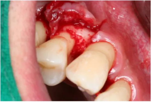
46세 남성 환자로 우측 상악 제1소구치의 동요도와 동통을 주소로 내원하였다. 만성 치주염으로 진단하고 치석제거 및 치 근 활택술을 시행하였으나 계속적인 탐침시 출혈과 2도의 치아 동요도 및 8mm의 근심측으로 깊은 탐침깊이를 보였다(Fig. 1).
치주 골내낭 치료를 위한 재생요법으로 탈단백 우골이식을 계획하고 전층판막을 거상하였다. 치근활택술과 육아조직을 소파한 후 치관측으로 1벽성, 치근단측으로 2벽을 가지는 깊은 골내 결손부를 확인하였다(Fig. 2). 골결손부 내로 탈단백 우 골(BBP®)을 이식하고 1주 후에 봉합을 제거하였다(Fig. 3, 4).
골이식 6개월 후 탐침깊이가 5 mm로 감소되고 치아 동요도도 줄어들었다. 방사선 사진상으로도 초진시와 비교해서 골 이식 부위에 방사선 불투과성이 증가된 것이 관찰된다(Fig. 5).
Figure 1. Preoperative periapical view Figure 2. Flap elevation Figure 3. Bovine bone material(BBPⓇ) grafting
Figure 4. suture Figure 5. Radiographic comparison between initial and 6month later
<2-3 벽성 골내 결손부>
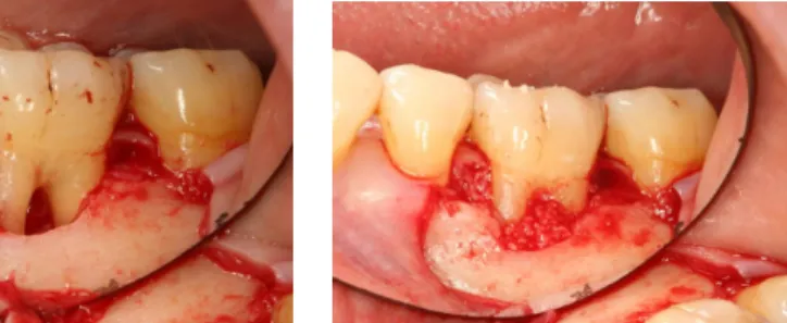
44세 남성으로 저작시 좌측 2소구치의 동통을 주소로 내원 하신 분으로 방사선 사진 상으로는 원심측으로 깊은 골결손부 가 관찰되고 임상적으로 원심측으로 10 mm의 탐침깊이와 2 도의 치아 동요도를 보여주고 있었다. 치석제거 및 치근활택 술후 재생요법으로 탈단백 우골이식을 계획하였다(Fig. 6).
전층 판막을 거상하고 육아조직을 소파한 뒤 원심부의 골결손 부 뿐 아니라 근심부의 골결손부까지도 확인할 수 있었다 (Fig. 7). 결손부내로 치주 재생을 도모하기 위해 탈단백 우골 (BBP®)을 충전하고 봉합하였다(Fig. 8, 9). 골이식 6개월 후 에 초진시와 비교했을 때 통증과 동요도가 사라졌고 탐침깊이 가 5 mm로 감소되었으며 방사선 사진상으로도 현저한 골재
생을 보여주었다(Fig. 10).
<3-벽성 골내 결손부>
45세 여성 환자로 하악 좌측 1대구치의 부종과 통증을 주소 로 내원하신 분으로 치주기원의 치주 농양소견을 보였다(Fig.
11). 치아의 동요도는 없었고 원심측으로 8 mm의 탐침 깊이 를 보였다(Fig. 12).
치은 열구를 통해 배농과 투약 후 통증과 부종은 경감되었 지만 깊은 치주낭은 개선되지 않았다(Fig. 13). 이를 해소하기 위해 골이식을 계획하고 전층판막을 거상하였다(Fig. 14). 탈 단백 우골(BBP®)를 이식하고 collagen membrane를 덮은 뒤 봉합하였다(Fig. 15, 16).
Figure 6. Preoperative periapical
radiograph Figure 7. Palatal aspect after flap
elevation Figure 8. Bovine bone material
(BBPⓇ)grafting
Figure 9. Suture Figure 10. Radiographic evaluation after 6month later
Figure 11. Clinical photo at 1st visit Figure 12. Periapical radiograph
shows distal bone defect Figure 13. After drainage
탈단백 우골이식 6개월 후에 임상적으로 8 mm의 탐침깊이 가 3 mm로 감소되었고 치주 농양의 재발도 없었다. 방사선 사진 비교에서도 하악 좌측 1대구치 원심측 골결손부에 현저 한 골재생을 보여주고 있다(Fig. 17).
<I급 이개부 병변>
45세 남성으로 하악 우측 1대구치 부근에서 저작시 불편감 을 주소로 내원하였고 임상검사시 협측 치근이개부에 수직적 으로 10 mm의 수평적으로 2 mm의 탐침깊이를 보이고 있었 Figure 14. Flap elevation Figure 15. Bovine bone material(BBP®)
grafting and collagen membrane coverage
Figure 16. Suture
Figure 17. Radiographic comparison between initial and 6month later
Figure 18. Initial clinical photo and periapical radiograph Figure 19. Flap elevation
Figure 20. Bovine-bonematerial (BBP®) grafting Figure 21. Radiographic comparison between initial and 6month later
다(Fig. 18). 전층판막을 거상하고 육아조직을 소파하였을 때 원심치근 부위로 심한 치조골 파괴를 보였으며 치근 분지로도 골파괴 양상을 확인할 수 있었다(Fig. 19). 재생요법으로 결손 부 내로 탈단백 우골(BBP®)을 충전하고 봉합하였다(Fig. 20).
1주 후에 봉합을 제거하였고 골이식 6개월 후에 초진시와 비 교하였다(Fig. 21).
임상적으로 저작시 불편감 사라졌고 10 mm의 수직적 탐침 깊이는 4 mm로 감소되었다. 방사선 사진상으로도 원심치근 과 이개부에 골밀도의 증가를 관찰할 수 있었다.
<II급 이개부 병변>
55세 여성 환자로 하악 좌측 1대구치에 비외과적 치주치료 후에도 계속되는 둔통을 호소하였다. 협측 치근 이개부로 탐 침하였을 때 수직적으로 8 mm 수평적으로 5 mm 탐침깊이를 나타내어 2급 분지부 병변으로 진단하고 재생요법으로 탈단 백 우골 이식을 계획하였다. 전층판막을 거상하고 육아조직을 소파한 뒤 치조골 파괴양상을 확인할 수 있었다(Fig. 22, 23).
재생요법으로 결손부 내로 탈단백 우골(BBP®)을 충전하고 봉 합하였다(Fig. 24, 25). 봉합은 1주 후에 제거되고 6개월 후에 임상적, 방사선학적으로 평가하였다(Fig. 26). 임상적으로 골 이식 전과 비교할 때 4 mm의 정상 탐침깊이를 회복하였고 방 사선학적으로도 치근 이개부로 골밀도의 증가를 확인할 수 있 었다.
고찰
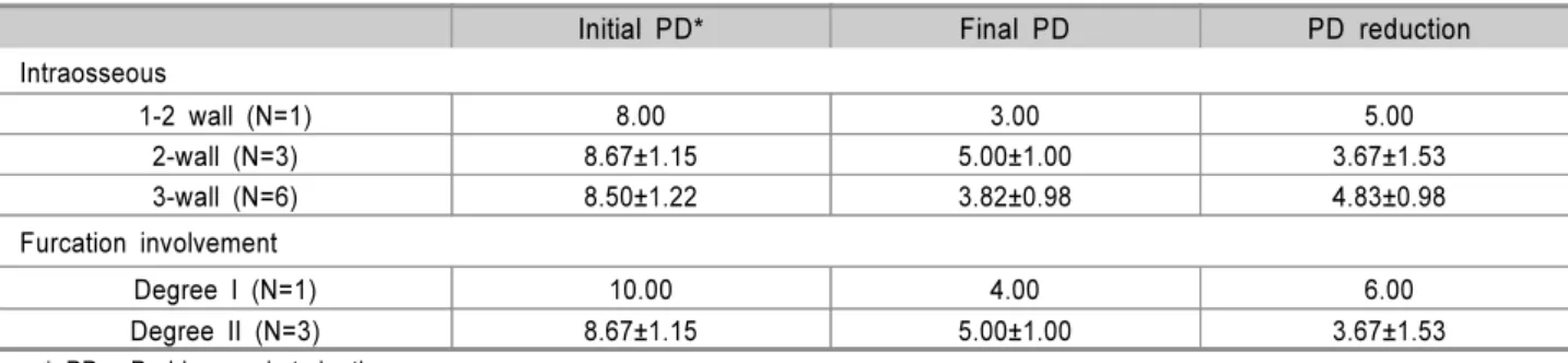
골이식의 목적으로 탐침깊이의 감소, 임상적 부착 획득, 골 결손부 내로의 골 충전, 새로운 치조골과, 백악질, 치주인대 의 재생을 들 수 있다4). 본 연구에서 사용된 증례는 1, 2-벽성 증례가 1개, 2-벽성 증례가 3개, 3-벽성 증례 6개와 1급 이개 부 증례 1개, 2급 이개부 증례 3개이고 각각의 임상적 탐침깊 이의 감소는 Table 1에서 나타내고 있다.
전반적으로 1벽성보다는 2, 3벽성 결손부에서 임상적인 지 표들의 상당한 개선을 보였다. 또한 하악 대구치의 1-2급 치 근 이개부 병변에서도 유사한 만족스러운 결과를 보였다. 이 것은 치주 재생에 있어서 잔존 골벽의 수가 많을수록 유리하 다는 이전의 연구들과도 일치한다8,9,10).
Reynold11) 등의 연구와 2005년 치주 재생에 대한 American Academy of Periodontology(AAP)의 Position paper12)는 치주 골내낭을 해결하기 위한 치료법으로서 골이 식은 단순한 판막 소파술에 비해서 더 나은 임상적인 개선을 이룬다고 결론 내렸다. 본 연구에도 증례들이 통계적인 의미 를 부여할 만큼 충분하지는 않지만 전반적으로 골이식 후에 상당한 임상적 지표의 개선를 보이고 있다. 2002년 Kalpidis 와 Ruben13)의 단순한 판막 소파술, GTR, Emdogain®들을 이 용한 치주 재생술(EGR)에 대한 메타 분석과 비교할 때 본 연 Figure 22. Preoperative view Figure 23. Flap elevation Figure 24. Bovine bone material(BBP®)
grafting
Figure 25. Suture Figure 26. Radiographic comparison between initial and 6month later
구에서 얻어진 탈단백 우골(BBP®)을 이용한 골이식의 결과는 단순한 판막 소파술보다 우수하고 Emdogain®을 사용한 재생 술식에 견줄 만한 탐침깊이의 감소를 보여주었다(Table 2).
하지만 진정한 치주 재생의 정도는 재진입(re-entry)을 통한 정확한 재생량을 확인할 수는 없었기에 거의 모든 증례에서 방사선학적으로 골밀도의 개선을 확인하는 수준에 그쳤다.
참고문헌
1. Papapanou PN, Wennstrὅm JL. The angular bony defect as indicator of further alveolar bone loss. J Clin Periodontol 1991;18:317-322.
2. Papapanou PN, Tonetti MS. Diagnosis and epidemiology of periodontal osseous lesions Periodontol 2000 2000;22:8-21.
3. Carnevale G, Kandahl WB. Osseous resective surgery.
Periodontol 2000 2000;22:59-87.
4. Brunsvold MA, Mellonig JT. Bone grafts and periodontal regeneration. Periodontol 2000 1993;1:80-91.
5. Sculean A, Berakdar M, Chiantella GC, Donos N, Arweiler NB, Brecx M. Healing of intrabony defects following treat- ment with a bovine-derived xenograft and collagen membrane. A controlled clinical study. J Clin Periodontol 2003;30:73-80.6.
6. Berglundh T, Lindhe J. Healing around implants placed in bone defects treated with Bio-Oss. An experimental study in the dog. Clin Oral Impl Res 1997; 8:117-124.
7. Mellonig J. Human histologic evaluation of a bovine-derived xenograft in the treatment of periodontal osseous defects. Int J Periodontics Restorative Dent 2000;20:1-29.
8. Goldman H, Cohen DW. The infrabony pocket: classi- fication and treatment. J periodontol 1958;29:272-291.
9. Prichard J. A Technique for treating intrabony pockets based on alveolar process morphology. Dent Clin North Am. 1960:85:1.
10. Tonetti M, Pini-Prato G, Cortellini P. Periodontal re- generation of human intrabony defect. IV. Determinent of the healing response. J periodontol 1993;64:934-940.
11. Reynolds MA, Aichelmann-Reidy ME, Branch-Mays GL, Gunsolley JC. The effect of bone replacement grafts in the treatment of periodontal osseous defects. A systematic review. Ann Periodontol 2003;8:227-265.
12. American Academy of Periodontology. Posotion paper.
Periodontal regeneration. J Periodontol 2005;76:1601-1622.
13. Kalpidis CD, Ruben MP. Treatment of intrabony perio- dontal defects with enamel matrix drivative: A literature re- view J Periodontol 2002;73:1360-1376.
Degree I (N=1) 10.00 4.00 6.00
Degree II (N=3) 8.67±1.15 5.00±1.00 3.67±1.53
* PD : Probing pocket depth
Table 2. Comparison of BBP® Graft to Other Treatment Regimens for Intrabony Defect (Kalpidis and Ruben13))
Treatment* Study Probing depth reduction (mm)
OFD 8 3.1±1.0
EGR 12 4.0±0.9
GTR 6 5.1±0.9
Bovine bone(BBP®) graft 14 cases 4.2±1.3
* OFD = open flap debradement
EGR = EMD(enamel matrix derivative) guided regeneration GTR = guided tissue regeneration