대한치주과학회지 : Vol. 37, No. 1, 2007
Impact of nonsurgical periodontal therapy on oral malodor and microbial load of saliva
Myung-Jin Kim, Sung-Jo Kim, Jeom-Il Choi, Ju-Youn Lee
*Department of Periodontology, School of Dentistry, Pusan National University
I. INTRODUCTION
Breath malodor affects a large proportion of population and may be the cause of a sig- nificant social and psychological problem1). It can be caused by a number of factors, both of intra- and extra-oral origins. Among oral etiological factors, breath malodor may result from periodontal diseases, poor salivary flow, improper dental restorations, excessive micro- bial colonization of the tongue, and unclean denture2,3). Extraoral etiological factors include upper and lower respiratory tract abnormal conditions, gastrointestinal and neurologic dis- orders, various systemic diseases and certain drugs4). In several studies, however, about 90%
of breath malodor have an intraoral origin, mostly gingivitis, periodontitis and/or tongue coating5-7).1)
The principal components of breath malodor are volatile sulphide compounds (VSCs), espe- cially hydrogen sulphide (H2S), methyl mercap- tan (CH3SH) and dimethyl sulphide [(CH3)2S] or compounds such as butyric acid, propionic acid, putrescine, and cadaverine2). These are made from the proteolytic degradation predominantly by anae- robic gram-negative bacteria on plaque, tongue coatings, saliva, blood and epithelial cells8).
There are various methods to assess breath malodor. The main approaches are organoleptic ratings, gas chromatography, and sulphide monitoring. Organoleptic ratings assessed by human judges still are the golden standard9). But, this method raises several problems in- cluding low intra- or inter-examiner reprodu- cibility (reliability) and lack of official stand- ards6). Although, gas chromatography allowing detection of all possible odorous component
* Corresponding author : Ju-Youn Lee, Department of Periodontology, School of Dentistry, Pusan National University, 1-10, Ami-Dong, Seo-Ku, Pusan, 602-739, Korea (E-mail : heroine@pusan.ac.kr)
* This work was supported for two years by Pusan National University Research Grant
seems to be an objective method, it also has disadvantages. In addition, the portable sulfide monitor, a more practical VSCs measuring de- vice, detects hydrogen sulphide and methyl mercaptan, some of the most common VSCs found in malodor5).
Another method to measure breath malodor is the microbial approach. Paryavi-Gholami et al10). reported that breath malodor is associated with increased levels of bacteria in the saliva.
Also, El-Halabi et al.11) used microbial counts of bacteria in saliva as a diagnostic test for oral malodor and concluded that microbial quantitative technique for assessing oral mal- odor may be useful as a clinical and research tool. Although the actual bacterial species that cause or contribute to oral malodor are not clearly identified in quantitative method, we assume that the change in the breath malodor can be evaluated indirectly by measuring sali- vary microbial load.
Whereas conventional periodontal therapy is performed in a quadrant-wise or sextant-wise manner for 4-6 weeks, 1-stage full-mouth disinfection presented by Quirynen et al. is completed within 24 hours12). They reported significant improvements both in clinical and microbiological aspects13). Original full mouth disinfection protocol included the additional use of chlorhexidine. To reduce the number of pe- riodontopathic bacteria, chlorhexidine is still the gold standard14). However periodontitis is chronic inflammatory disease and oral status of periodontitis patients(wide interdental spaces and deep periodontal pockets) can not be changed dramatically. Moreover tongue coating that is the main predisposing factor for hal- itosis is present for the whole life. In addition,
chlorhexidine has some side effects (bad taste, tongue and tooth discoloration etc.)15). Therefore, the long-term use of antiseptics in periodontal patients for the purpose of treat- ment of halitosis can not be recommended. Due to these limitations, we excluded the use of antiseptics. The present study aims to examine the impact of full-mouth nonsurgical perio- dontal therapy on breath malodor by measuring VSCs level and microbial load of saliva.
II. MATERIALS AND METHODS
1. Patient selection
Twenty-three subjects, who visited the de- partment of periodontology at Pusan National University Hospital, presenting moderate perio- dontitis were randomly allocated to test and control group. Smokers and other medically compromised patients were excluded. Patient who have less than 20 teeth, or untreated ca- ries teeth were also excluded. All subjects had not received antibiotics or non-steroidal an- ti-inflammatory drugs at least one month prior to the experience. All participants were given a thorough explanation about this experiments and had signed an informed consent form.
2. Treatment procedure
At baseline, all patients instructed oral hy- giene instruction with tooth and interdental brushing including tongue cleansing. Oral hy- giene was controlled and reexplained on every weeks. The control group were given oral hy- giene instruction only. Oral hygiene instruction included interdental brushing and tongue
cleansing. The test group were treated with 1-stage full mouth scaling and root planing in- cluding oral hygiene instruction performed by one practitioner within 24 hours. At baseline and 4 weeks later, VSCs were measured with a portable sulfur monitor (Halimeter®, Interscan Corp., CA, USA). Saliva samples were cultured in aerobically and anaerobically on non-se- lective media to calculate the mean number of colony forming units (CFU) per ml.
3. VSCs measurement
The monitor was adjusted to zero point on ambient air before each measurement. A dis- posable flexible drinking straw was connected to the factory-supplied tubing and inserted in the patient mouth. Each subject was instructed to put his slightly-opened mouth over the straw so that the tube extended about 3cm be- hind the incisors, and to breath through his nose during measurement. Three measurements of the peak were taken, and the mean of these values were determined in parts per billion (ppb) sulphide equivalents5).
4. Microbiological culture
At baseline and 4 weeks later, saliva sample was obtained by placing a sterile cotton swap in the patient's mouth for 60 seconds. The tip of the swap was collected in sterile test tube containing 3ml of reduced transport fluid (RTF)16) for culturing.
All samples were transferred to the labo- ratory, stored in a refrigerator, and processed in less than 1 hours. Serial 10-fold dilutions were prepared in RTF. For all samples, dilution
10-3 was plated by means of spreading onto brucella blood agar plates, supplemented with hemine (5mg/ml), menadione (10mg/ml), and 5%
sterile sheep blood. After five days of aerobic and anaerobic (90% N2, 5% CO2, and 5% H2) incubation at 37℃, the total number of colony forming units (CFU)/ml was counted.
5. Statistical Analysis
The statistical analysis was conducted with SPSS (version 12k for windows, SPSS Inc., Chicago, America). To evaluate patients factors (age, bone loss) between control and test group, independent t-test was applied. The change in VSCs level and anaerobic/(aero- bic+anaerobic) ratio of CFU/ml between base- line and 4 weeks after were examined by means of Wilcoxon signed rank test for intra-group change. To evaluate relationship of these pa- rameters between two groups, Wilcoxon rank sum test was used for inter-group differences.
The correlation between VSCs level and anae- robic/(aerobic+anaerobic) ratio of CFU/ml was tested by Spearman correlation coefficient. The threshold level for significance was p≤0.05.
III. RESULTS
1. Descriptive statistics and periodontal status of examined population
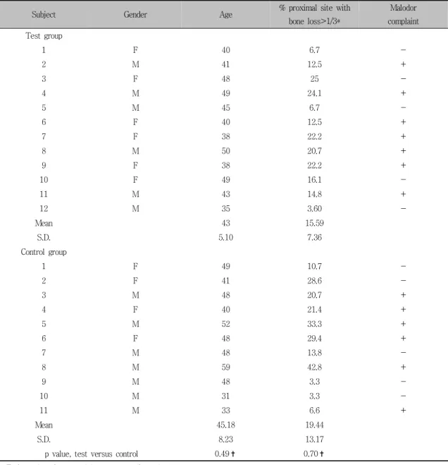
Descriptive statistics and periodontal status of examined population at baseline are shown in Table 1. At base line, there are no statistically significant differences in age and percentage of proximal site with bone loss ≥ 1/3 between the control group and the test group (p>0.05).
Subject Gender Age % proximal site with bone loss>1/3*
Malodor complaint Test group
1 F 40 6.7 -
2 M 41 12.5 +
3 F 48 25 -
4 M 49 24.1 +
5 M 45 6.7 -
6 F 40 12.5 +
7 F 38 22.2 +
8 M 50 20.7 +
9 F 38 22.2 +
10 F 49 16.1 -
11 M 43 14.8 +
12 M 35 3.60 -
Mean 43 15.59
S.D. 5.10 7.36
Control group
1 F 49 10.7 -
2 F 41 28.6 -
3 M 48 20.7 +
4 F 40 21.4 +
5 M 52 33.3 +
6 F 48 29.4 +
7 M 48 13.8 -
8 M 59 42.8 +
9 M 48 3.3 -
10 M 31 3.3 -
11 M 33 6.6 +
Mean 45.18 19.44
S.D. 8.23 13.17
p value, test versus control 0.49✝ 0.70✝
* Estimated on intra-oral long-cone radiographs
✝ Not significant by independent t-test (p>0.05)
Table 1. Descriptive statistics and periodontal status of examined population at baseline
2. The changes of VSCs level and Ratio of anaerobic bacterial loads to total bac- terial load at baseline and 4 weeks later
In both groups, the changes in VSCs level are depicted in Figure 1. VSCs levels of all participants reduced at 4 weeks later without
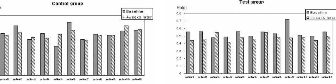
exception. But the test group have shown the more reduction. The number of CFU after aero- bic and anaerobic culturing decreased in both groups. But, the statistical differences were not significant. With regards to the ratio of anae- robic bacterial loads to total bacterial loads, more reduction have been detected in the test
Figure 1. Changes in VSCs values (expressed as parts per billion, ppb) of each group. VSCs levels of all participant reduced at 4 weeks later without exception. But the more reduction could be detected in test group.
Figure 2. Changes in the ratio (anaerobic CFU/total CFU) of each group. More reduction have been detected in the test group.
group (Figure 2).
In table 2, the changes of VSCs level and bacterial loads per treatment group were presented. There are no statistically significant difference of mean of VSCs levels between two groups at baseline (p=0.166, Wilcoxon rank sum test). After 4 weeks, the VSCs levels are sig- nificantly decreased in both groups (control:
p=0.003, test: p=0.002, Wilcoxon signed rank test). In comparison of two groups, the test group showed significantly greater reduction in VSCs levels (p=0.001, Wilcoxon rank sum test).
In the number CFU of aerobic and anaerobic culture, there are no statistically significant changes (p>0.05, Wilcoxon signed rank test).
But, the ratio of anaerobic bacterial loads to total bacterial loads were significantly reduced
at 4 weeks later in the test group (p=0.010, Wilcoxon signed rank test). The reduction of VSCs level(p=0.001) and the ratio of anaerobic CFU(p=0.049) were significantly greater in the test group (Wilcoxon rank sum test).
3. Correlation between VSCs level and the ratio of anaerobic bacterial loads to total bacterial loads
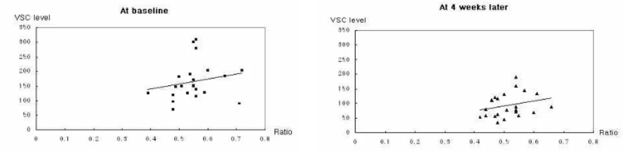
The correlation of VSCs levels and the ratio of anaerobic bacterial loads to total bacterial loads were evaluated at baseline and 4 weeks later (Figure 3). As shown in scatter plots, the correlation were low at each time (p>0.05, by Spearman correlation coefficient).
Figure 3. Scatter plots of correlation between VSCs level and ratio of anaerobic bacterial loads to total bacterial loads(anaerobic CFU + anaerobic CFU) at baseline and 4 weeks later. The correlation was low(at baseline: p=0.090, at 4 weeks later: p=0.149 by Spearman correlation coefficient)
Baseline 4-weeks later
Test Control Test Control
VSCs level
180.83±58.55 148.45±65.99 78.33±30.43 108.55±45.26
*
✝ ✝
254.17±95.24 232.82±124.62 146.75±94.60 196.27±77.22
CFU /ml
Aerobic
292.42±104.74 297.18±166.02 135.17±80.97 248.91±122.86
Anaerobic
0.54±0.06 0.56±0.09 0.48±0.04 0.55±0.05
Ratio
*
✝
* statistically significant difference between test and control group at 4 weeks later, tested by Wilcoxon signed rank test (VSCs level: p=0.085, Ratio: p=0.012).
✝ statistically significant change between baseline and 4-weeks later, tested by Wilcoxon rank sum test (p<0.05).
Table 2. Means of VSCs level and anaerobic/aerobic ratio in saliva at the baseline and 4 weeks later.
IV. DISCUSSION
The relationship between halitosis and perio- dontal disease has been an interesting subject.
There is also general agreement that the VSCs content of exhaled mouth air and the concen- tration of VSCs precursors increases with the severity of periodontal disease17,18). Perry et al.19) said volatile sulphide compounds (VSCs) is not only the principal components of breath malodor, but also have toxicity direct to gin-
gival epithelium and accelerate gingivitis and periodontitis. Thus, the objective evaluation and treatment of breath malodor have im- portant meaning in periodontal treatment.
Two treatment approaches can be used to re- duce breath malodor: debriding the tongue and tooth surfaces by physical means and reducing the bacterial loads by chemical agents. In chemical agents, the chlorhexidine rinses has been proven efficacy in the treatment of oral malodor by reducing bacterial load14,21). Bosy et
al.15) report that large and significant reduc- tions in VSCs levels, organoleptic scores, and positive BANA test scores were measured fol- lowing a one week with chlorhexidine rinse.
But, the long term use of chlorhexidine has some side effects, and is not recommended.
The our study aims to examine the impact of full-mouth nonsurgical periodontal therapy on breath malodor by measuring VSCs level and microbial load of saliva. The observation that the VSCs levels were significantly reduced in both groups emphasizes the impact of the oral hygiene care including interdental brushing and tongue cleansing on breath malodor. This im- provement in VSCs level agree with the pre- vious observation of Tonzeitich et al.22). However, the nonsurgical periodontal treatment resulted in a dramatic improvement in the VSCs levels. This observation also coincides with other studies23). They explained the contribution of periodontitis to malodor: the shift towards a more gram-negative microbiota which produces hydrogen sulfide and methyl mercaptan, the increase in crevicular fluid carrying metabolic products, the increased salivary putrefaction due to a higher concentration of disintegrated epithelial cells and the bleeding tendency of periodontal tissues which provides essential substrates for odor production. Thus, for pa- tients with periodontitis, periodontal therapy can dramatically reduce bad breath.
The impact of full mouth disinfection on the microbial load was reported by Quirynen et al.13). In adult periodontitis group, the original full mouth disinfection always resulted in sig- nificant reductions in total CFU/ml. of perio- dontal pocket, tongue, buccal mucosa and saliva. In our study, although there was no
statistically significant difference, the number of CFU of saliva was reduced by the full mouth periodontal therapy and oral hygiene improvement. However, the ratio of anaerobic CFU to total CFU was statistically significant decreased in the test group.
The correlation between the ratio of micro- bial load of saliva and VSCs level was not clearly proven in this study. If the reduction of VSCs is not directly effected by the ratio of microbial load of saliva, what contributed to the reduction of VSCs levels in our study? We suspect that other compound factors, such as periodontal pocket depth reduction, gain of at- tachment level, and so on, seemed to have an effect on oral malodor.
Based on these observations, it is logical that breath malodor caused by oral bacteria will be more reduced by full-mouth nonsurgical pe- riodontal therapy with oral hygiene care than oral hygiene care only. This hypothesis was substantiated by the clinical outcome of the periodontal therapy as proven earlier. It was due to the faster and larger reduction in orga- noleptic ratings in patients who were treated with full-mouth disinfection within 24 hours12,24).
In this study, as oral hygiene care including tongue cleansing was controlled and reexplained on every weeks, we did not consider oral hy- gienic variables such as plaque index, tongue coatings and so on. Oral hygiene care including tongue brushing can be performed by patients themselves, but, periodontal treatment should be performed by dentist.
We can conclude that 1-stage nonsurgical periodontal therapy is effective in breath mal- odor treatment of moderate periodontitis
patient. Since we had small samples size and short term result, there were some limitation in this study. Further studies are needed for the better understanding of the effect of non- surgical periodontal therapy to the oral malodor and microbial loads of saliva.
V. CONCLUSION
The purpose of our study is to examine the impact of full-mouth nonsurgical periodontal therapy on breath malodor by measuring VSCs level and microbial load of saliva. In our study, the VSCs levels were significantly reduced in both groups, but the greater reduction could be detected in the test group. On the other hand, the ratio of anaerobic CFU to total CFU was statistically significant decreased only in the test group. However, the correlation between the ratio of microbial load of saliva and VSCs was not clearly proven in this study. Thus, we have concluded that the 1-stage nonsurgical periodontal therapy results in more significant reduction in VSCs level more than oral hygiene care only does. And we can conclude that 1-stage nonsurgical periodontal therapy is ef- fective in breath malodor treatment of moder- ate periodontitis patient.
VI. REFERENCES
1. van Steenberghe D. Breath malodor.
Current Opin Periodontol 1997;4:137-143.
2. Tonzetich J. Production and origin of oral malodor: A review of mechanisms and methods of analysis. J Periodontol 1977;48:
13-20.
3. Kostelc JG, Preti G, Zelson PR, Brauner L,
Baehni P. Oral odors in early experimental gingivitis. J Periodont Res 1984;19:303 -312.
4. Attia EL, Marshall KG. Halitosis. Can Med Assoc J 1982;126:1281-1285.
5. Rosenberg M, Septon I, Eli I, et al.
Halitosis measurement by an industrial sulphide monitor. J Periodontol 1992;62:487 -489.
6. Rosenberg M, McCulloch CA. Measurement of oral malodor: Current methods and future prospects. J Periodontol 1992;63:776-782.
7. Delanghe G, Ghyselen J, Bollen C, et al.
An inventory of patients' response to treatment at a multidisciplinary breath odor clinic. Quintessence Int 1999;30:307-310.
8. Persson S, Edlund MB, Claesson R, Carlsson J. The capacity of subgingival microbiota to produce volatile sulfur com- pounds in human serum. Oral Microbiol Immunol 1989;4:169-172.
9. Greenman J, Duffield J, Spencer P. Study on the organoleptic intensity scale for measuring oral malodor. J Dent Res 2004;83:81-85.
10. Paryavi-Gholami F, Manah GE, Turge BF.
Oral malodor in children and volatile sulfur compound-producing bacteria in saliva:
preliminary microbiological investigation.
Pediatr Dent 1999;21:320-324.
11. El-Halabi, Minah GE, Turng B-F, Zhang M.: Correlation Between Volatile Sulfur Levels and Odorigenic Bacteria in Saliva. J Dent Res 1999;78(Spec. Issue):290, ab- stract#1478
12. Quirynen M, Bollen CM, Vandekerckhove BNA, et al. Full-versus partial-mouth dis- infection in the treatment of periodontal infections. short-term clinical and micro-
biological observations. J Dent Res 1995;74:1459-1467.
13. Quirynen M, Mongardini C, Pauwels M, et al. One Stage full- vs. partial mouth dis- infection in the treatment of patients with chronic adult or early onset periodontitis.
Part II: Long-term impact on microbial load. J Periodontol 1999;70:646-656.
14. Jones CG. Chlorhexidine: Is it still the gold standard? Periodontol 2000 1997;1555-1562.
15. Flotra L, Gjermo P, Rolla G, Waehaug J.
Side effects of chlorhexidine mouth washes.
Scand J Dent Res 1971;79:119-125.
16. Syes SA, Loesche WJ. Survival of human dental plaque flora in various transport media. Appl Microbiol 1972;24:120-127.
17. Tonzeitich J. Oral malodor: An indicator of health status and oral cleanliness. Int Dent J 1978;28:309-319.
18. Yaegaki K, Sanada K. Periodontal disease and precursors of oral malodorous component.
J Dent Health 1989;39:733-741.
19. Ratcliff PA, Johnson PW. The relationship between oral malodor, gingivitis, and
periodontitis. A review. J Periodontol 1999;70:
485-489.
20. Loesche WJ, Kazor C. Microbiology and treatment of halitosis. Periodontol 2000 2002;28:256-279.
21. Bosy A, Kulkarni GV, Rosenberg M, McCulloch CA. Relationship of oral malodor to periodontitis: evidence of independence in discrete subpopulations. J Periodontol 1994;65:37-46.
22. Tonzeitich J, Ng SK Reduction of malodor by oral cleansing procedures. Oral Surg Oral Med Oral Pathol 1976;42:172-181.
23. Quirynen M, Mongardini C, van Steenberghe D. The effect of a 1-stage Full-mouth Disinfection on oral malodor and microbial colonization of the tongue in periodontitis patients. A pilot study. J Periodontol 1998;69:374-382.
24. Bollen CML, Mongardini C, Papaioannou W, Quirynen M. The effect of a full-mouth disinfection on different intra-oral niches.
Clinical and microbiological observations. J Clin Periodontol 1998;25:56-66.
- 국문초록 -
구취와 타액내의 미생물 수에 대한 비외과적 치주치료의 영향
김명진, 김성조, 최점일, 이주연
부산대학교 치과대학 치주과학교실연구 목적 : 구취의 주된 화학적 요소는 휘발성 황 화합물 (VSCs)들이다. 이들은 치태, 설태, 타액과 혈액, 상피세포의 혐기성 그람 음성 세균에 의한 단백분해 변성으로 만들어 진다. 또한 구취는 치주질환과 타액내의 세균의 증가를 야기하는 불량한 구강 위생상태에서 기인한다고 보고되고 있다. 이 연구의 목적은 VSCs를 측정 함으로 구취에 대한 비외과적 치주 치료의 영향을 평가하고, 타액내의 세균 수를 측정함으로써 VSCs와의 상관 관계를 살펴보고자 하는 것이다.
연구 대상 및 방법 : 중등도의 만성치주염을 가지는 23명의 참가자들이 대조군과 실험군으로 분류되었다. 대 조군은 구강위생교육 (치간청결기구와 혀세정 포함)만을 시행하였고 실험군은 구강위생교육과 함께 24시간 내 에 전악 치석제거술과 치근활택술을 시행하였다. 치료 전과 치료 4주 후에 VSCs 농도를 Halimeter®(Interscan Corp., CA, USA)를 사용하여 측정하였고 타액 시료는 혐기성과 호기성 조건에서 배양되었다.
결과 : 4주 후에 두 군 모두에서 휘발성 황화합물의 농도가 유의하게 감소하였고, 실험군에서의 감소량이 통 계적으로 유의하게 더 크게 나타났다. 타액 내 세균의 배양에서 총세균수에 대한 혐기성 세균의 비율이 실험군 에서만 통계적으로 유의하게 감소하였다. 그러나 세균의 비율과 휘발성 황화합물의 농도와의 상관관계는 낮게 나타났다. 즉 구취의 감소는 타액 요인 보다는 치주치료에 의한 다른 복합적인 인자의 작용에 의한 것으로 추 론할 수 있었다.
결론 : 본 연구는 중등도의 치주염환자에서 구강위생교육만으로도 구취의 주요 화학적 요소인 휘발성 황화 합물의 농도를 감소시킬 수 있으나 비외과적 치주치료를 동반하였을 때 더욱 큰 효과를 얻을 수 있는 것으로 해석된다. 그러나 실험 참가자의 수가 적었고 장기간의 분석이 이루어지지 않았으므로 앞으로 더 많은 연구가
구취에 대한 치주치료의 영향을 이해하기 위해서 필요할 것으로 사료된다.2)
주요어 : volatile sulphide compounds, moderate periodontitis, nonsurgical periodontal therapy, microbial load of saliva