大韓放射線훌훌學會훌훌 Vol. XlX. No. 3. 1983
- Abstract -
춰1 장암의 내시경적 역행성 담 훼판 조영술소켠
광주 기독병원 방사선과
전 현 우·박 병 란 · 김 병 근 광주 기독병원 내과
박 홍 배
Endoscopic Retrograde Cholangiopancreatography(ERCP)
in Pancreatic Cancer
Hyun Woo Chun, M.D., Byoung Lan Park, M.D., and Byoung Geun Kim, M.D.
Department of Radiology
,
Kwangju Christian HospitalHong Bae Park, M.D.
Department of Internal Medicine, Kwangju Christian Hospital
Endoscopic Retrograde Cholangiopancreatography( E RCP) is one of the important diagnostic methods for pancreatic cancer. I t has an essential role in the early detection of pancreatic cancer.
The ERCP findings of 35 cases of pathologically proven pancreatic cancer during the period of five and a half years from April 1977 through November 1982 at Kwangju .Christian Hospital were studied. The results were as follows:
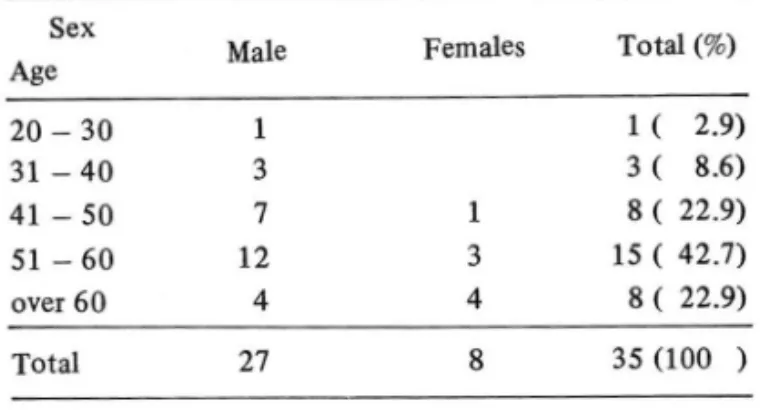
1. The ratio of male of female was 3.4: 1, and most of the patients were over 40 years of age (88.5%).
2. The locations of the lesions were the head in 28 cases (80%), the body in 7 cases (20%), and the tail in 3 cases (8.6%).
3. The ERCP findings of pancreati,c cancers were as follows;
1) Encasement or obstruction of common bile duct
,
1 g cases (51.4%).2) Obstruction of pancreatic duct, 16 cases (45.7%).
3) I rregular stenosis of pancreatic duct, 8 cases (22.3%).
4) Double duct sign, 7 cases (20%).
5) Diffuse narrowing of pancreatic duct, 2 cases (5.7%).
1. 서 토:
내시경적 역햇성 닦 쉐관 조영숭 (Endoscopic Ret ro.
grade Ch이angio Pancreatography: 이하 E.R.C.P 로 이 논문은 1983 년 8월 30 일에 채택되었음.
lI!ã함) 은 1968 년 Mc-Ctme등이 처음으로 섭이지장 유두 부에 내시경적 삽관을 시행하여 춰1 관조영에 성공한 이래 큰 성과를 옹리고 있다.
웨장암이나 만성 춰l장염과 갇은 만성웨장 질환의 진단 은 임상적으로 힘든 분야의 하나이며 특히 조기진단이 거의 불가능하였는데 E.R.C.P를 실시하므로 퀘 장암의
- 575-
진만에 많은 발전을 이룩하게 되었다.
최근에는 웨장암의 진단 목적으로 주업압을 증가시켜 웨 장의 본관 분지는 물론 세분지 및 실질을 조영하는 궤 설질 조영 볍 (Endoscopic Ret rograde Parenchyrnograp'
hy of the Pancreas : 이하 E.R.P.P. 로 略함) 은 춰1 장암의 침윤 범위를 영확하게 나타내서 암의 진행범위 및 절제 가능성 여부의 판정에 효과적인 방볍으로써 취l 장암 진단에 도웅을 주고 있다
U.
대상 및I) 검사 대상
t:H~내 는; t=I
1977 년 4월부터 1982 년 11월까지 광주기독병원에서 담 웨관계 질환을 의심하여 E.R.C.P. 및 E.R.P.P ,를 시행한 후 개복수술과 생겸소견으로 확진된 퀘장암 35 例에 대한 방사선학적 소견을 중심으로 고창하였다.
2) 검사 방법
시술전에는 공복으로 Atropine 0.5 mg과 Demer이 50 mg 으후 전처치하였고 위내 유포성 정액 제거제인 Gascon 2 teaspoon 을 주고 Xylocaine 액 분무로 인 후부 를 국소 마취시키고 내시경을 투엽한 후 Buscopan 으로 심 이 지 조l을 저 긴 장성 으로 만든후에 내 시 경 o!ympus
]f -B2을 사용하여 삽관을 시행한 후 수용성 조영제를 주입하고 투시하에 처l 위를 바꾸어가며 촬영을 설시하였 다 E.R.P.P. 는 E.R.C.P. 와 같은 조작 과정을 통 하여 상관후 투시하여 조영제를 약 3 kgjcm2의 압력으 로 주입시 웨관의 본관 세분지 및 실칠이 조영되는 것을 촬영하였다.
m.
성 적총 35 例의 춰1 장암 환자의 남·녀비는 3.4 : 1 이었고 연 령별 분포는 40세 이후가 88.5 %를 차지하고 병력기 간은 첫 증상이 발생하여 병원에 요기까지 1 개월-6개
월간이 85.7 %를 차지하였고 주요한 임상증상은 체중
감소 (85.7 %). 소화불량 (80 %). 황딸 (70 %). 전신쇠 약 (80 %). 동통 (74.3 %) 등이 있다 (Table
r.
ll.m).간 기능검사 소견으로는 71.4 %에서 혈통 Alkaline Phosphatase의 상송을. 77.1 %에서 혈청 Bilirubin의 상승을, 68.6 %에서 SGOT상승을, 65.7 %에서 SGPT 의 상송을 볼 수 있었다 (Table
N) .
침범부위를 보면 웨장 두부를 침벙한 것이 28 例 (80
Table I. Age and Sex distribution of the patients.
Sex Tota1 (%)
Age Male Fema1es
20 - 30 1 1 ( 2.9)
31 - 40 3 3 ( 8.6)
41 - 50 7 1 8 ( 22.9)
51 - 60 12 3 15 ( 42.7)
over 60 4 4 8 ( 22.9)
Total 27 8 35 (100 )
Table 11. Duration of Symptoms.
Duration Within 1 month 1 - 6 months 6 - 12 months over 12 months Total
No. of patients (%) 14 ( 40 ) 16 ( 45.7) 3 ( 8.6) 2 ( 5.7) 35 (100 )
Table III. Major symptoms.
Symptoms No. of patients (%)
Weight loss 30 (85.7)
Indigestion 28 (80.0)
General weakness Abd.ominal pain Jaundice
28 (80.0) 28 (74.3) 24 (70.0)
Table IV. Abnormal Lab. findings.
Elevated Bilirubin Elevated Alk P’tase Elevated SGOT Elevated SGPT
No. of patients (%) 27 (77.1) 25 (71.4) 24 (68.6) 23 (65.7)
%), 체부플 침범한 것이 7 例 (20 %). 마부를 침범한 것이 3 例 (8.6%)이었다 (Table V) .
궤 장암의 E.R.C.P. 소견을 보면 (Table
V
J.)1) 총 담관의 협 착 또는 폐쇄 (EnCasement or obstr- uction of Common bi le duct) 가 18 例 (51.4 %) 로 가
Location Head Body Tail
Table V. Location of the lesions.
No. of patients (%) 28 (80.0)
7 (20.0) 3 ( 8.6)
-576-
Tab1e VI. Findings of ERCP.
Findings No. of patients (%)
Encasement or obstruction ofCBD 18 (51.4) Obstruction of pancreatic duct 16 (45.7) Irregu1ar stenosis of pancreatic duct 8 (22.3)
Doub1e duct sign 7 (20.0)
Difωse narrowing of pancreatic duct 2 ( 5.7)
장 많았다 (Fig. l).
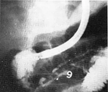
2) 춰1 관의 폐쇄 (obstruction of Pancreatic duct) 가 16 例(45.7 %) (Fig.2).
3) 웨관의 볼규칙한 협소화 (Stenosis of Pancreatic duct )가 8 例 (22.8%) (Fig.3).
4) 웨관의 협착 또는 폐쇄에 총 담관의 협착이 동반되 는 Double duct sign이 7 例 (20%) (Fig.lJ.
5) 혜관의 전반적인 협소화 (diffuse narrowing of Pa.
ncreatic duct ) 가 2 例 (5.7%) 이 었다 (Fig .4).
Fig. 1. Double duct sign.
Fig. 3. Short irregular stenosis of pancreatic duct at head.
Fig. 4. Diffuse narrowing of pancreatic duct.
N.
고 안웨장악성 종양의 년령별 분포에서 평균 60-65 세이 며 1) 84.4 %가 40 - 50 세에서 분포를 보고하고 있는 데 2) 저자의 경우는 40-50 세에서 65.7 %로 가장 많 았다.
성별비는 남·여가 1.6 : 1 석 2 : 1-7 : 1 4 , 5) 로 보고 되고 있다. 저자의 경우는 남여가 3.4: 1 정도였다.
병력기간 즉 첫 증상이 나타나서 병원에 오기까지 평 균 6-7개월, 75 %가 6 개월 이내로 보고 6) 되고 있 S며 저자의 경우도 6 개월 이내가 85.7 %로 대부분이 었다. 임상 증상에서 체중감소 (92 %), 소화불량 (82 o/ol Fig. 2. Abrupt obstruction of pancreatic duct at tail. 황달 (82 %), 통통 (72 %), 전산쇄 약 (64 %) 로 보고되
η
ζ 니
고 있으나 7) 저자의 경우 체중감소 (85.7%), 소화불량 (80 %), 전신쇠약 (80 %), 복통(74.3%) , 황달 ( 70
%) 의 순으로 나타났다.
검사소견으로 Gu며onsson등8) 의 예에서는 혈청 bil- irubin 치 가 증가한 경 우가 55 %, Alkaline Phosphatase 증가가 82 %, sGOT증가는 64 %였으며 Fit zgerald등
9) 의 예에서는 Alkaline Phospha tase 의 증가가 85 %
sGOT 의 증가가 63 %에서 보였는데, 저자의 경우 bil- irubin 증가는 77.1 %, AIKaline Phosphatase 증가는 71.4 %, sGOT 증가는 68_6 %, sGOT의 증가는 65.7
%에서 나타났다.
Goswits 10) 은 웨 장 두부에 서 (60~70%) , 체부 (20
~30%) , 미부에서는(1 0 %)를 나타내고, 저자의 경우 는 쉐장 두부에 (80 %) , 체부에 (20 %), 미부에 ( 8" 6
%) 의 비 슷한 발생 빈도를 나타냈다.
퀘장암의 E.R.C.P. 의 소견을 챔本 11)은 @ 폐쇄형 CObstructive Type) @협 착형 ( S t enosing Type) @경 화 협소형 (narrowing Type) CV이상 분지형 ( abnormal
branching Type) 으로 구분했고 궤 장암 37 例를 이에따 라 분류할 때 폐쇄형이 26 例로써 가장 많았고 협착성 이 10 例, 경화 협소형이 l 例였다고 하였다 12) 반면 박등13)의 연구에서는 쉐장암 16例를 분류해 보면 폐 쇄형 5 例, 협착형 7例, 경화 협소형 1 例, 쉐 장암에 의해 춰1 장관이 보이지 않는 경우가 3 例였다.
궤장암의 80 %는 웨관의 상피세포에서 발생하므로 가 장 흔히 나타내는소견은
@당관의 결손, 협착, 폐쇄등이며 쥐꼬리 모양의 협착
。1 걷~;<l
--, o.
@종%에 의한 웨관, 담관의 전위.
@가장 중요한 웨관의 협착, 폐쇄가 있으면서 총담관
장 두부암 또는 총담관 전이에 의한 것으로 생각된다.
2) 궤관의 폐쇄 (obstruction of Pancreatic duct) 는 16 例 (45.7 %) .
3) 웨관의 불규칙한 협소화 (Stenosis of Pancreatic duct) 가 8 例 (22.8 %) .
4)해관의 혐착 또는 폐쇄에 총담관의 협착이 동반되 는 Double duct si맑은 7 例(20 %) •
5) 웨관의 전반적인 협소화 (며 ffuse narrowing of Pa- ncreatic duct) 가 2 例(5.7 %) 의 소견을 보였다-
취1 장암의 진단에 있어 E.R.C.P. 의 진단율은 Free- ny등 16) 삼입된 경우에 95~97 %인 반면 CT 는 83
~88 %, PTC 의 경 우 78~83 %의 진단율, MOSSI7) 등 은 E.R.C.P.는 88
%
CT 가 76 %의 진단율,반면Hatifield 18)등은 26 例의 퀘장안에서 E.R.C.P. 의 진 단율이 65 %, 세포진 양성융이 54 %이 었으며 종합 진단 율은 92 %였다고 함을 볼 때 웨장임의 진단에 E.R.C.P.
는 중요한 진단 방법이 되며 최근에 웨섣질 조영 법 (E.R.P.P.)의 발전으로 암의 진행 범위 및 절제가능 성 여부의 유무판정에 효과적인 방법으로 기대되고 있다 19,æ,21,22,23) 결국 E.R.C.P. 는 궤장암의 중요한 진 단겸사 방법의 하나이다.
V. 결 론
저자들은 1977 년 4월부터 1982 년 11월까지 광주 기독병원에서 개복수술과 생겸결과로 확정된 35 역의 춰1 장암 환자의 내시경적 역행성 담, 웨관 조영술의 소견을 관참하여 다음과 같은 결론음 내 렸다.
1 • 남녀의 비율은 3.4 : 1 이고 연령은 40세 이삭이 88.5 %를 차지했다.
에 협착이 동반되는 double duct sign. 2 . 발생장소는웨장두부에 80 %, 체부에 20 %,미부
@춰l 장 실질과 춰1관 분지의 충만 결손 (field ddect). 에 8.6 %로 발생하였다.
@ 종양 괴사에 의한 공동의 소견을 나타낸다
Freeny 15) 등에 의 하면 웨 장암 8 명 에 대 하여 총담관 의 협 착 또는 폐쇄 (Encasement or obstruction of co-
mmon Rile duct) 가 6 例(50 %) 였고 Double duct sign 이 4 例 (33.3%), 결정성 경화협소형 (nodular na. rrowing) 가 1 例(8.3 %), 쥐꼬리 모양의 협 착성 (rat- tailed Stenosing Type) 例 (8.3 %) 였음을 보고하였 다. 저자의 경우든 35명의 쉐장암 환자에서
E.
R.C.P‘ 소견을 보면 아래와 같다.1 ) 총 담관의 협 착 또는 폐쇄 (Encasement or obst- rution of Common bile duct) 는 18 例(51.4 %)로 춰l
3. 웨장암의 내시경적 역행성 담웨관 조영숭 소견을 분류해 보면
1) 총 담관 협 착 또는 폐쇄 (Encasement or obst r- uction of Common bile duct ) 기. 18 例 (51.4 %) 로 가 장많았다.
2) 쉐관의 폐쇄 (obstruction of pancreatic duct) 가 16 f깨 (45.7 %) .
3) 웨관의 불규칙한 협소화 ( Steno외 s of pancreat- ic duct) 가 8 例 (22.8 %) .
4) 궤관의 협착 또는 폐쇄에 총담관의 협착이 동반 되 는 Double duct sign 이 7 例(20 %).
- 578-
5) 춰1 관의 전반적인 협소화 (diffuse narrowing of Pancreatic duct) 가 2 例(5.7 %) 였다.
REFERENCE5
1. Banks PA, Janowitz H D: Carcinoma of the pancreas. practice of medicine 7:40.7978.
2. 김 권, 김수태 : 웨장질환의 임상적 고철,외과학회 지 17 : 825-834,1975.
3. 이창수, 박홍길, 낌수태 : 웨장 외분비선 악성종양 에 관한 임상적 고찬. 외과학회지 22 : 794-807,
1980
4. Moldow R
,
Connslly R: Epidermiology of pancreatic cancer in Connecticut. Gastroenterology 55:677-686, 7968. 7968.
5. 5almon PA: Carcinoma of the pancreas and extra- hepatic biliary system. 5urgery 60:554-565
,
7966.6. Berk, JE: Diagnosis of carcinoma of the pancreas.
Arch Intern Med 68:525, 7947.
7. Howard JM
,
Jordan GL JR: Cancer ofthe pancreas.Current problem cancer 2:7,7977.
8. Guidonsson B, Livstone EM, 5piro HM: Cancer of the pancreas: Diagnostic accuracy and survival statistics. Cancer42:2494-2506
,
7978.9. Fitzgerald P J. F ortner J G
,
Watson RC et al: The value of diagnostic aids in detecting pancreas cancer.Cancer 47;‘868, 7978.
10. Goswitz jT: Carcinoma of the pancreas. A comprise review of 773 cases emphasizing in adequency of our diagnostic technique. Ohio 5tate Med journal 57:7255,7967.
11. 홈本圭志, 光놈 夫等 : 內視鏡的 隊臨管造影 (EPC) lζ J: 7.> g¥1품 17) 該斷. Gastroenterological Endoscμ
py 19: 430, 1974
12. 최흥재 쉐장질환의 내시경 진단. 대한의학 협회지 21 : 754- 762,1978
13. 박충식, 박병란, 낌병근 등 :내시경적 역행성 담춰1 관의 조영술의 임 상적 및 방사선학적 고찰. 대한방 사션학회지 17 : 492 - 499, 19i1l
14. 한만청 :훼장질환의 방사션학적 진단,대한의학협회 지 21 : 763-768.1978
15. Freeny PC
,
Bilbao MK,
Katon RM: “Blind" evalua- tion of endoscopic retrograde cholangi’'oPancreato- graphy (ERCP) in the diagnosis of pancreatic carci- noma: The “'double duct" and other signs. Radio- logy 734:347-352\ 7980.16. Freeny PC, Ball TJ: Rapid diagnosis of pancreatic carcinoma. Radiology 727:627-633
,
7978.17. Moss AA, Federle M, 5hapiro HA et al: The com- bined use of computed tomography and endoscopic retrograde cholangiopancreatography in the assess- ment of suspected pancreatic neoplasm: A blind
c/inical evaluation. Radiology 734: 754-773
,
7980 18. Hatifield ARW,
5mith A,
Wilkins R et al: Assessmentof endoscopic retrogradecholangiopancreatography and pure pancreatic juice cytology in patients with pancreaticdisease. Gut 77:74.7976.
19. 박홍배 혜장암의 내시경적 역행성 담궤관 조영술 및 춰1 실질 조영법의 비교소견. 대한 소화기영 학회 지 11 : 74, 1979
20. 최영청, 조정봉, 박홍배 등 . 내시경적 역행성 담춰1 관 조영술 및 조영술 361 예. 대한 내과 학회지 23
: 1013-1019,1980
21. 정상셜, 송영택, 검희규 : 쉐장암과 심이지장 팽대 부 주변부암에 대한 영상적 고찰. 외과학회지 21 :
471-478,1979
22. 김인원, 이승노, 한만청 등 : 내시경적 역행성 담궤 관 조영슬에 관한 고찰. 대한방사선학회지 15 :427
-433, 1979
23. Yoshimato 5
,
Ohnishi R,
Dol S et al: Endoscoplc retrograde pancreatic Parenchymography'. Radio- logy 747:279-222,
7987.- 579 -